Glaucoma Recognition and Segmentation
Using Feed Forward Neural Network and
Optical physics
Vijayan T, Ashutosh Singh
Asst. Professor, School of Electronics Engineering, Bharath University, Chennai, Tamil Nadu, India
P.G. Student, School of Electronics Engineering, Bharath University, Chennai, Tamil Nadu, India
ABSTRACT: Glaucoma is the silent thief of sight, glaucoma has no warning and symptom it is the leading cause of blindness . Glaucoma is of various type but the most common is open- angle glaucoma slowly develop without any symptom, side vision is affected first and progress leads to central vision and then total loss of sight .This paper proposed disc and cup segmentation technique for glaucoma detection using feed forward network. In optic disc segmentation and optic cup segmentation , center surround statistics are used to classify each superpixel as disc or non disc. The segmented optic disc and optic cup are then used to compute the cup to disc ratio, this cup to disc is then compared with threshold value, this threshold value is already been calculated from healthy eye ,the retinal fundus image is put into the process the algorithm to calculate the CDR , calculating is not the end of experiment we should know the stage of glaucoma. The feed forward neural network is used to detect weather the glaucoma disease is in initial or final stage, or disease can be cured or not. The proposed segmentation method have be evaluated in a database of 20 images.
KEYWORDS: Glaucoma disease, Steps to calculate CDR, optic cup segmentation, optic disc segmentation.
I. INTRODUCTION
Glaucoma is a disease that causes damage to your eye's optic nerve and gets worse over time. It's often associated with a buildup of pressure inside the eye. Glaucoma tends to be inherited and may not show up until later in life. The increased pressure, called intraocular pressure, can damage the optic nerve, which transmits images to the brain. If damage to the optic nerve from high eye pressure continues, glaucoma will cause permanent loss of vision. Without treatment, glaucoma can cause total permanent blindness within a few years. Because most people with glaucoma have no early symptoms or pain from this increased pressure, it is important to see your eye doctor regularly so that glaucoma can be diagnosed and treated before long-term visual loss occur[1]. If you are over age 40 and have a family history of glaucoma, you should have a complete eye exam with an eye doctor every one to two years. If you have health problems such as diabetes or a family history of glaucoma or are at risk for other eye diseases, you may need to visit your eye doctor more frequently. In India 95% of patient are unaware of glaucoma disease until it has reached its advanced stage. Glaucoma usually occurs when pressure in your eye increases. This can happen when eye fluid isn't circulating normally in the front part of the eye. Normally, this fluid, called aqueous humor, flows out of the eye through a mesh-like channel. If this channel becomes blocked, fluid builds up, causing glaucoma. The direct cause of this blockage is unknown, but doctors do know that it can be inherited, meaning it is passed from parents to children. Less common causes of glaucoma include a blunt or chemical injury to the eye, severe eye infection, blockage of blood vessels in the eye, inflammatory conditions of the eye, and occasionally eye surgery to correct another condition. Glaucoma usually occurs in both eyes, but it may involve each eye to a different extent.
rendering it unsuitable for widespread screening. The third method assessment of the damage to the head of the optic nerve is the most reliable but requires a trained professional and is time-consuming, expensive and highly subjective. Glaucoma is characterized by a vertical elongation of the optic cup, a white area at the center of the optic nerve head, or optic disc. This elongation alters the cup-to-disc ratio (CDR) but does not normally affect vision[1]-[2]. The computerized technique developed to measures the CDR from two-dimensional images of the back of the eye. The technique uses an algorithm that divides the images into hundreds of segments called superpixels and classifies each segment as part of either the optic cup or the optic disc. The cup and disc measurements can then be used to compute the CDR.
Fig 1 Major structure of optic cup and optic disc. The region between cup and disc is neuroretinal rim.
II. STEPSTOCALCULATECDR
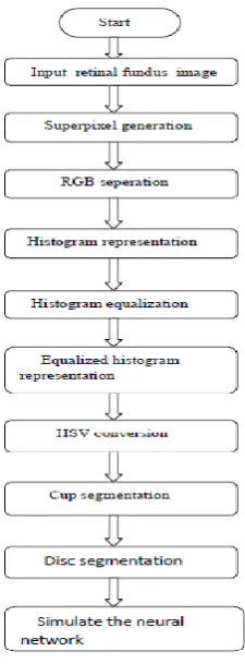
The segmentation of optic disc and optic cup done in a particular steps, those steps can be explain with a flow diagram .
The block diagram is explained as follows, each block has its own important in problem solving. A Retinal Fundus Image
Retinal fundus images are the images of interior parts of eye including the retina , optic disc, fundus. The fundus images are used by ophthalmologist for the detection of any eye disease[3]. In our work we use fundus images to detect the glaucoma disease and also the the stage of disease.
B Superpixel Generation
Superpixel are greater than normal pixel, these pixel have same color and contrast, we easily get the required information from image, superpixel have wide application in defence field, traffic, disease detection . In our proposed work superpixel is generated using SLIC algorithm.
C RGB Separation
RGB model is color model which is formed by the combination of three color called red, green, blue , these are the primary color by the combination of these color other color are formed ,when these color are mixed they formed a broad array of colors[4][6] . The main purpose of this model is to represent the image on digital media like computer, television. In propose work we increase the sharpness of image so that image can be easily analysis.
D Histogram Representation
Histogram represent as a graphical representation of tonal distribution of digital image , it plots number of pixel for each tonal value, by the histogram the viewer can easily get the tonal distribution at a glance. Histogram is widely used in digital camera, disease detection. Horizontal axis of histogram represented tonal distribution and vertical axis represent number of pixel.
E Histogram Equalization
This method is used to increase the global contrast of image especially when image having close contrast. This method widely used in x-ray technique for better view of bone structure, in thermal imaging also this technique is used. Histogram gives a realistic view to a dull image and help in extracting required information.
F HSV Conversion
In RGB separation we do not get the required brightness or contrast so HSV conversion is required, it help in giving different maturity to image, each superpixel is so clear to mark the cup and disc on fundus image.
G Cup Segmentation
For the detection of glaucoma cup segmentation is very important, cup is interior part of eye through which light travel inside the eye, to detect glaucoma the cup diameter is calculated to find out the CDR.
H Disc Segmentation
Disc is the white part of the eye, when glaucoma spread the diameter of disc start decreasing, with the help of disc diameter we can calculate CDR. Disc and cup segmentation are the important steps to detect glaucoma[1][2][3]. G Neural Network
These are interconnected neural which transfer information, after calculating CDR we feed the information to feed forward neural network that compare the CDR with threshold and open up a dialogue box to so stage of glaucoma .
III. OPTICDISCSEGMENTATION
Segmentation of disc is very important in for the diagnosis of glaucoma disease. Segmentation include finding of disc pixel this can be done by superpixel generation then histogram representation to extract useful data from image to calculate CDR. Some approaches have been proposed for disc segmentation, these two approaches are template based method and deformable model based method and some time circular Hough transformation is also used to for optic disc segmentation [1][2][4]. Both the template and deformable model based methods are based on edge characteristic. The above methods are sensitive to poor initilization, so to overcome the problem we propose superpixel classification based method .With the help of superpixel classification method we easily extract the useful data if the image is not clear.
A. SLIC Superpixel
are very slow and require large memory to work. SLIC is fast, memory efficient and to use with only one parameter i.e the number of desired superpixel k.
To produce roughly equally sized superpixels, on a regular grid space by S= √N/k. The centers are moved to seed locations corresponding to the lowest gradient position.
This paper focuses on automatic glaucoma using CDR from 2-D fundus images.
This concept can be used for detection of brain cancer , lung cancer and for blood vessel segmentation[7] .In this we compute center surround statistic from superpixels and unify them with histogram for disc and cup segmentation ,based on segmented cup and disc ,CDR is computed for glaucoma screening ,this CDR then compared with threshold value ,after comparison we easily detect the stage of glaucoma[17,18]
IV. OPTICCUPSEGMENTATION
Cup segmentation from 2-D fundus image without depth information is a tough task Pallor is the region to determine cup segmentation[12][13] .Like disc segmentation, fewer method have been purposed for cup segmentation and the most common are level set based approach and threshold based method these method are based on pallor, the main challenge in cup segmentation is to determine the cup boundary when the pallor are weak. Liu et al. proposed a method in which a potential set of pixels belonging to cup region is first derived based on the reference color obtained from a manually selected point. Next, an ellipse is fit to this set of pixels to estimate the cup boundary.
A Cup to Disc Ratio
CDR is an important indicator for glaucoma screening, after computing disc and cup , various feature can be computed ,we compare the threshold and CDR ,if CDR is greater than threshold ,it is glaucomatous, other-wise ,healthy.CDR is an important indicator for glaucoma screening computed as
CDR= VCD/VDD
Here, VCD –vertical cup diameter VDD- vertical disc diameter
B. Feed Forward Neural Network
Artificial neural networks are generally presented as systems of interconnected "neurons" which can compute values from inputs, and are capable of machine learning as well as pattern recognition thanks to their adaptive nature[8][9]. The output disc and cup segmentation are then pass to neural network , this along CDR and threshold value compute the result and show the stage of glaucoma ,if the eye is healthy it open a dialogue box and show eye is healthy and same for glaucomatous eye.
V. EXPERIMENTALRESULT
Our experiment uses 15 images from 15 different subject eye and IOP have been measured we use a SLIC algorithm to generate superpixel. The calculation of CDR is with good accuracy is quite tough, the glaucoma screening with segmented disc and cup depend on the accuracy of CDR there are some image related to our experiment these retinal fundus images are used for glaucoma screening.[19,20]
Fig 4 Input retinal fundus images
A Superpixel generation
The superpixel generation from above image using SLIC algorithm .
Fig 5 Superpixel generation from data base
B RGB Sepration
C Histogram representation
When superpixel is generated histogram representation is the next step, the histogram representation help in extracting useful data from image, in case there is any change in color or contrast it help in adjusting all such things.[21]
Fig.7 Red, green ,blue histogram representation
D Histogram Equalization
Fig 8 Equalization of red, green, blue channel
E Equalized Histogram Representation
Fig 9 Equalized histogram for RGB
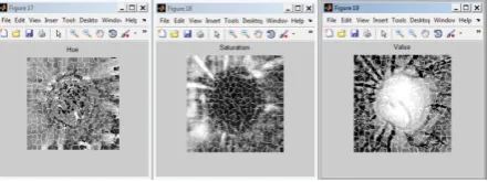
F HSV Conversion
Fig 10 Hue, saturation, value conversion
G Optic disc and optic cup segmentation
Fig 11 Cup and disc segmentation of retinal fundus image
H Output
The proposed work help in detecting glaucoma in large number of patient with 95% of accuracy, this method not only detect but also tell the stage of disease. There is the experimental result , the complete experiment is done with advanced metlab tool.
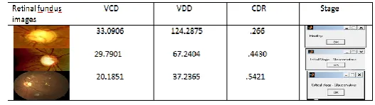
A Compare Output
Fig 13 Compare output of various image
VI. CONCLUSION
The proposed work glaucoma detection using feed forward neural network show how easily we can detect the disease and find out the stage of disease, this proposed work can be applicable to large quantity of images , center surrounding statistic helps in determination of cup to disc ratio , some time its hard to determine cup and disc segmentation due to the presence of blood vessels. The modelling approach helps to identify cup and disc segmentation . The method correctly identified 80% of glaucoma cases and 85% of normal cases. Although the proposed method is not error-free, the results indicated that it can be useful for analysis of the optic disc in glaucoma examination.
REFERENCES
1 Jun Cheng*, Jiang Liu, Yanwu Xu, Fengshou Yin, Damon Wing Kee Wong, Ngan-Meng Tan, Dacheng Tao,Ching-Yu Cheng, Tin Aung, and Tien Yin Wong superpixel classification based optic disc and optic cup segmentation for glaucoma screening , IEEE TRANSACTIONS ON MEDICAL IMAGING, VOL. 32, NO. 6, JUNE 2013
2 J. Liu, D. Wong, J. Lim, H. Li, N. Tan, and T. Wong, ―Argali—Anautomatic cup-to-disc ratio measurement system for glaucoma detectionand analysis framework,‖ in Proc. SPIE Med. Imag., 2009,
3 Dandona L, Dandona R, Mandal P, et al. Angle-closure glaucoma in an urban population in southern India. The Andhra Pradesh Eye Disease Study. Ophthalmology. 2000;107:1710–1716.
4 Vijaya L, George R, Arvind H, et al. Prevalence of angle-closure disease in a rural southern Indian population. Arch Ophthalmol. 2006;124:403–409.
5 Quigley HA. Number of people with glaucoma worldwide. Br J Ophthalmol. 1996;80:389–393.
6 Merickel, M.B., Wu, X., Sonka, M., Abramoff, M.D.: Optimal segmentation of the opticnerve head from stereo retinal images. In: Proc. SPIE, vol. 6143, pp. 61433B-1–61433B-8.SPIE, Bellingham (2006).
7 Hatanaka, Y., Nakagawa, T., Hayashi, Y., Fujita, A., Kakogawa, M., Kawase, K., Hara, T.,Fujita, H.: CAD scheme to detect hemorrhages and exudates in ocular fundus images.
8 Quigley, H., Broman, A.: The number of people with glaucoma worldwide in 2010 and2020. British J. Ophthalmology 90, 262–267 (2006). 9 Chrastek, R., Wolf, M., Donath, K., Niemann, H., Paulus, D., Hothorn, T., Lausen, B.,\Lammer, R., Mardin, C.Y., Michelson, G.: Automated segmentation of the optic nervehead for diagnosis of glaucoma. Medical Image Analysis 9, 297–314 (2005).
10 P. L. Rosin, D. Marshall, and J. E. Morgan, ―Multimodal retinal imaging: New strategies for the detection of glaucoma,‖ in Proc. ICIP.
11 R. Bock, J. Meier, G.Michelson, L. G. Nyl, and J. Hornegger, ―Classifyingglaucoma with image-based features from fundus photographs,‖in Proc. DAGM, 2007, pp. 355–364.
12 D. Wong, J. Liu, J. Lim, X. Jia, F. Yin, H. Li, and T. Wong, ―Level setbased automatic cup-to-disc ratio determination using retinal fundusimages in argali,‖ in Proc. EMBC, 2008, pp. 2266–2269.
13 G. Michelson, S. Wrntges, J. Hornegger, and B. Lausen, ―The papillaas screening parameter for early diagnosis of glaucoma,‖ DeutschesAerzteblatt Int., vol. 105, pp. 34–35, 2008.
14. Sharmila S., Jeyanthi Rebecca L., Saduzzaman M., "Biodegradation of domestic effluent using different solvent extracts of Murraya koenigii", Journal of Chemical and Pharmaceutical Research, ISSN : 0975 – 7384, 5(2) (2013) PP.279-282.
15. B. Fulkerson, A. Vedaldi, and S. Soatto. Class segmentation and objectlocalization with superpixel neighborhoods. In International Conferenceon Computer Vision (ICCV), 2009.
16. Beula Devamalar P.M., Thulasi Bai V., Srivatsa S.K., "Design and architecture of real time web-centric tele health diabetes diagnosis expert system", International Journal of Medical Engineering and Informatics, ISSN : 1755-0661, 1(3) (2009) PP.307-317.
17 Greg Mori. Guiding model search using segmentation. In IEEE International Conference on Computer Vision (ICCV), 2005.
18. Niranjan U., Subramanyam R.B.V., Khanaa V., "Developing a Web Recommendation System Based on Closed Sequential Patterns", Communications in Computer and Information Science, ISSN : 1865-0929, 101() (2010) PP.171-179.
20. Y. Boykov and M. Jolly. Interactive Graph Cuts for Optimal Boundary& Region Segmentation of Objects in N-D Images. In InternationalConference on Computer Vision (ICCV), 2001.
[22Anbuselvi S., Jeyanthi Rebecca L., Sathish Kumar M., Senthilvelan T., "GC-MS study of phytochemicals in black gram using two different organic manures", Journal of Chemical and Pharmaceutical Research, ISSN : 0975 – 7384, 4(2) (2012) PP. 1246-1250.
[23] D.Prakash and Dr. A.Mukunthan, A Theoretical Study of Internal Pressure And Free Volume for Single Molecule of a Sample Liquid, International Journal of Innovative Research in Science, Engineering and Technology, ISSN: 2319-8753,pp 7252-7257, Vol. 2, Issue 12, December 2013
[24] Dr. A. Mukunthan & Ms.S.Sudha, FTIR Spectroscopic Features of Blood Serum of Diseased and Healthy Subjects (Animals), International Journal of Innovative Research in Science, Engineering and Technology, ISSN: 2319-8753, pp 2035-2040 Vol. 2, Issue 6, June 2013
[25] Dr. A.Mukunthan, A Survey of Applications of Laser in Dermatology – Medical Physics, International Journal of Innovative Research in Science, Engineering and Technology, ISSN: 2319-8753 ,pp 33-36, Vol. 1, Issue 1, Nov 2012
[26] Dr. M. Ganesan, Mergers and Acquisitions, International Journal of Innovative Research in Science, Engineering and Technology, ISSN: 2319-8753,pp 9081-9085, Vol. 3, Issue 2, February 2014
[27] Dr.G.Brindha, A Study on Latest Management Governance Techniques in Indian Companies, International Journal of Innovative Research in Science, Engineering and Technology, ISSN: 2319-8753,pp 284-292, Vol. 2, Issue 1, January 2013
[28] D.Kalaivani, Mrs.M.Indirani & Dr.A.Mukunthan, A Theoretical Study of Primary Nucleation Kinetics of L-Histidine Bromide: Semi Organic Optical Single Crystal, International Journal of Innovative Research in Science, Engineering and Technology, ISSN: 2319-8753,pp 4192-4197, Vol. 2, Issue 9, September 2013