| WORMBOOK GENE AND GENOME REGULATORY MECHANISMS
Repressive Chromatin in
Caenorhabditis elegans
:
Establishment, Composition, and Function
Julie Ahringer*,†,1and Susan M. Gasser‡,§,1
*The Gurdon Institute and†Department of Genetics, University of Cambridge CB2 1QN, United Kingdom,‡Friedrich Miescher Institute for Biomedical Research (FMI), 4058 Basel, Switzerland, and§Faculty of Natural Sciences, University of Basel, 4056, Switzerland ORCID IDs: 0000-0002-7074-4051 (J.A.); 0000-0003-3610-9123 (S.M.G.)
ABSTRACT Chromatin is organized and compacted in the nucleus through the association of histones and other proteins, which together control genomic activity. Two broad types of chromatin can be distinguished: euchromatin, which is generally transcriptionally active, and heterochromatin, which is repressed. Here we examine the current state of our understanding of repressed chromatin in
Caenorhabditis elegans, focusing on roles of histone modifications associated with repression, such as methylation of histone H3 lysine
9 (H3K9me2/3) or the Polycomb Repressive Complex 2 (MES-2/3/6)-deposited modification H3K27me3, and on proteins that recognize
these modifications. Proteins involved in chromatin repression are important for development, and have demonstrated roles in nuclear
organization, repetitive element silencing, genome integrity, and the regulation of euchromatin. Additionally, chromatin factors
participate in repression with small RNA pathways. Recent findings shed light on heterochromatin function and regulation in
C. elegans, and should inform our understanding of repressed chromatin in other animals.
KEYWORDSC. elegans; chromatin; heterochromatin; histone methylation; H3K9me; H3K27me; WormBook
TABLE OF CONTENTS
Abstract 491
Histone Methyltransferases and Recognition of
Modifications 492
The C. elegans Genome and the Distribution of Heterochromatin 496
The Spatial Organization of Repetitive DNA 497
The Nuclear Lamina and Chromatin Association 498
Phenotypes of Chromatin Repressor Mutants 498
Histone H3K9 Methyltransferases 498
H3K9me Readers 499
Continued
Copyright © 2018 by the Genetics Society of America doi:https://doi.org/10.1534/genetics.117.300386
Manuscript received October 11, 2017; accepted for publication November 18, 2017 Available freely online through the author-supported open access option.
1Corresponding authors: The Gurdon Institute and Department of Genetics, University of Cambridge, Cambridge, United Kingdom CB2 1QN. E-mail: j.ahringer@gurdon.
CONTENTS,continued
CEC-4- and H3K9me-Mediated Anchoring at the
Nuclear Periphery 500
H3K9 Demethylases 501
Phenotypes Caused by the Loss of H3K9
Methyltransferases or H3K9me Readers 502
PRC2/H3K27me3 and Interactions with MES-4/
H3K36me3 504
PRC2 in Reprogramming and Maintenance of
Cell Fate 505
Chromatin Regulators and Small RNA Pathways 506
Perspectives 507
E
UKARYOTIC DNA is organized and compacted in the nucleus through its association with histones and non-histone proteins, forming a complex called chromatin. The N-terminal tails of all histones, as well as the C-terminal tails of histones H2A and H2B, are subject to post-translational mod-ifications that selectively impact many aspects of nuclear function. The most common histone modifications include methylation, acetylation, ubiquitination, sumoylation, and phosphorylation.Two broad classes of chromatin, euchromatin and hetero-chromatin, can be distinguished based on protein composi-tion, characteristic post-translational modifications on histones, and transcriptional activity. Euchromatin is either potentially or actively transcribed, and is enriched for RNA polymerase, histone tail acetylation, and trimethylation on histone H3 lysines 4 and 36 (H3K4me3 and H3K36me3). In contrast, heterochromatin tends to be transcriptionally repressed and is associated with histone methylations such as H3K9me3 or the Polycomb-deposited modification H3K27me3. The proteins that recognize these modified histones and the association of heterochromatin with the nuclear envelope help hold chromatin in a compact conformation, and promote the spread of the repressed chromatin state (Eskelandet al.2007; Towbinet al. 2010; Elgin and Reuter 2013; Simon and Kingston 2013; Wiles and Selker 2016). Despite its heritable nature, heterochromatin is nonetheless dynamically regulated. Moreover, its tendency to aggregate and create repressive subnuclear compartments means that it can also indirectly influence the organization of euchromatin and gene expression (Francastel et al. 2000; Sextonet al.2007).
Although there are many types of repressed chromatin, reflecting a range of modifications and ligands, heterochro-matin was traditionally split into two classes that largely reflect the properties of the underlying DNA. On one hand, constitutive heterochromatin covered regions of the genome that are repeat rich and gene poor, and that are kept in a silent state throughout cell division and cell differentiation by
H3K9me2/3 and its ligand, Heterochromatin Protein 1 (HP1) (Eissenberg and Elgin 2014; Saksouk et al.2015; Wanget al.2016). Facultative heterochromatin, on the other hand, encompasses genes that are potentially active, such as those with spatial, temporal, or other types of context-specific expression. Its hallmark is H3K27me3, which is deposited by Polycomb Repressive Complex 2 (PRC2), and which defines a pathway that maintains transcriptional repression (Wiles and Selker 2016).
Recentfindings suggest that these classical distinctions are inadequate to describe the complexity of heterochromatin types. For instance, chromatin immunoprecipitation (ChIP) analyses inCaenorhabditis eleganshave shown that much of the H3K9me3-marked chromatin coincides with H3K27me3 (Liuet al.2011). In mammals, H3K9me3 and H3K27me3 are negatively correlated when scored for coincidence on the same histone tail, yet some genomic regions carry both mod-ifications, as detected by ChIP. Additionally, cooperation be-tween H3K9 and H3K27 methylation in heterochromatin formation has been reported (Hawkins et al. 2010; Boros et al. 2014; Schwammle et al. 2016). Finally, H3K23me2 has been reported to coincide independently with K9 and K27 methylation on histone H3 tails in C. elegans (Vandamme et al.2015; Sidoli et al.2016), much like the trimethylation of H4K20 in mammals, which accumulates on both facultative and constitutive heterochromatin during cel-lular senescence (Nelson et al. 2016). Here, we focus on histone H3K9 and H3K27 methylations, as they are the best-understood heterochromatin marks, and are involved in genetically distinct but highly conserved pathways of tran-scriptional repression.
Histone Methyltransferases and Recognition of Modifications
contain a conserved catalytic domain called SET, which stems fromSu(var)3-9,Enhancer of zeste, andTrithorax, thefirst HMTs known to carry this domain (Tschierschet al.1994). The SET domain contains a S-adenosylmethionine (SAM)-binding site and a catalytic center (Yeates 2002). The C. elegansgenome encodes 38 SET domain-containing, putative HMTs (Andersen and Horvitz 2007). A loss-of-function mu-tant has been isolated for 30 of these, of whichfive are
es-sential for viability (Andersen and Horvitz 2007; Ni et al. 2012). The preferred substrates of most HMTs have not yet been identified inC. elegans, apart from:SET-25andMET-2/ SETDB1, which target H3K9 (Bessleret al.2010; Towbinet al. 2012); MES-2/EZH2, which modifies H3K27 (Bender et al. 2004);SET-1andSET-4, which modify H4K20 (Vielleet al. 2012); andMET-1andMES-4, which are responsible for H3K36 methylation (Bender et al. 2006; Furuhashi and Kelly 2010;
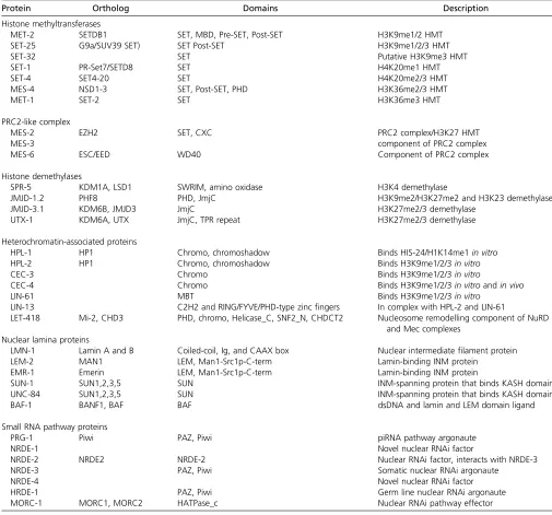
Table 1 Chromatin proteins discussed in this review
Protein Ortholog Domains Description
Histone methyltransferases
MET-2 SETDB1 SET, MBD, Pre-SET, Post-SET H3K9me1/2 HMT
SET-25 G9a/SUV39 SET) SET Post-SET H3K9me1/2/3 HMT
SET-32 SET Putative H3K9me3 HMT
SET-1 PR-Set7/SETD8 SET H4K20me1 HMT
SET-4 SET4-20 SET H4K20me2/3 HMT
MES-4 NSD1-3 SET, Post-SET, PHD H3K36me2/3 HMT
MET-1 SET-2 SET H3K36me3 HMT
PRC2-like complex
MES-2 EZH2 SET, CXC PRC2 complex/H3K27 HMT
MES-3 component of PRC2 complex
MES-6 ESC/EED WD40 Component of PRC2 complex
Histone demethylases
SPR-5 KDM1A, LSD1 SWRIM, amino oxidase H3K4 demethylase
JMJD-1.2 PHF8 PHD, JmjC H3K9me2/H3K27me2 and H3K23 demethylase
JMJD-3.1 KDM6B, JMJD3 JmjC H3K27me2/3 demethylase
UTX-1 KDM6A, UTX JmjC, TPR repeat H3K27me2/3 demethylase
Heterochromatin-associated proteins
HPL-1 HP1 Chromo, chromoshadow Binds HIS-24/H1K14me1in vitro
HPL-2 HP1 Chromo, chromoshadow Binds H3K9me1/2/3in vitro
CEC-3 Chromo Binds H3K9me1/2/3in vitro
CEC-4 Chromo Binds H3K9me1/2/3in vitroandin vivo
LIN-61 MBT Binds H3K9me1/2/3in vitro
LIN-13 C2H2 and RING/FYVE/PHD-type zincfingers In complex with HPL-2 and LIN-61
LET-418 Mi-2, CHD3 PHD, chromo, Helicase_C, SNF2_N, CHDCT2 Nucleosome remodelling component of NuRD and Mec complexes
Nuclear lamina proteins
LMN-1 Lamin A and B Coiled-coil, Ig, and CAAX box Nuclear intermediatefilament protein
LEM-2 MAN1 LEM, Man1-Src1p-C-term Lamin-binding INM protein
EMR-1 Emerin LEM, Man1-Src1p-C-term Lamin-binding INM protein
SUN-1 SUN1,2,3,5 SUN INM-spanning protein that binds KASH domain
UNC-84 SUN1,2,3,5 SUN INM-spanning protein that binds KASH domain
BAF-1 BANF1, BAF BAF dsDNA and lamin and LEM domain ligand
Small RNA pathway proteins
PRG-1 Piwi PAZ, Piwi piRNA pathway argonaute
NRDE-1 Novel nuclear RNAi factor
NRDE-2 NRDE2 NRDE-2 Nuclear RNAi factor, interacts with NRDE-3
NRDE-3 PAZ, Piwi Somatic nuclear RNAi argonaute
NRDE-4 Novel nuclear RNAi factor
HRDE-1 PAZ, Piwi Germ line nuclear RNAi argonaute
MORC-1 MORC1, MORC2 HATPase_c Nuclear RNAi pathway effector
TheC. elegansgenome contains 38 SET domain proteins, 6 amino oxidase-type putative histone demethylases, 14 jmjC domain proteins, 67 putative histone mark readers (bearing either a chromodomain, Tudor, MBT, PHD, or WD-40 domain), 27 argonaute domain proteins, and an as yet undetermined number of nuclear lamina-associated
proteins [for a more complete survey of nuclear envelope proteins see Dobrzynskaet al.(2016)]. See text for references and types of data supporting these definitions. In the
Rechtsteineret al.2010). One cannot exclude the possibility that these HMTs (summarized in Table 1 and Table 2) modify lysines in other proteins as well.
Histone modifications can directly alter nucleosome– nucleosome or nucleosome–DNA interactions by changing the charge of the highly basic histone tail or disrupting contact sites between DNA and the nucleosomal core par-ticle. Alternatively, specific histone modifications can create binding sites for proteins that specifically recognize a given modified amino acid. These“readers”of post-translational histone modifications can in turn alter the chromatin com-paction state, or recruit additional transcriptional regula-tors or chromatin-modifying enzymes. A growing list of structural motifs have been shown to recognize modified histones, the most common being Bromo, Chromo, Tudor, malignant brain tumour (MBT), plant homeodomain (PHD)PHD, WD40 repeat (40 amino acid terminating in Trp-Asp) 14-3-3, and BRCT (BRCA1 C Terminus) do-mains (Taverna et al. 2007). C. elegans has 67 proteins containing such domains, which are predicted to be readers of histone modifications (Towbin et al.2012; Gonzalez-Sandovalet al.2015). Histone H3 tail methylations alone are known to be recognized by Chromo, MBT, PWWP (Pro-TrpTrp-Pro motif) or Tudor domains, as well as by special-ized WD40 repeat structures (Margueron et al.2009; Xu et al.2010; Khorasanizadeh 2011).
The C. elegans Genome and the Distribution of Heterochromatin
The 100-Mbp genome ofC. elegans is separated intofive autosomes and an X chromosome (C. elegansSequencing
Consortium 1998). Hermaphrodites are diploid for all six chromosomes, whereas males havefive pairs of autosomes and only a single X chromosome. The regulation and char-acteristics of the autosomal chromosomes differ from the X chromosome, in part due to dosage compensation in the soma, which represses gene expression approximately two-fold on the two hermaphrodite X chromosomes to match expression on the single male X. Here, we do not cover dosage compensation, but refer the reader to reviews of this subject (Meyer 2010; Stromeet al.2014).
Like most nematodes,C. eleganschromosomes are holo-centric (Albertson and Thomson 1982; Maddoxet al.2004). That is, centromere activity is distributed along chromo-somes at sites of incorporation of a variant histone H3 CENP-A instead of being focused in a single, heritable cen-tromeric site (Gassmannet al.2012). The distal arms and central regions of thefive autosomes have different charac-teristics. Most meiotic recombination occurs in the distal arm regions, where genes are on average longer and have larger introns. Genes in central regions are generally more highly expressed and show higher evolutionary conservation (C.
elegans Sequencing Consortium 1998). These differences
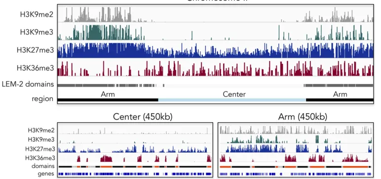
are reflected in the distributions of chromatin modifications. For example, average levels of modifications associated with
gene activity are higher in central regions (Liuet al.2011). Di- and trimethylation of H3K9, hallmarks of constitutive het-erochromatin, are both predominantly found on distal chro-mosome arms, as is H3K9me1, although they have distinct distributions, reflecting distinct modes of recruitment of the relevant HMTs (Gu and Fire 2010; Liuet al.2011) (J. Padeken, P. Zeller, and S. M. Gasser, unpublished results; Figure 1). Nonetheless, the distal arms are not devoid of transcribed genes, and those found interspersed among heterochromatic domains carry the same histone modifications as actively tran-scribed genes in central chromosomal domains (Liu et al. 2011).
In contrast to constitutive heterochromatin, facultative het-erochromatin carries H3K27 trimethylation, the most abundant histone methylation mark as measured by mass spectrometry in C. elegans. H3K27me3 is found on 67% of histones in embryos (Vandamme et al.2015), and maps both to distal arms and central chromosome regions (Liuet al.2011; Figure 1).
Pro-filing early embryos and L3 larvae, levels were found to be higher on chromosome arms and higher on the X chromosome than on autosomes. On distal chromosome arms, H3K27me3 often colocalizes by ChIP sequencing with H3K9me3, as it does on large integrated arrays of transgenes, whereas in central regions of autosomes H3K27me3 is found without H3K9me3 (Meisteret al. 2010; Liuet al. 2011) (Figure 1). Consistent with a role in transcriptional repression, the genome-wide dis-tribution of H3K27me3 is anticorrelated with RNA levels and the presence of RNA polymerase (Liuet al.2011).
WhileC. eleganshas the repressive histone methyl marks and the ligands that are present in other organisms, it lacks 5-methyl cytosine on DNA and the 5meC-binding proteins that repress transcription in vertebrates. A very low level of adenine N(6)-methylation (6mA) has been reported on DNA inC. elegans(Greer et al.2015), yet given that 6mA is the
most abundant RNA modification, and no dedicated adenine methyltransferase for DNA has been identified, it is unclear whether 6mA-DNA has any physiological significance.
The Spatial Organization of Repetitive DNA
Roughly 20% of theC. elegansgenome is repetitive DNA (Hillier et al.2008; Hubleyet al.2016), including tandem repeats and sequences derived from DNA or RNA transposons. These repeti-tive elements are enriched in the distal arm regions, and are generally marked by nucleosomes bearing H3K9me2 and/or H3K9me3 (C. elegans Sequencing Consortium 1998; Gerstein et al.2010; Zelleret al.2016; McMurchyet al.2017). Among repetitive sequences, those derived from DNA transposons are particularly abundant (covering 12.6% of the genome). Some DNA transposons can be activated, yet only a small minority of elements encode full-length transposases (Bessereau 2006; Hubleyet al.2016). LTR-containing and LTR-free sequences de-rived from RNA transposons (retrotransposons) cover1% of the genome, but there are few full-length elements, and none appear to be active under wild-type conditions (Bessereau 2006; Hubley et al.2016).
In organisms with localized centromeres, pericentric do-mains contain large arrays of tandem satellite repeats, which can occupy up to 1 Mbp in vertebrates (Plohl et al. 2008). Difficulties in sequencing and mapping these regions unam-biguously have meant that the exact extent and composition of repetitive domains are unclear for most vertebrate genomes, yet pericentric satellite sequences are estimated to account for 5–10% of the human genome (Schueler and Sullivan 2006).
2016). This is reminiscent of the dispersed nature ofC. elegans centromeres. The shortness of the repeat clusters has allowed for high-quality sequencing of nearly allC. elegansrepetitive DNA (C.
elegansSequencingConsortium 1998; Hillieret al.2005).
Addi-tionally, 71% of the repeats are uniquely mappable with 50-bp single-end reads. Interestingly, several microsatellite families have been shown to be enriched in CENP-A, suggesting that they may be associated with centromere function (Subirana et al. 2015), although centromere function in worms is independent of H3K9 methylation (Towbin et al. 2012; Ho et al. 2014; Garrigueset al.2015; Zelleret al.2016).
The Nuclear Lamina and Chromatin Association
The mapping of chromatin regions associated with proteins of the nuclear lamina has shown that the central and distal arm regions ofC. eleganschromosomes have distinct distributions with respect to the nuclear envelope (Ikegami et al.2010; Towbinet al.2012; Gonzalez-Aguileraet al.2014).C. elegans expresses a single lamin protein (LMN-1), that forms a stable meshwork underlying the nucleoplasmic side of the nuclear envelope together with a number of well-characterized lamin-associated proteins (Dobrzynska et al. 2016). These lamin-associated factors include the LEM domain proteins
LEM-2(MAN1) andEMR-1(Emerin), the SUN domain
pro-teinsUNC-84andSUN-1, the accessory protein BAF, and four KASH domain proteins that form a bridge to the cytoskeleton (Bank and Gruenbaum 2011, see Table 1 for abbreviations). While this is undoubtedly a nonexhaustive list, this set of proteins and functions is conserved across animal species.
The genome-wide mapping profiles ofLEM-2,LMN-1, and
EMR-1 all show strong enrichment on distal chromosome
arms, indicating that these regions are associated with the nuclear envelope (Ikegamiet al.2010; Towbinet al.2012; Gonzalez-Aguilera et al. 2014; Gonzalez-Sandoval et al. 2015). A detailed analysis of LEM-2 binding showed that these domains are not continuous along the chromosome arms, but are interspersed with gaps that bear expressed genes (Ikegami et al. 2010), which are thought to extend inwards from the peripheral lamin- or LEM-2-associated do-mains. The LEM-2-associated domains are enriched for H3K9-methylated histones, and their perinuclear positioning is largely dependent on this mark, which is specifically rec-ognized by a nuclear envelope-associated chromodomain protein, CEC-4 (Towbin et al. 2012; Gonzalez-Sandoval et al.2015). The LEM-2-bound regions are not always tran-scriptionally silent, and those with gene expression are enriched for the HP1 homologHPL-2(Garrigueset al.2015).
Phenotypes of Chromatin Repressor Mutants
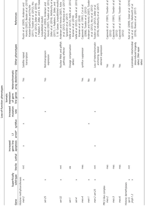
InC. elegans, the development of the vulva has proven to be a powerful system to identify regulators of cell fate, as it is dis-pensable for survival and defects are easily observable (Horvitz and Sternberg 1991). Many genes with presumed roles in chromatin repression, including those encoding proteins that
generate or bind methylated H3K9, were originally identified based on their synthetic multivulval (synMuv) phenotype (Fay and Yochem 2007). Single mutants of synMuv genes have a normal vulva, but double mutants between synMuv genes of different genetic classes develop extra vulvae, arguing for a partial functional redundancy between synMuv gene classes. The synMuv genes were found to encode proteins with a wide range of chromatin-modifying activities, including enzymes that deposit or remove methylation and acetylation on histone tails, ligands for the methylated residues, and nucleosome remodelers. Interestingly, ectopic vulval development in syn-Muv mutants was shown to be due to a failure to repress expression of a single gene,lin-3/EGF, in the epidermis (Cui et al.2006). Of note, most synMuv genetic interactions occur between genes that encode different biochemical activities (e.g., between an acetyltransferase and a deacetylase). This func-tional redundancy reflects the inherent complexity found in chromatin regulatory mechanisms, and argues for redundancy in repression mechanisms. Although synMuv genes were iden-tified based on their roles in the repression of vulval develop-ment, most are widely expressed, and the single mutants often show pleiotropic developmental defects and genetic interac-tions in nonvulval processes. Common phenotypes among them are impaired fertility, altered regulation of repetitive transgenes, L1 arrest at high temperature, and ectopic expres-sion of germ line genes in somatic tissues (Table 2, references therein). We refer readers to chapters on developmental roles of chromatin factors and the synMuv genes for more detailed information (Cui and Han 2007; Fay and Yochem 2007).
Histone H3K9 Methyltransferases
Over the years, a number ofC. elegansgenes have been pos-tulated to encode HMTs that target H3K9me [e.g., MET-2,
SET-9, SET-26, SET-25, and SET-32; (Bessler et al. 2010; Towbin et al. 2012; Greer et al. 2014; Ni et al. 2016; Kalinavaet al.2017)]. However, it has been shown recently that the vast majority of H3K9me1, me2, and me3 depend on two key enzymes:MET-2, which is able to deposit me1 and me2 on H3K9, andSET-25, the major, if not only, HMT that deposits H3K9me3 in somatic cells of larvae and embryos (Towbin et al. 2012). In a double knockout for these two SET domain genes, embryos and L1 stage larvae lacked all detectable H3K9me when analyzed by mass spectrometry (Towbinet al.2012; Garrigueset al.2015). Moreover, immu-nofluorescence (IF) of embryos, L2 larvae, and dissected go-nads of the double mutant showed no H3K9me signal (Towbin et al.2012; Hoet al.2014; Garrigueset al.2015; Zelleret al. 2016). Thus, if other H3K9-modifying HMTs exist in worms, either they are expressed under very select conditions or in very few cells, or else their activity requiresSET-25orMET-2. Based on the lower limit of detection by the mass spectroscopy method used, we estimate that,5% of embryonic histone H3K9 methylation is retained in aset-25 met-2double mutant.
provide a low level of H3K9me3 methylation in the germ line. Two recent studies found that set-32mutants are transge-nerationally sterile and had reduced H3K9me3 on nuclear RNA interference (RNAi) targets in young adults (Kalinava et al.2017; Spracklinet al.2017). However, at most genomic locations, H3K9me3 levels were as low inmet-2 set-25double as in met-2 set-25; set-32 triple mutants (Kalinava et al. 2017), confirming previous studies reporting loss of detect-able H3K9 methylation inmet-2 set-25mutants, both in go-nads and somatic cells (Towbinet al.2012; Garrigueset al. 2015; Zelleret al.2016).MES-2, the EZH2-like H3K27 meth-yltransferase, was also initially suggested to modify H3K9 in the germ line (Bessleret al.2010). However, the alterations detected were likely due to antibody cross-reactivity between methylated H3K27 and H3K9. Using validated H3K9me3 an-tibodies, mes-2 mutant germ lines have normal levels of H3K9me3 staining, whilemet-2 set-25double-mutant germ lines have none (Hoet al.2014; Zelleret al.2016).
MET-2, the C. eleganshomolog of mammalian SETDB1/
ESET, was first described as a potential transcriptional re-pressor based on its synMuv phenotype (Poulinet al.2005; Andersen and Horvitz 2007).SET-25is less conserved, yet its catalytic SET domain shares 28.8% identity and 44.6% sim-ilarity in protein sequence with mammalian EHMT1/G9a, as well as 27.9% identity and 45.7% similarity with Suv39h1/2, although SET-25 lacks both the chromodomain found in Suv39h and the Ankyrin repeats present in G9a. Because the relative abundance of other common H3 tail methylation marks did not change in the set-25 met-2mutant (Towbin et al. 2012), it is likely that these enzymes are specific for histone H3K9. However, one cannot exclude the possibility that the HMTs modify nonhistone targets as well.
Using afluorescent heterochromatin reporter that allows the quantification of transcriptional silencing, nuclear posi-tion, and nucleosome modifications, it was shown thatMET-2
works together with SET-25 to silence constitutively expressed promoters, and to tether silent transgene arrays and endogenous repeat sequences at the nuclear envelope (Towbinet al.2012).SET-25was shown to be essential for all H3K9me3 in embryos and L1 larvae, and to maintain
20% of wild-type levels of H3K9me1 and me2 in met-2
mutants.MET-2, on the other hand, is the main H3K9 mono-and dimethyltransferase, mono-and it can compensate forSET-25
to maintain wild-type levels of H3K9me1 and me2 inset-25
embryos and L1 larvae (Towbinet al.2012). Dissecting their individual contributions to chromatin localization using null alleles, it was shown that MET-2is sufficient to confer an-choring of integrated transgene arrays and endogenous het-erochromatin at the nuclear envelope, whileSET-25activity
in a met-2 mutant was sufficient to anchor the integrated
transgene arrays only.
There is evidence that bothMET-2andSET-25associate with their own enzymatic products in the nucleus. Impor-tantly, whereas a highly overexpressed MET-2-GFP fusion protein is primarily cytoplasmic (Towbinet al., 2012), and a MET-2-mCherry fusion expressed as a single-copy gene
from the met-2 promoter gives a spotty nuclear signal (M. Guidi and S. M. Gasser, unpublished results). Moreover, the nuclearMET-2ChIP pattern in adults is very similar to that of its product H3K9me2 (McMurchyet al.2017).SET-25is exclusively nuclear, and it binds to H3K9me3 in a SET domain-independent manner, marking heterochromatin- and H3K9me3-enriched foci (Towbinet al.2012). In other words, once SET-25 trimethylates H3K9, it either recognizes its product or else binds another reader that recognizes this mark, such that it remains associated with the chromatin that it modified. The association ofSET-25with silent chromatin means that it can act to extend methylation to nearby histone H3 tails, ensuring the spread or potentially self-maintenance of heterochromatic domains.
H3K23me2 was recently shown to be strongly associated with H3K27- and H3K9-methylated heterochromatin in C. elegans(Vandammeet al.2015; Sidoliet al.2016). Meth-ylated forms of H3K23 are also found in mouse, where the levels of modification seem to correlate with H3K27 methyl-ation in a Suz12 (PRC2) mutant (Schwammle et al. 2016). Hints regarding its function are suggested by the affinity of the mammalian HP1b chromodomain for H3K23me1/2/3 in vitro(Liuet al.2010) and the apparent affinity ofC. elegans
HPL-1for H3K23me1/2 in binding assaysin vitro(Vandamme et al.2015). This association suggests that H3K23 methylation may function in transcriptional repression, although functional analysis requires identification of the HMT and demethylase that are responsible for the deposition and removal, respectively, of this mark. Interestingly, the H3K9me2 and H3K27me2 histone demethylaseJMJD-1.2appears to act on H3K23me2, asjmjd-1.2
mutants show increased H3K23me2 by IF and recombinant
JMJD-1.2 can demethylate H3K23me2in vitro (Linet al.
2010). Intriguingly, this interaction appears to be conserved in mouse (Liuet al.2010).
H3K9me Readers
Given that lysine methylation does not mask the positive charge of the side chain, but instead deposits a bulky adduct, this modifi -cation is thought to work primarily through the recruitment of specific ligands or readers. The prototype H3K9me reader is Dro-sophilaHP1a, which has been shown to bind H3K9me through its chromodomain (Clark and Elgin 1992; Nielsen et al. 2002; Fischle et al.2003). HP1a is essential for centromeric satellite heterochromatin compaction and silencing (Lachner et al. 2001; Nakayamaet al.2001). Additionally, HP1 proteins contain a C-terminal chromo-shadow domain that contributes to dimer-ization, and a hinge region that binds RNA and promotes HP1 association with chromatin (Muchardtet al.2002; Meehanet al. 2003; Maison and Almouzni 2004; Kelleret al.2012; Eissenberg and Elgin 2014). So far,fiveC. elegansproteins (Table 1) have been shown to recognize methylated H3K9, although only two,
MBT motifs (Koester-Eiserfunke and Fischle 2011; Greeret al. 2014; Garrigueset al.2015; Gonzalez-Sandovalet al.2015).
HPL-1andHPL-2are theC. elegansorthologs of HP1, as they bear both a chromodomain and a chromoshadow domain, sep-arated by a less well-conserved hinge region (Couteau et al. 2002). Mutants lacking hpl-1 are phenotypically wild-type, whereashpl-2mutants have pleiotropic defects including loss of repression of a heterochromatic reporter, slow growth, abnor-mal germ line development and sterility, somatic expression of germ line genes, and a synMuv phenotype (Couteauet al.2002; Cousthamet al.2006; Schottet al.2006, 2009; Simonetet al. 2007; Meisteret al.2011; Petrella et al.2011; Towbinet al. 2012; McMurchyet al.2017). While the two orthologs clearly have different functions,HPL-1is partially redundant with
HPL-2, as evidenced by enhanced sterility and growth defects of double mutants (Schottet al.2006).HPL-1andHPL-2were also shown to promote fertility and vulval development syn-ergistically withHIS-24, an H1 linker histone, which, when methylated on K14, has been shown byin vitroassays to be a target ofHPL-1recognition (Studenckaet al.2012).
HPL-1 and HPL-2 GFP fusion proteins are both widely
expressed, yet the two fusions localize to different nuclear foci (Couteauet al.2002; Schottet al.2006).LIN-13, a multi-Zn-finger protein, was shown to be essential for the localiza-tion of HPL-2::GFP into foci and to physically interact with
HPL-2, but had no effect on HPL-1::GFP (Coustham et al. 2006). HPL-1::GFP binds integrated transgene arrays, while HPL-2::GFP does not (Towbinet al.2012). Additional studies of the HPL-2::GFP fusion showed that its foci became more peripheral upon disruption of euchromatic regulators such as the TIP60/NuA4 remodeling complex (Grant et al. 2010). Because it is unclear if the HPL-1::GFP and HPL-2::GFP fu-sions are fully functional, these localization studies should be repeated with HPL-1- and HPL-2-specific antibodies. None-theless, it is clear that these two HP1 proteins do not have identical binding sites or functions.
In vitro, HPL-2 can bind all three methylated forms of H3K9 as well as H3K27me3, butin vivomapping by ChIP-chip in embryos showed that it binds primarily on the distal arms of autosomes in a pattern that correlates well with that of H3K9me1 and me2, but not with H3K9me3 or H3K27me3 (Garrigueset al.2015). TheHPL-2signal is reduced, but not lost, in themet-2 set-25double mutant, indicating thatHPL-2
can associate with chromatin independently of H3K9 meth-ylation (Garrigueset al.2015), for example through binding another protein, methylation mark, or RNA. Genetic analyses also indicate thathpl-2has roles independent of H3K9 meth-ylation, becausehpl-2mutants have stronger sterility pheno-types at high temperature than met-2 set-25 mutants (Garrigueset al.2015). Intriguingly,HPL-2is found at many expressed genes, even in the distal arms of autosomes, and 94% of HPL-2-bound genes on chromosome arms also show binding by the nuclear envelope protein LEM-2(Garrigues et al.2015). This suggests that neither association withHPL-2
nor association with the nuclear envelope is sufficient to re-press transcription.
CEC-3/EAP-1 is a chromodomain-containing protein that can bind to all methylated forms of H3K9in vitro. Its associ-ation with germ line chromatin in vivodepends onMET-2, suggesting that it binds H3K9me1 or me2 (Greeret al.2014). Loss ofCEC-3suppresses the transgenerational sterility ob-served in worms lacking the H3K4 demethylase SPR-5. Strains lacking SPR-5 accumulate H3K4me2 and lose H3K9me3 over multiple generations (Katz et al. 2009; Greeret al.2014). Interestingly,met-2mutants also display transgenerational sterility, and the loss of MET-2enhances
spr-5 sterility, while the loss ofSET-25 has no effect (Katz
et al.2009). Given thatspr-5mutants also accumulate high levels of H3K9me2 at several tested targets (Katzet al.2009), it may be that an imbalance between H3K9me2 and H3K4me2 leads to transgenerational sterility in spr-5 mu-tants. Interaction between these two methylation events is also suggested by thefinding that loss of the H3K4 methyl transferase SET-2 rescues hpl-2 somatic defects (Simonet et al.2007).
LIN-61is an MBT domain-containing protein that recog-nizes H3K9me2 and me3 (Harrison et al. 2007; Koester-Eiserfunke and Fischle 2011). Likehpl-2,lin-61is a synMuv gene, and mutants lackingLIN-61have a reduced brood size (Harrisonet al.2007; Koester-Eiserfunke and Fischle 2011).
LIN-61has also been shown to form a complex withHPL-2
andLIN-13(Wuet al.2012). Bothhpl-2andlin-61are hy-persensitive to ionizing radiation (Johnsonet al.2013; McMurchyet al.2017), contribute to the repression of trans-gene arrays (Towbinet al.2012), and play roles in cell type-specific control of gene expression (Studenckaet al.2012; Zhenget al.2013). Specifically, mutations inCEC-3,HPL-2, orLIN-61alleviate the tissue-specific repression ofunc-4, a transcription factor expressed in a subset of ventral cord (VC) neurons. Loss of the H3K9 methyl-transferaseMET-2,HPL-2,
LIN-61, orCEC-3leads to the ectopic expression ofunc-4in all VC neurons, while loss of theJMJD-2histone demethylase reduces this unscheduled expression (Zheng et al. 2013). Such results underscore the partial redundancy found among H3K9me ligands (Schottet al.2006; McMurchyet al.2017), and suggests that H3K9 methylation is involved in the proper repression of some cell type-specific promoters (Zelleret al. 2016).
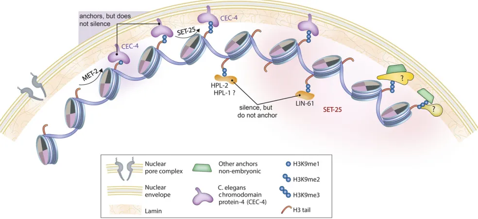
CEC-4- and H3K9me-Mediated Anchoring at the Nuclear Periphery
yeast and in worms, where it ensures the peripheral tethering of H3K9-methylated chromatin throughout early develop-ment (Gonzalez-Sandovalet al.2015). The chromodomain ofCEC-4binds mono-, di-, and trimethylated H3K9 with
af-finities similar to mammalian HP1b, and with high specifi c-ity: out of 188 methylated histone peptides tested, only histone H3 tail peptides bearing K9 methylation, and, more weakly, H3K37me2, were bound. Because the latter modifi -cation has never been detected inC. elegans, the H3K9me is probably the most important ligand. Indeed, ablation of two conserved aromatic residues in the CEC-4 chromodomain was sufficient to compromise the tethering of heterochroma-tin at the nuclear envelope inC. elegansembryos (Gonzalez-Sandovalet al.2015). ThisCEC-4-H3K9me interaction is the
first chromodomain–ligand interaction shown to be neces-sary for chromatin positioning in the interphase nucleus in any species (Figure 2).
Although dependent on CEC-4, perinuclear heterochro-matin tethering in embryos is independent ofHPL-1,HPL-2,
and LIN-61and of H3K27me3 (Figure 2). Indeed, the
tethering of large transgene arrays is independent of their transcriptional state, although it requires H3K9 methylation (Gonzalez-Sandoval et al. 2015). Consistently, the loss of
CEC-4had very little effect on gene expression under unchal-lenged growth conditions. Nonetheless,CEC-4 was needed for a full response to the ectopic expression of a cell fate regulator in pregastrulation embryos (Gonzalez-Sandoval et al. 2015). Following challenge by the induction of the
myogenic transcription factor HLH-1 before gastrulation, wild-type embryos all arrest as differentiated masses of mus-cle cells. In thecec-4mutant, on the other hand,25% of the embryos fail to arrest with a muscle-like phenotype, even though the induction ofHLH-1leads to muscle-specific pro-tein expression. In other words, in the absence of CEC-4, other tissue lineages were not efficiently repressed, arguing that perinuclear anchorage helps restrict gene expression to that of the HLH-1-induced lineage (Gonzalez-Sandovalet al. 2015).
In another study, it was shown thatCEC-4, H3K9 methyl-transferases, andLEM-2contribute to the condensed perinu-clear status of the dosage-compensated X chromosomes in terminally differentiated postmitotic cells of adult animals (Snyderet al.2016). This result suggests that nuclear orga-nization, and specifically anchoring of chromosomal regions to the nuclear lamina, may affect dosage compensation. Nonetheless, during normal development,cec-4embryos de-velop into fertile adults.
H3K9 Demethylases
Histone methyl marks are removed by demethylases that generally fall into two structural classes (Kooistra and Helin 2012). One class contains amine oxidases, of which LSD1 (a homolog of theC. elegansH3K4 demethylaseSPR-5) is the founding member. These have been shown to be able to demethylate mono- and dimethylated lysine residues, but
they are unable to act on trimethylated lysine. The second class contains the Jumonji C (JmjC) domain, which can act on all three methylation states.
C. eleganscontains 13 JmjC domain-containing proteins (Klose et al. 2006), of which two (JMJD-2/JMJD2a and
JMJD-1.2/CeKDM7a) are involved in H3K9 demethylation
(Whetstineet al.2006; Kleine-Kohlbrecheret al.2010; Lin et al.2010). IF of meiotic chromosomes showed that deple-tion ofjmjd-2by RNAi led to increased H3K36me3 on the X chromosome and increased H3K9me3 on the autosomes (Whetstine et al. 2006). Reduction of JMJD-2 also led to replication stress, as indicated by a replication checkpoint-dependent increase in germ cell apoptosis, slow DNA repli-cation fork progression (incorporation of Cy3-dUTP), and an accumulation ofRAD-51foci in the germ line, indicative of DNA breaks (Whetstine et al.2006; Blacket al. 2010). In-terestingly, these germ line phenotypes could be rescued by deletion of the H3K9me readerHPL-2, suggesting that mis-targeting ofHPL-2might be responsible for the phenotypes (Blacket al.2010). Loss ofJMJD-2would presumably lead to an accumulation of H3K9me, which may facilitate ectopic
HPL-2binding. Although H3K9me2 or me3 have been shown
to correlate with late replication in other organisms (Schwaiger et al. 2010; Lubelskyet al.2014), it is not yet clear if H3K9 methylation directly influences replication tim-ing inC. elegans. Alternatively, perturbations in H3K9me lev-els may lead to replication stress that arrests replication (Zelleret al.2016).
JMJD-1.2/CeKDM7a is a bispecific H3K9me2/H3K27me2
demethylase (Kleine-Kohlbrecheret al.2010; Linet al.2010). Recombinant JMJD-1.2 has been shown to demethylate H3K9me2 and H3K27me2 in vitro, and jmjd-1.2 mutants have increased levels of H3K9me2 and H3K27me2, by west-ern blot analysis (Kleine-Kohlbrecheret al.2010; Linet al. 2010). ChIP of JMJD-1.2has shown binding at H3K4me3-marked promoters, which are depleted for H3K9me2 and H3K27me2, and deletion ofjmjd-1.2leads to decreased ex-pression of a subset of tested targets (Lin et al.2010). This may be due to the local acquisition of heterochromatic marks, although this has not yet been demonstrated.JMJD-1.2 ap-pears to have a role in the nervous system, as a JMJD-1.2
transgene is predominantly expressed in neurons and jmjd-1.2mutants display movement defects (Kleine-Kohlbrecher et al.2010).
Phenotypes Caused by the Loss of H3K9 Methyltransferases or H3K9me Readers
C. elegansprovides an opportunity to characterize the effects of a complete loss of H3K9 methylation during development of a multicellular organism, given thatmet-2 set-25mutant embryos, larvae, and germ lines lack detectable H3K9 meth-ylation, and are viable and fertile at 15 or 20°(Towbinet al. 2012; Ho et al. 2014; Garrigues et al. 2015; Zeller et al. 2016). Two recent studies investigated the consequences of an absence of H3K9 methylation or of interacting
heterochro-matin factors including H3K9me readers (Zelleret al.2016; McMurchyet al.2017). In adults, the heterochromatin fac-tors studied were HPL-2and LIN-61, the multi-zinc finger proteinLIN-13(Melendez and Greenwald 2000; Coustham et al. 2006; Harrison et al. 2007; Koester-Eiserfunke and Fischle 2011; Wu et al. 2012; Garrigues et al. 2015), and
LET-418, an Mi-2 homolog that is part of the repressive NuRD (Nucleosome remodeling and histone deacetylase) and MEC (Mi-2 and MEP-1) complexes (von Zelewsky et al. 2000; Unhavaithayaet al.2002; Passannanteet al.2010).
Unlike the situation in other metazoans, chromosome segregation is normal in the absence of H3K9 methylation (Zelleret al.2016), possibly due to the holocentric nature of worm chromosomes. However, the loss of the MET-2 and
SET-25 HMTs shares some phenotypes with the loss of the
studied heterochromatin factors, such as the transcription of repetitive elements, an accumulation of DNA damage, re-duced fertility, and increased germ cell apoptosis (Zeller et al.2016; McMurchyet al.2017). Not surprisingly, loss of the histone modification itself often showed more pro-nounced phenotypes than loss of a single reader.
Similar to other organisms, in C. elegansH3K9me2 and me3 are enriched on repetitive elements, with the majority of repetitive elements being marked by one or both modifi -cations. Importantly, a detailed analysis of the two marks shows that they have different distributions: H3K9me2 is more significantly associated with a subset of DNA transpo-sons and satellite repeats, while H3K9me3 was more prom-inent on retrotransposons and a second subset of DNA transposons (Zelleret al.2016; McMurchyet al.2017). With respect to genes, H3K9me3 is enriched on pseudogenes and silent cell type-specific genes, whereas H3K9me2 marks genes independently of their transcriptional activity (Ho et al. 2014; Garrigues et al. 2015; Zeller et al. 2016). H3K9me2, but not H3K9me3, is also associated with telo-meres (McMurchy et al.2017), although it is not required for their interaction with the nuclear envelope (Ferreiraet al. 2013).
The genomic distributions ofHPL-2,LIN-13,LIN-61,MET-2, andLET-418are strikingly similar and highly correlated with H3K9me2, but not H3K9me3 (Garrigues et al. 2015; McMurchy et al.2017). This begs the question of whether there are readers with a selective affinity for H3K9me3. While no direct binding studies have been reported to date,
SET-25colocalizes with repetitive arrays bearing H3K9me3, but not those with H3K9me2, suggesting that it might di-rectly or indidi-rectly associate with the me3 mark (Towbin et al.2012). ForHPL-1, its definitive binding specificity and endogenous genomic distribution remain to be determined. Preliminary evidence indicates thatHPL-1can recognize all three methylated states of H3K9in vitro(W. Fischle, personal communication), and it bound methylated H3K23 in a pep-tide pull-down assay (Vandammeet al.2015).
of the methyltransferasesMET-2andSET-25, or ofHPL-2or
LIN-61, derepressed heterochromatic reporters (Towbin
et al. 2012) and endogenous genes (Gonzalez-Sandoval et al. 2015). Mutants lacking H3K9me or any of the four studied heterochromatin proteins (HPL-2, LIN-61, LIN-13, or LET-418) derepressed genes and repetitive elements of all classes and families, including satellite repeats, simple repeats, and RNA and DNA transposons (Zelleret al.2016; McMurchy et al. 2017). The majority of derepressed se-quences are enriched for the relevant histone marks or chro-matin factors in wild-type animals, suggesting that the effect is direct.
The observed derepression of repetitive elements is temper-ature-dependent, with many more repeat families being tran-scribed at 25°than at 20°(Zelleret al.2016). This temperature dependence is particularly pronounced for tandem repeats. Temperature effects were also reported for the silencing of re-petitive elements by the nuclear RNAi pathway (Niet al.2016), and for phenotypes of null alleles of other heterochromatin mutants and small RNA pathway factors (e.g., Batista et al. 2008; Wang and Reinke 2008; Schottet al. 2009). Whether these reflect temperature-dependent hyperactivity of RNA po-lymerase II at 25°, a heat-stress response, or a sensitivity of other factors to heat is unknown. Interestingly, a recent report sug-gests that wild-typeSET-25activity may be temperature sensi-tive, and that its effect in silencing repetitive sequences can be transgenerationally inherited (Klosinet al.2017). However, loss
ofset-25does not derepress endogenous micro- or mini-satellite
repeats (J. Padeken and S. M. Gasser, unpublished results). The detection of reproducible but low levels of repeat element transcription inmet-2 set-25mutant embryos argues for a broad misregulation of RNA polymerase II-mediated transcription caused by the loss of H3K9 methylation (J. Padeken, P. Zeller, and S. M. G., unpublished results). For simple repeats, transcriptional start sites are not yet mapped, but it is possible that cryptic transcription factor binding sites become exposed by the loss of repressive het-erochromatin. Importantly, the loss of individual HMTs,i.e., singlemet-2andset-25mutants, shows that different classes of repeats become expressed in the two mutants, consistent with their differential effects on H3K9me marks (J. Padeken, P. Zeller, and S. M. G., unpublished results).
The genes that carry H3K9 methylation in wild-type larvae and adults are generally pseudogenes or silent tissue-specific genes, and these too are derepressed inmet-2 set-25double mutants (Zeller et al. 2016; McMurchy et al. 2017). The expressed repetitive elements are highly enriched for full-length DNA transposases and LTR-containing elements de-rived from RNA transposons, which carry functional Pol II promoter sequences (McMurchy et al.2017). Nonetheless, few elements marked by H3K9 methylation become ex-pressed in its absence, suggesting that other mechanisms pre-vent repetitive element expression. For instance, loss of the nuclear RNAi component nrde-2 leads to derepression of many elements not affected by the loss of H3K9 methylation (McMurchy et al.2017). Nuclear RNA degradation
mecha-nisms may be involved in repeat repression, as exosomes have been shown to play a role in heterochromatic silencing in other organisms (Shinet al.2013; Sugiyamaet al.2016; Tuckeret al.2016). There may also be functional redundancy in repetitive element silencing among the heterochromatin factors studied, given that the mutants show redundancy in the promotion of fertility (McMurchyet al.2017). In addition to transcriptional control, H3K9 methylation may prevent the movement of nonautonomous transposons or inhibit homol-ogous recombination between repeats.
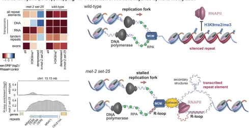
A striking phenotype associated with the loss of heterochro-matin over repeat elements is the loss of genomic integrity. Increased germ line apoptosis in met-2 set-25 mutants was shown to becep-1/p53-dependent and therefore linked to the DNA damage response (Zeller et al. 2016; McMurchyet al. 2017). Activation of the DNA damage response pathway con-tributes to the sterility of heterochromatin factor mutants, as sterility is partially suppressed by mutation of cep-1/p53, al-though this is not the case formet-2 set-25(Zelleret al.2016; McMurchy et al. 2017). Interestingly, resilencing of the MI-RAGE1 DNA transposon by RNAi partially restored fertility in
hpl-2,let-418,and lin-13mutants, potentially by suppressing
expression of its transposase activity (McMurchyet al.2017). The expression of repetitive sequences inmet-2 set-25 mu-tant worms led to an increase in the formation of RNA:DNA hybrids or R-loops, a pathological annealing of RNA with DNA that generates a ssDNA loop where the RNA is bound (Zelleret al.2016) (Figure 3). R-loops are the most common cause of replication fork-associated DNA damage (Aguilera and Garcia-Muse 2012). The RNA:DNA hybrids map specifi -cally to the tandem repeats and DNA transposons that are derepressed by the loss of H3K9 methylation and, like tran-scription, the levels of RNA:DNA hybrids are enhanced at 25° vs. 20°, and are barely detectable at 15°, a temperature at which fertility defects are also suppressed (Zelleret al.2016). The appearance of R-loops at repeat elements correlates with frequent small insertions and deletions at these sites in both somatic cells and the germ line, as detected by whole-genome sequencing (Zeller et al. 2016). Enhanced levels of Rad51 foci, indicative of spontaneous DNA damage, were observed in the germ lines of the met-2 set-25 double mutant, as in heterochromatic regions in Drosophila lacking the H3K9 HMT Su(var)3-9 (Peng and Karpen 2009). Importantly,
met-2 set-25mutant embryos and larvae are not
hypersensi-tive to ionizing radiation, but are sensihypersensi-tive to replication stress induced by hydroxyurea (Zelleret al.2016).
endonuclease partially rescues the fertility defect of K9me readers (McMurchyet al.2017). The difference in hypersen-sitivities between H3K9me-deficient worms andhpl-2or lin-61mutants, suggests that the roles ofHPL-2andLIN-61in double-strand break repair may be independent of H3K9 methylation. Indeed,HPL-2binds chromatin both in the pres-ence and abspres-ence of H3K9 methylation (Garrigues et al. 2015), and in mammals the recruitment of HP1 proteins to sites of damage is H3K9me-independent (Luijsterburget al. 2009; Baldeyronet al.2011).
PRC2/H3K27me3 and Interactions with MES-4/ H3K36me3
The conserved PRC2 complex catalyzes methylation of lysine 27 of histone H3 and functions in the maintenance of tran-scriptional repression in metazoans (Steffen and Ringrose 2014; Piunti and Shilatifard 2016). TheC. elegansPRC2-like complex is composed ofMES-2,MES-3, andMES-6(Bender et al.2004). The SET domain proteinMES-2, an ortholog of EZH2, provides H3K27 methylation activity. MES-6 is an ortholog of ESC/EED, andMES-3is a novel protein.
The genes encoding PRC2 components were initially
identi-fied through genetic screens for genes required for germ cell development (Capowskiet al.1991).mes-2,mes-3, andmes-6
mutants are maternal-effect sterile; homozygous mutants de-rived from heterozygous mothers are viable and fertile, but their progeny are sterile because germ cells die. The activities and functions of these genes are primarily germ line-specific. So-matic development of mes-2, mes-3, and mes-6 mutants is overtly normal, although weak somatic defects in the expression of Hox genes has been observed (Capowskiet al.1991; Ross and Zarkower 2003). At least one somatically active H3K27 HMT exists because somatic H3K27 methylation is present inmes-2
mutants, but it has not yet been identified (Benderet al.2004). In addition to components of PRC2, the messcreens also identifiedmes-4, which encodes a germ line-expressed H3K36 HMT with functions in both the germ line and in early embryos as a maternal product (Capowski et al. 1991; Bender et al. 2006). MES-4 is a nuclear receptor binding SET domain (NSD)-type HMT, that also bears PHD and post-SET domains, and methylates H3K36me in a transcription-independent man-ner. (Benderet al.2006; Furuhashi and Kelly 2010; Rechtsteiner et al.2010). In addition toMES-4,C. elegansalso containsMET-1,
an ortholog of the transcription-coupled H3K36 HMT SET-1
(Andersen and Horvitz 2007; Furuhashi and Kelly 2010; Rechtsteineret al.2010). The loss ofMES-4also results in phe-notypes in somatic cells of L1 larvae derived from homozygous adults (D. Cabianca and S.M.G., unpublished results). Mass spectrometry of histones isolated from L1 larvae shows that the loss of MET-1 eliminates 95% of the H3K36me3, while the full complement of me1/me2 is retained. RNAi depletion
ofMES-4inmet-1mutants reduces H3K36me1/me2 and the
residual H3K36me3 in an additive manner (D. Cabianca and S. M. Gasser, unpublished results).
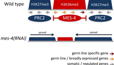
Studies of PRC2 andmes-4mutants revealed an intimate relationship between H3K27 and H3K36 methylation. Genome-wide profiling in embryos and L3 larvae showed that H3K36me3 and H3K27me3 occupy mutually exclusive do-mains on autosomes, consistent with thefinding that meth-ylation of H3K36 inhibits EZH2 activity (Schmitges et al. 2011; Yuan et al. 2011; Gaydoset al. 2012). Additionally, the X chromosome has higher levels of H3K27me3 and lower levels of H3K36me3 than autosomes (Liuet al.2011; Gaydos et al.2012). Intriguingly, despite their marking different ge-nomic regions, gene expression profiling in the germ line showed that loss of eitherMES-4or PRC2 had similar conse-quences: reduced expression of germ line genes and in-creased expression of somatic and X chromosome genes (Gaydos et al. 2012). This was explained by showing that these two marking systems functionally antagonize each other. Loss ofMES-4causes spreading of H3K27me3 to germ line genes, and a concomitant reduction of H3K27me3 on somatic and X chromosome genes (Gaydos et al. 2012). Therefore, H3K27me3 marking by PRC2 and H3K36me3 by
MES-4 cooperate to ensure correct gene expression in the germ line.
The chromatin modifications generated by MES-4 and PRC2 are transgenerationally inherited.MES-4marks genes expressed in the germ line with H3K36me2/3, and then ma-ternally contributedMES-4maintains H3K36me2/3 marking of these genes in the early embryo independently of scription, providing an epigenetic memory of germ line tran-scription to progeny (Furuhashi et al. 2010; Rechtsteiner et al. 2010). Similarly, H3K27 methylation generated by PRC2 in the germ line is inherited by progeny (Gaydos et al. 2014). Together, H3K27 methylation and maternal PRC2 components provide a memory of transcriptional re-pression (Gaydoset al.2014).
A recent study used patterns of chromatin states in early embryos and L3 larvae to investigate domain properties of the C. elegansgenome, defining two types of chromatin domain: active and regulated (Evans et al.2016). It was found that active domains are associated with H3K36me3 and regulated domains with H3K27me3, which overlap the mutually exclu-sive patterns of these modifications noted by Gaydoset al. (2012). The domains separate genes of different types: active domains contain genes expressed in the germ line and broadly expressed across development and cell types, whereas regulated domains predominantly contain genes un-der spatial, temporal, or conditional control, lacking germ line expression. Regulated expression is consistent with the repressive role of H3K27me3 in facultative heterochromatin in other organisms. The locations of H3K36me3- and H3K27me3-marked domains in undifferentiated early em-bryonic cells were similar to those in differentiated L3 larvae, indicating that domain positions are a core property of the genome. The mechanism of domain definition is not yet un-derstood, but appears to involve interactions between PRC2 andMES-4(Gaydoset al.2012; Evanset al.2016) (Figure 4). Additionally, thefinding that border regions between active and regulated domains contain long intergenic regions enriched for transcription factor binding suggests that tran-scriptional activity may play a role in defining the boundaries of active and regulated domains (Evanset al.2016). A future challenge will be to investigate possible tissue-specific do-main regulation by analyzing histone mark distribution in individual tissues rather than whole animals.
PRC2 in Reprogramming and Maintenance of Cell Fate
PRC2 is important for the maintenance of a repressed state, and this may be due in part to inhibition of developmental plasticity. Early evidence for this inC. elegans came from a study showing that loss ofmes-2in early (undifferentiated) embryos causes prolonged sensitivity to ectopic expression of developmental regulators that can cause cell fate transforma-tions (Yuzyuket al.2009). This study also showed that loss of
mes-2causes a change in chromosome conformation
sugges-tive of a loss of compaction. Studies in germ cells similarly found that loss of PRC2 components makes germ cells sus-ceptible to conversion to a somatic fate when challenged by
expression of cell type-inducing transcription factors (Patel et al.2012). Sensitivity to somatic conversion was also ob-served in response to increased Notch activity (Seelket al. 2016). Notch appears to act by antagonizing PRC2 repression of numerous genes, including the H3K27me3 demethylase
UTX-1. These studies support a role for PRC2 in preventing inappropriate responses to regulatory inputs, perhaps by in-creasing the barrier to response. Consistent with this role, the H3K27me2/3 demethylaseJMJD-3.1is necessary for a nat-ural C. elegans transdifferentiation event, where a hindgut cell is transformed into a motor neuron (Zurynet al.2014).
Chromatin Regulators and Small RNA Pathways
In addition to classical RNAi that carries out post-transcriptional silencing in the cytoplasm,C. elegansalso has a nuclear RNAi pathway (called the Nrde pathway) that directs transcriptional silencing, whereby small RNAs provide sequence specificity that directs heterochromatin assembly at target loci and downregu-lation of RNA polymerase activity (Guang et al.2008, 2010; Burkhartet al.2011; Buckleyet al.2012; Guet al.2012; Mao et al.2015). Although the precise mechanism of repression is
still being worked out, a series of experiments showed that argonaute proteins bound to small interfering (siRNAs) (NRDE-3 in the soma or HRDE-1 in the germ line) recruit
NRDE-1, NRDE-2, and NRDE-4 to target loci, leading to an accumulation of H3K9me3 in a MET-2- and SET-25-dependent manner, and the stalling of RNA polymerase II (Guanget al. 2008, 2010; Burkhart et al. 2011; Buckley et al. 2012; Gu et al.2012; Maoet al.2015) (Figure 5). Recently,SET-32was also shown to affect HRDE-1-dependent H3K9 methylation (Kalinavaet al.2017; Spracklinet al.2017), andMORC-1has been implicated as a downstream effector needed to maintain H3K9me3 atHRDE-1targets (Spracklinet al.2017; Weiseret al. 2017). Additionally, it was found that MES-2-dependent H3K27me3 is also induced at Nrde targets (Maoet al.2015), although the relationship between PRC2 and Nrde silencing is not yet known. The results support a model whereby the small RNA-targeted Nrde pathway represses transcription by trigger-ing the generation of repressed chromatin. Interesttrigger-ingly, mu-tants affecting nuclear RNAi processes often show progressive sterility over generations (Table 2).
The Nrde pathway is also engaged by the piwi-interacting RNA (piRNA) pathway, a small RNA pathway active in the
germ line, which is important for the repression of transpos-able elements as well as endogenous genes (Asheet al.2012; Bagijn et al.2012; Leeet al.2012) (Figure 5). piRNAs are nuclear-encoded 21-nucleotide RNAs (starting with a U) that are bound by the argonautePRG-1in the cytoplasm (Ruby et al.2006; Batista et al.2008; Das et al.2008; Wang and Reinke 2008). These direct the generation of secondary siRNAs that become bound by the germ line Nrde argonaute,HRDE-1, to engage the Nrde pathway in transcriptional silencing (Ashe et al.2012; Buckleyet al.2012; Luteijnet al.2012). Silencing of a piRNA pathway reporter was shown to require the H3K9 HMTsSET-25,MET-2, andSET-32, as well as the heterochro-matin proteinsHPL-2,LIN-61, andLET-418(Asheet al.2012; McMurchyet al.2017). In adult worms, bothprg-1mutants and mutants lacking these heterochromatin proteins derepress a common spectrum of repetitive elements, suggesting that these may work together in the germ line (McMurchyet al.2017) (Figure 5).
Interestingly, loss of the Nrde pathway leads to the de-repression of a different set of repetitive elements than loss of the piRNA pathway or of the above subset of heterochromatin factors (Kalinavaet al.2017; McMurchyet al.2017). Retro-transposons are more affected innrde-2mutants, whereas a bias for DNA transposons was observed forprg-1and hetero-chromatin mutants. Moreover, redundancy appears to under-lie at least part of this apparent separation of function, because nrde-2 genetically interacts with let-418, lin-13,
and hpl-2. Additionally,nrde-2;let-418 double mutants
de-repress many more repetitive elements than either single mutant (McMurchyet al.2017) (Figure 5). Consistent with redundancy, we note that although the Nrde pathway in-duces H3K9 methylation, endogenous Nrde targets are still repressed in the absence of H3K9 methylation, indicating that H3K9 methylation is not necessary for repression by the Nrde pathway (Kalinava et al.2017; McMurchy et al. 2017). The observed complexity and redundancy of silencing mechanisms underscores the importance of repeat element repression for genome stability.
Perspectives
Heterochromatin fulfills many important functions in the genome of complex organisms. In this respect,C. elegansis no exception, as indicated by the diverse physiological de-fects of mutants lacking the enzymes that deposit heterochro-matic marks or the proteins that recognize them. Repressive chromatin is essential for nuclear organization, genome do-main structure, repression of repetitive elements, genome stability, and regulation of protein-coding genes. Cross talk and redundancy between the proteins and pathways in-volved has been observed, but these interactions are poorly understood. Furthermore, the specificity of most HMTs and histone mark readers remains to be determined, as does the nature and importance of the interactions observed between heterochromatin and small RNA interference mechanisms. Above all, the role of heterochromatin in controlling or
shap-ing developmental programs is still an open question, and the genetics and rapid development ofC. elegansare particularly useful for its investigation. As demonstrated in the past, C. elegansis an excellent organism for gene expression studies, given its genetic flexibility, rapid developmental time, and well-defined cell differentiation pathways.
Acknowledgments
We thank Jan Padeken, Peter Zeller, Anna Mattout, Jennifer Harr, and Tessa Gaarenstroom for critical reading of the article. S.M.G. thanks the Swiss National Science Founda-tion and the Novartis Research FoundaFounda-tion for continued support. J.A. was supported by a Wellcome Trust Senior Research Fellowship (101863).
Literature Cited
Agger, K., P. A. Cloos, J. Christensen, D. Pasini, S. Roseet al., 2007 UTX and JMJD3 are histone H3K27 demethylases involved in HOX gene regulation and development. Nature 449: 731–734. Aguilera, A., and T. Garcia-Muse, 2012 R loops: from transcription byproducts to threats to genome stability. Mol. Cell 46: 115–124. Albertson, D. G., and J. N. Thomson, 1982 The kinetochores of
Caenorhabditis elegans. Chromosoma 86: 409–428.
Andersen, E. C., and H. R. Horvitz, 2007 TwoC. eleganshistone methyltransferases repress lin-3 EGF transcription to inhibit vul-val development. Development 134: 2991–2999.
Ashe, A., A. Sapetschnig, E. M. Weick, J. Mitchell, M. P. Bagijn
et al., 2012 piRNAs can trigger a multigenerational epigenetic memory in the germline ofC. elegans. Cell 150: 88–99. Bagijn, M. P., L. D. Goldstein, A. Sapetschnig, E. M. Weick,
S. Bouasker et al., 2012 Function, targets, and evolution of
Caenorhabditis eleganspiRNAs. Science 337: 574–578. Baldeyron, C., G. Soria, D. Roche, A. J. Cook, and G. Almouzni,
2011 HP1alpha recruitment to DNA damage by p150CAF-1 promotes homologous recombination repair. J. Cell Biol. 193: 81–95.
Bank, E. M., and Y. Gruenbaum, 2011 Caenorhabditis elegansas a model system for studying the nuclear lamina and laminopathic diseases. Nucleus 2: 350–357.
Batista, P. J., J. G. Ruby, J. M. Claycomb, R. Chiang, N. Fahlgren
et al., 2008 PRG-1 and 21U-RNAs interact to form the piRNA complex required for fertility inC. elegans. Mol. Cell 31: 67–78. Bender, L. B., R. Cao, Y. Zhang, and S. Strome, 2004 The MES-2/ MES-3/MES-6 complex and regulation of histone H3 methyl-ation inC. elegans. Curr. Biol. 14: 1639–1643.
Bender, L. B., J. Suh, C. R. Carroll, Y. Fong, I. M. Fingermanet al., 2006 MES-4: an autosome-associated histone methyltransfer-ase that participates in silencing the X chromosomes in the
C. elegansgerm line. Development 133: 3907–3917.
Bessereau, J.-L., 2006 Transposons inC. elegans(January 18, 2006), WormBook, ed. TheC. elegansResearch Community, WormBook, doi/10.1895/wormbook.1.70.1, http://www.wormbook.org. Bessler, J. B., E. C. Andersen, and A. M. Villeneuve,
2010 Differential localization and independent acquisition of the H3K9me2 and H3K9me3 chromatin modifications in the
Caenorhabditis elegans adult germ line. PLoS Genet. 6: e1000830.
Boros, J., N. Arnoult, V. Stroobant, J. F. Collet, and A. Decottignies, 2014 Polycomb repressive complex 2 and H3K27me3 cooper-ate with H3K9 methylation to maintain heterochromatin protein 1alpha at chromatin. Mol. Cell. Biol. 34: 3662–3674.
Buckley, B. A., K. B. Burkhart, S. G. Gu, G. Spracklin, A. Kershner
et al., 2012 A nuclear argonaute promotes multigenerational epigenetic inheritance and germline immortality. Nature 489: 447–451.
Burkhart, K. B., S. Guang, B. A. Buckley, L. Wong, A. F. Bochner
et al., 2011 A pre-mRNA-associating factor links endogenous siRNAs to chromatin regulation. PLoS Genet. 7: e1002249.
C. elegans Sequencing Consortium, 1998 Genome sequence of the nematodeC. elegans: a platform for investigating biology. Science 282: 2012–2018.
Capowski, E. E., P. Martin, C. Garvin, and S. Strome, 1991 Identification of grandchildless loci whose products are required for normal germ-line development in the nematode
Caenorhabditis elegans. Genetics 129: 1061–1072.
Clark, R. F., and S. C. Elgin, 1992 Heterochromatin protein 1, a known suppressor of position-effect variegation, is highly con-served in Drosophila. Nucleic Acids Res. 20: 6067–6074. Coustham, V., C. Bedet, K. Monier, S. Schott, M. Karali et al.,
2006 The C. elegansHP1 homologue HPL-2 and the LIN-13 zincfinger protein form a complex implicated in vulval devel-opment. Dev. Biol. 297: 308–322.
Couteau, F., F. Guerry, F. Muller, and F. Palladino, 2002 A hetero-chromatin protein 1 homologue inCaenorhabditis elegansacts in germline and vulval development. EMBO Rep. 3: 235–241. Cui, M., and M. Han, 2007 Roles of chromatin factors inC. elegans
development (May 3, 2007), WormBook, ed. The C. elegans
Research Community, WormBook, doi/10.1895/wormbook.1.139.1,
http://www​.wormbook.org.
Cui, M., J. Chen, T. R. Myers, B. J. Hwang, P. W. Sternberget al., 2006 SynMuv genes redundantly inhibit lin-3/EGF expression to prevent inappropriate vulval induction inC. elegans. Dev. Cell 10: 667–672.
Das, P. P., M. P. Bagijn, L. D. Goldstein, J. R. Woolford, N. J. Lehrbach et al., 2008 Piwi and piRNAs act upstream of an endogenous siRNA pathway to suppress Tc3 transposon mobil-ity in theCaenorhabditis elegansgermline. Mol. Cell 31: 79–90. Dobrzynska, A., P. Askjaer, and Y. Gruenbaum, 2016 Lamin-binding proteins inCaenorhabditis elegans. Methods Enzymol. 569: 455–483. Eissenberg, J. C., and S. C. Elgin, 2014 HP1a: a structural chromo-somal protein regulating transcription. Trends Genet. 30: 103–110. Elgin, S. C., and G. Reuter, 2013 Position-effect variegation, het-erochromatin formation, and gene silencing in Drosophila. Cold Spring Harb. Perspect. Biol. 5: a017780.
Eskeland, R., A. Eberharter, and A. Imhof, 2007 HP1 binding to chromatin methylated at H3K9 is enhanced by auxiliary factors. Mol. Cell. Biol. 27: 453–465.
Evans, K. J., N. Huang, P. Stempor, M. A. Chesney, T. A. Downet al., 2016 StableCaenorhabditis eleganschromatin domains sepa-rate broadly expressed and developmentally regulated genes. Proc. Natl. Acad. Sci. USA 113: E7020–E7029.
Fay, D. S., and J. Yochem, 2007 The SynMuv genes ofCaenorhabditis elegansin vulval development and beyond. Dev. Biol. 306: 1–9. Ferreira, H. C., B. D. Towbin, T. Jegou, and S. M. Gasser,
2013 The shelterin protein POT-1 anchorsCaenorhabditis ele-ganstelomeres through SUN-1 at the nuclear periphery. J. Cell Biol. 203: 727–735.
Fischle, W., Y. Wang, S. A. Jacobs, Y. Kim, C. D. Allis et al., 2003 Molecular basis for the discrimination of repressive methyl-lysine marks in histone H3 by Polycomb and HP1 chro-modomains. Genes Dev. 17: 1870–1881.
Francastel, C., D. Schubeler, D. I. Martin, and M. Groudine, 2000 Nuclear compartmentalization and gene activity. Nat. Rev. Mol. Cell Biol. 1: 137–143.
Furuhashi, H., and W. G. Kelly, 2010 The epigenetics of germ-line immortality: lessons from an elegant model system. Dev. Growth Differ. 52: 527–532.
Furuhashi, H., T. Takasaki, A. Rechtsteiner, T. Li, H. Kimuraet al., 2010 Trans-generational epigenetic regulation of C. elegans
primordial germ cells. Epigenet. Chromatin 3: 15.
Garrigues, J. M., S. Sidoli, B. A. Garcia, and S. Strome, 2015 Defining heterochromatin inC. elegansthrough genome-wide analysis of the heterochromatin protein 1 homolog HPL-2. Genome Res. 25: 76–88.
Gassmann, R., A. Rechtsteiner, K. W. Yuen, A. Muroyama, T. Egelhoferet al., 2012 An inverse relationship to germline tran-scription defines centromeric chromatin in C. elegans. Nature 484: 534–537.
Gaydos, L. J., A. Rechtsteiner, T. A. Egelhofer, C. R. Carroll, and S. Strome, 2012 Antagonism between MES-4 and Polycomb repressive complex 2 promotes appropriate gene expression in
C. elegansgerm cells. Cell Rep. 2: 1169–1177.
Gaydos, L. J., W. Wang, and S. Strome, 2014 Gene repression. H3K27me and PRC2 transmit a memory of repression across generations and during development. Science 345: 1515–1518. Gerstein, M. B., Z. J. Lu, E. L. Van Nostrand, C. Cheng, B. I. Arshinoff
et al., 2010 Integrative analysis of the Caenorhabditis elegans genome by the modENCODE project. Science 330: 1775–1787.
Gonzalez-Aguilera, C., K. Ikegami, C. Ayuso, A. de Luis, M. Iniguez
et al., 2014 Genome-wide analysis links emerin to neuromus-cular junction activity in Caenorhabditis elegans. Genome Biol. 15: R21.
Gonzalez-Sandoval, A., B. D. Towbin, V. Kalck, D. S. Cabianca, D. Gaidatzis et al., 2015 Perinuclear anchoring of H3K9-methylated chromatin stabilizes induced cell fate in C. elegans
embryos. Cell 163: 1333–1347.
Grant, J., C. Verrill, V. Coustham, A. Arneodo, F. Palladinoet al., 2010 Perinuclear distribution of heterochromatin in develop-ingC. elegansembryos. Chromosome Res. 18: 873–885. Greer, E. L., S. E. Beese-Sims, E. Brookes, R. Spadafora, Y. Zhu
et al., 2014 A histone methylation network regulates transgenerational epigenetic memory inC. elegans. Cell Rep. 7: 113–126.
Greer, E. L., M. A. Blanco, L. Gu, E. Sendinc, J. Liuet al., 2015 DNA methylation on N6-adenine inC. elegans. Cell 161: 868–878. Greer, E. L., B. Becker, C. Latza, A. Antebi, and Y. Shi, 2016
Muta-tion ofC. elegansdemethylase spr-5 extends transgenerational longevity. Cell Res. 26: 229–238.
Gu, S. G., and A. Fire, 2010 Partitioning theC. elegansgenome by nucleosome modification, occupancy, and positioning. Chromo-soma 119: 73–87.
Gu, S. G., J. Pak, S. Guang, J. M. Maniar, S. Kennedy et al., 2012 Amplification of siRNA inCaenorhabditis elegans gener-ates a transgenerational sequence-targeted histone H3 lysine 9 methylation footprint. Nat. Genet. 44: 157–164.
Guang, S., A. F. Bochner, D. M. Pavelec, K. B. Burkhart, S. Harding
et al., 2008 An argonaute transports siRNAs from the cyto-plasm to the nucleus. Science 321: 537–541.
Guang, S., A. F. Bochner, K. B. Burkhart, N. Burton, D. M. Pavelec
et al., 2010 Small regulatory RNAs inhibit RNA polymerase II during the elongation phase of transcription. Nature 465: 1097– 1101.
Harrison, M. M., X. Lu, and H. R. Horvitz, 2007 LIN-61, one of two
Caenorhabditis elegans malignant-brain-tumor-repeat-containing proteins, acts with the DRM and NuRD-like protein complexes in vulval development but not in certain other biological processes. Genetics 176: 255–271.