Iris Pattern Recognition Using Image
Processing and Soft Computing Approach
Sanket Dan1, Manisha Barman2, J Paul Choudhury3
Dept. of Information Technology,Kalyani Govt Engineering College, Kalyani, Nadia, India Dept. of Information Technology,Kalyani Govt Engineering College,Kalyani, Nadia, India Dept. of Information Technology, Kalyani Govt Engineering College, Kalyani, Nadia, India
ABSTRACT: Iris recognition is a method of identifying people based on unique patterns within the ring-shaped region surrounding the pupil of the eye. The iris usually has a brown, blue, gray, or greenish color , with complex patterns that are visible upon close inspection. Because it makes use of a biological characteristic, iris recognition is considered a form of biometric verification. The work presented in this thesis involved developing an ‘open-source’ iris recognition system in order to verify both the uniqueness of the human iris and also its performance as a biometric. For determining the recognition performance of the system two databases of digitized grayscale eye images were used. The iris recognition system consists of an automatic segmentation system that is based on the Hough transform, Radial suppression and is able to localize the circular iris and pupil region, excluding eyelids and eyelashes, and reflections. Iris segmentation is a key step in the iris recognition system. The conventional methods of iris segmentation are based on the assumption that the inner and outer boundaries of an iris can be taken as circles. However, we investigate the iris boundaries in the CASIA-IrisV3 database, and find that the actual iris boundaries are not always circular. In order to solve this problem, a new approach for iris segmentation based on radial- suppression edge detection is implemented in our project work. In the radial-suppression edge detection, a non-separable wavelet transform is used to extract the wavelet transform modulus of the iris image.We use a method of radial non-maxima suppression to retain the annular edges and simultaneously remove the radial edges. Next, a thresholding operation is utilized to remove the isolated edges and produce the final binary edge map. Based on the binary edge map, a self-adaptive method of iris boundary detection is proposed to produce final iris boundaries. Experimental results demon- strata that the proposed iris segmentation is desirable. Results of the self-adaptive inner boundary have been taken as a feature to be classified by linear neural network and simultaneously by fuzzy logic.
KEYWORDS: Radial-suppression edge detection, wavelet functions, Radial non-maxima suppression, edge thresholding.
I. INTRODUCTION
trust biometric security systems over time.Iris recognition systems capture an image of an individual's eye, the iris in the image is then segmented and normalized for feature extraction process. The performance of iris recognition systems highly depends on segmentation and normalization.
Phases of proposed iris segmentation and iris inner boundary detection
II. RADIAL-SUPPRESSION EDGE DETECTION
Edge detection is initial step to object recognition. It is necessary to point out the true edge to get the best result from the matching process.
A. METHOD OF EDGE DETECTION
The edge detection methods [4,5] that have been published mainly differ in the types of smoothing filters that are applied and the way the measures of edge strength are computed. As many edge detection methods rely on the computation of image gradients, they also differ in the types of filters used for computing gradient estimates in the x- and y-directions.The commonly used detection operators are Roberts, Sobel, Hueckel, Prewitt, Kirsch and Laplacian operators. Laplacian of Gaussian (LOG), is better than Prewitt and Sobel in case of noisy image. In this paper, classified and comparative study of edge detection algorithms are presented. Experimental results prove that Boie-Cox, Shen- Castan and Canny operators are better than Laplacian of Gaussian (LOG).Canny developed a conceptual approach of edge detection. An optimal detector has been derived which can be approximated by the first derivative of a Gaussian function. Wavelet transform has been found to be a remarkable tool to analyse the singularities including edges. Thus wavelet transform has been established for edge detection. Some researcher proved that [15, 16] the maxima of the modulus of wavelet transform can detect the segmentations of irregular structure. Disadvantage of wavelet transform is it can detect the noises (such as edges of eyelash and edges of texture of iris). In case of iris segmentation these noises are invalid and may result in inaccuracy in iris segmentation. So we use a method of radial-suppression edge detection based on the non-separable wavelet transform to detect high quality edge maps of iris images and segment iris accurately.
i. Selection of wavelet functions and calculation of the modulus of the wavelet transform
In Mallatetal.work[15,16] they used these parable wavelet transform. The two-dimensional separable wave- let transform of an image at scale s and in orientation k is defined as
(x, y) * Q ,
k k
s s
w I I k=1,2, (The two directional wavelets) (1)
Qk
sCan be construct as 1 (x, y) Q (x, y)
x
AND
2 (x, y)
Q (x, y) y
(2)
Calculation of wavelet transforms modulus
Estimating the pupillary center
Radial non-maxima suppression
Where (x, y) is a separable spline scaling function. It can be shown that the two-dimensional separable wavelet transform gives the gradient of I(x, y) smoothed by (x, y) at scales s
1
2 2
(x, y) (x, y)
(x, y)
(x, y) (x, y) s s
s
s
w I x
I s w I y
=s (I*s)(x, y)
(3)
The modulus and phase angle of the gradient vector are
1 2 2 2
MsI(x, y) | WsI(x, y) || Ws I(x, y) (4)
1
2
W (x, y) arctan | |
W (x, y) s s s I B I
The separable wave- lets are anisotropic and prefer certain directions (vertical and horizontal directions as well as diagonal) [17]. While, in the case of the iris segmentation, it is required to determine the approximately annular iris boundary, which demands detecting edges reflecting various orientations. Therefore, the separable wavelets are unsuitable for detecting edges in iris images. We need a new wavelet to overcome the limitations of separable wavelets.
1
o cos cos
cos cos o 1 2 3 sin sin sin sin sin sin sin sin p p p p
2 3 1
1 1
( , ) 1 1
1 3 2
1 2
o p p o
p o o p
U
p o o p
o p p o
(6)
Where and are the arbitrary real numbers. From the previous centrally symmetric matrix, we may derive a class of the non-separable product wavelet filters below. The low-pass filter m0(z , z )1 2 is defined as follows:
, ,2 2
0 1 2 1 2 1 2 ( ) 1 2 ( ) 0
1
1
(z , z ) 1, z , z , z , z (z , z ) U ;
4 k k k k
N
T
n
m U C V
(7) 1 2z , z C C;
whereC{z :| z | 1} ,C{z :| z | 1} and three high pass filters mj(z , z )1 2 j=1,2,3 with respect to the above low-pass
filter m (z , z )0 1 2 are as follows:
, ,2 2
0 1 2 1 2 1 2 ( ) 1 2 ( )
1
1
(z , z ) 1, z , z , z , z (z , z ) U ;
4 k k k k
N
T J n
m U C V
(8) j=1,2,3 Where 0 (1,1,1,1) TV , 1 (1, 1,1, 1)
T
V , 2 (1,1, 1, 1)
T
V ,
3 (1, 1, 1,1)
T
V (9)
,
(k k)
U is centrally symmetric orthogonal matrix defined in
Eq.(6)and C(z , z )1 2 is the matrix of trigonometric
polynomial below
1
1 2
2
1 2
1 0 0 0 0 z 0 0 (z , z )
0 0 z 0 0 0 0 z z C (10)
Since them1and m2 are anti- symmetric filters, and anti-symmetric filters are suitable for detecting the local extreme
values as edge points[18], the modulus of the non-separable wavelet transform can be computed by
2 2
1 2
| m | | m I |
J j j
M I I (11)
ii. RADIAL NON-MAXIMA SUPPRESSION AND EDGE THRESHOLDING.
This method may be deviated far away from true location of pupillary center because in CASIA-IrisV3, images have eight bright spots in the pupil.
Estimating the approximate center point of edge ;we replace the bright spots with the values of points of the pupil. Then we perform radial non-maxima suppression technique.
Algorithm 1 (Estimating the papillary center)
Input: The iris image is Z(x, y)
1:xc⟵argminimumx (x, y) y
Z
;// (xc,yc) is a point of
the pupil.
2:yc⟵argminimum
x (x, y) x
Z
;3: for allxc50xxc50ANDyc50yyc50do
4: if Z(x, y) >ISthen // detect the bright spots near the point (xc,yc).
5: Z(x, y) ⟵ Z(xc,yc); //replace the intensity values of
bright spots
6:end if
7: end for
8: xd⟵argminimum
x y Z(x, y)
;
9:yd⟵argminimum
x (x, y) x
Z
;10: return (xd,yd)
Output: a point (xd,yd) of pupillarly center
The annular edges are potential edges of iris and radial edges are always nose. We propose Radial non-maxima suppression because it can retain the annular edges and remove the radial edges where the conventional method of non-maxima suppression detects all edges (annular edges and radial edges).So conventional method of non-non-maxima is not suitable for iris segmentation. To retain the annular edges and remove the radial edges we propose Radial non-maxima suppression.
Algorithm 2(Radial non-maxima suppression)
Input: m (the modulus image), (xd,yd) (the approximate
center point of edge).
1.For all point pnof the modulus image m do
2. Determine the radial direction of point pn; // the
direction from (xd,yd) to pn
3. Determine the two points P1, P2 in the eight pixel neighborhood of pnalong the radial direction;
4. if m(pn)> m(P1) AND m(pn)> m(P2) then
5.q(pn)⟵ m(pn);
6. else
7. q(pn)⟵ 0;
8.end if
9. end for
We use “hysteresis” thresholding [1] with Cao’s method of edge length to further remove noises and detect the edge of the iris boundary.The conventional method of iris segmentation [2] is based on assumption that inner and outer boundary of iris can approximately be taken as circles. Therefore we use self-adaptive method to get inner circular border of iris.
Algorithm 3 (Self-adaptive inner boundary detection)
Input: a. r (the final binary edge map) b. l(array of the list of curves) c.( fi,go) (the range of diameter of the
inner boundary)
d. el(array of the lengths of the edges curves) 1: loop_inner ⟵ 0;
2: for all edge curve l[i] of array l AND their lengths el[i] of array el do
3: if l[i] is loop curve AND
i f <
1,2 [i]
maximum
p pl | p1 - p2| <gothen
4: go⟵ l[i];// get inner boundary
5: loop_inner⟵ 1; 6: end for
7: if loop_inner ⟵0 then
8: go ⟵HOUGH_ellipse(r, fi, go ) // elliptical Hough
transform(cannot get inner boundary because the edge curve corresponding to the pupil may be distorted by some noise)
9: end if Output: go(inner boundary)
Begin
Acquisition
Row and column centre detection
Modulas
Figure :1STEP WAYISES OUTPUT
Step 1:
From ...database out of 42 attributes, 3 attributes have been taken as input and 1 attribute has been used as output. The input attributes are Row-center, Column-center, and Radius.
Step 2:
The first input field is “Row-Center” comprises of three types as denoted by numerical value. The value 1 indicates Maximum value of Row-center from the 42 image dataset which is 187”, 0.5 indicates “Mid value of row center from the 42 image dataset which is 102, 0.1 indicates test “Minimum value of row center from the 42 image dataset which is
0”.
The second input fields is “Column _Centre” comprises of three types as denoted by numerical value. The value 1 indicates Maximum value of column center from the 42 image dataset which is 450”, 0.24 indicates “Mid value of row center from the 42 image dataset which is 108, 0.1 indicates “Minimum value of row center from the 42 image dataset which is 0”.
The third input fields is “Radius” comprises of three types as denoted by numerical value. The value 1 indicates Maximum value of radius from the 42 image dataset which is 116”, 0.27 indicates “Mid value of radius from the 42 image dataset which is 32, 0.1 indicates “Minimum value of Row-center from the 42 image dataset which is 0”.
Step 3:
The following data items (3 attributes per iris image) have been used as input. The input data items have been furnished in Table 1:-
Serial-No. Row-center Column-center Radius
eye.jpg 123 450 90 ss.jpg 150 447 85 1.jpg 127 167 116
2.jpg 148 150 44
3.jpg 156 133 50
4.jpg 169 193 32
5.jpg 155 183 47
6.jpg 169 179 41
Pupil extraction
END
Step 4:
Thus input variables have been classified based on their interim values, which have been furnished in table2, table3, table4, table5 respectively.
Radius-center type: Thus input variable supports 3 types of Radius-center type of the iris images. Each Radius-center type in a fuzzy set shown in figure 2. In case of center type fuzzy sets do not have overlap because the Radius-center of an iris image is different.
Table 2 Radius-center type classification
Column-center type: Thus input variable supports 3 types of center type of the iris images. Each column-center type in a fuzzy set shown in figure 2. In case of column-column-center type fuzzy sets do not have overlap because the column-center is different for each and every iris image.
Table 3 Column-center type classification
Radius type: Thus input variable supports 3 types of column-center type of the iris images. Each radius type in a fuzzy set shown in figure 4. In case of radius type fuzzy sets do not have overlap because the radius is different for each and every iris image.
Table 4 Radius type classification
Step 5:The fuzzy rules have been applied on the data to estimate the angiographic status of iris image using fuzzy rule base.
Step 6:Now Gaussian membership function has been applied to all input and Triangular membership function has been applied to output variables. membership function editor and rule editor and rule viewer using Matlab 2011b have been furnished figure 2 and 3 .
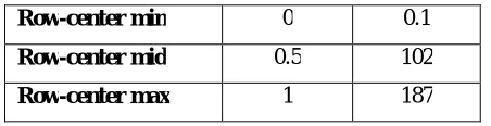
Row-center min 0 0.1
Row-center mid 0.5 102
Row-center max 1 187
Column _center min 0 0.1
Column _center mid 0.24 108
Column _center max 1 450
Radius min 0 0.1
Radius mid 0.27 32
Figure 2 membership function editor Figure 3 Fuzzy rule editor
III. RESULT
It has been observed that the Gaussian membership function is suitable for implementation of fuzzy rule uniqueness of iris image. The estimated status of iris image based on fuzzy rule base using gaussian membership function for all input and triangular membership function for output.
IV. CONCLUSION
Fuzzy rule base system using Gaussian membership function for input and triangular membership function has been generated which shows, estimation of row center of iris image which should lie between 0.5 to 1 has been found to be around 0.7. The value 0.7 is the expected result of this experiment. Also, found that estimation of column center of iris image which should lie between 0.24 to 1 has been found to be around 0.45. The value 0.45 is the expected result of this experiment and estimation of radius of iris image which should lie between 0.27 to 1 has been found to be around 0.46. The value 0.46 is the expected result of this experiment In other words, we also said that the value of row-center, column-center and radius should lie their range according to the numerical values.
REFERENCES
[1]W. Cao, R. Che, D. Ye, An illumination-independent edge detection and fuzzy enhancement algorithm based on wavelet transform for nonuniform weak illumination images, Pattern Recognition Letters 29 (3) (2008) 192–199.
[2] J. Canny, A computational approach to edge detection, IEEE Transactions on Pattern Analysis and Machine Intelligence 8 (6) (1986) 679–698. [3] L. Ma, T. Tan, Y.Wang, D. Zhang, Personal identification based on iris texture analysis, IEEE Transactions on Pattern Analysis and Machine Intelligence 25 (12) (2003) 1519–1533.
[4] D. Ziou and S. Tabbone (1998) "Edge detection techniques: An overview", International Journal of Pattern Recognition and Image Analysis, 8(4):537–559, 1998
[5] Lindeberg, Tony (2001), "Edge detection", in Hazewinkel,Michiel, Encyclopedia ofMathematics, Springer, ISBN 978-1-55608-010-4
[6] Mohamed Roushdy (2006), “Comparative Study ofEdge Detection AlgorithmsApplying on the Grayscale Noisy Image Using Morphological Filter”, GVIP Journal, Vol. 6, Issue 4
[7]Faulds, H, 1880. On the Skin-Furrows of the Hand. Nature, 22, pp. 605. [8]L. Flom, A. Safir. Iris recognition system.United States Patent, (4641349), 1987.
[9]J. Daugman, C. Downing, Epigenetic randomness, complexity, and singularity of human iris patterns, Proceedings of the Royal Society of London Series B: Biological Sciences 268 (2001) 1737–1740.
[10]Nalini K. Ratha, Jonathan H. Connell, Ruud M. Bolle, Enhancing security and privacy in biometrics-based authentication systems, IBM system (2001)