Stephen Higham BSc(Hons)MSc
Submitted in partial fulfilment for the degree of
Doctor of Philosophy
Department of Psychology
Lancaster University
All rights r e se r v e d INFORMATION TO ALL USERS
The q u a lity of this r e p r o d u c tio n is d e p e n d e n t u p o n the q u a lity of the c o p y s u b m itte d . In the unlikely e v e n t that the a u th o r did not sen d a c o m p l e t e m a n u scrip t
and there are m issing p a g e s , th e se will be n o t e d . Also, if m a teria l had to be r e m o v e d , a n o t e will in d ic a te the d e le t io n .
uest
P ro Q u est 11003684
Published by ProQ uest LLC(2018). C o p y rig h t of the Dissertation is held by the Author.
All rights r eserv ed .
This work is p r o te c te d a g a in s t u n a u th o riz ed c o p y in g under Title 17, United S ta tes C o d e M icroform Edition © ProQ uest LLC.
ProQ uest LLC.
7 8 9 East Eisenhow er Parkway P.O. Box 1346
In the first instance, my thanks go to Dr. Trevor Crawford for his overall supervision o f this thesis and everlasting assistance, vision, guidance and encouragement during the entirety of the work.
I am extremely grateful to Dr Edward B. Renvoize, for his inspiration, motivation, endless
advice and knowledge, and for providing guidance as consultant psychiatrist throughout the course o f the study.
I would also like to thank Julie Patel whose support and help as clinical psychologist was
greatly valued, particularly when it really mattered. Thanks also to Caroline O ’Brien, for her
kind thoughts and support over the past years.
I am grateful to Professor Brian Francis for his helpful comments with statistical aspects of the
work.
Many thanks go to Dr. Sandip Shaunak for his informative and thoughtful advice.
I would like to extend my thanks to Dr. Anar Suriya, for her help on psychiatric aspects and
recruiting patients. Sue Tetley, for her help in gathering the ‘Young Control’ data.
I wish to thank all The Lytham League of Friends and their associates, for their kindness, support throughout this research project and generosity with substantial funding. In particular
G eoff Stoppard, Audery Stappard, Brian Clarke, Edith Clarke, Sheila Kirrane, Joan Hutchinson, Isabel Lord, Peter Lord, Harry Cartmell and in remembrance Bill Hutchinson,
who sadly passed away in 2004.
My thanks go to Alison McAteer, Sylvia Lancaster, Karen Collins and Denise Linley on the
nursing staff in the Greenlands Day Hospital, Lytham, who were a pleasure to work with and I am very grateful for their spirit, help and support throughout this work.
I would also like to thank Maureen Kerr and Sylvia Stott at the Memory Clinic, Department of
Old Age Psychiatry at Lytham Hospital, Blackpool, Wyre and Fylde Community Health Services NHS Trust, who assisted with patient administration and helpful suppport.
Furthermore, I should also like to thank Clare Hannon and Sheila Whalley in the Department
o f Psychology at Lancaster University, who provided helpful administrative support.
I would like to thank Cyril and Doreen of the Alzheimer’s Disease Society who have been very
supportive during the study.
I should also like to extend my gratitude to all the Patients and members of the community, who volunteered so much of their time throughout this project.
Last but by no means least, I would like thank my children Tamaris and Elliot who kept
smiling all the way through, even when the going became tough, lighting up my life with joy,
love and happiness. I am indebted to Tracey my wife, my mother and the rest of the family,
The main purpose of this thesis was to investigate longitudinally, cognitive and eye movement
deficits in Alzheimer’s disease. A key aspect of the work was to examine the potential utility
o f saccadic eye movements in the diagnosis o f Alzheimer’s disease. Study I investigated
saccadic error rates and error correction in Alzheimer’s disease, other dementias and healthy
elderly control participants using reflexive and voluntary saccade paradigms, to identify salient
findings for further analysis. Study II explored the fixation offset effect in Alzheimer’s
disease, other dementias and healthy elderly control participants, to study the attention
(fixation) disengagement deficit previously reported in Alzheimer’s disease. Study III
examined the effects o f normal aging and disease, comparing Alzheimer’s disease patients and
other dementia types with healthy young adult control participants, healthy elderly control
participants and Parkinson’s disease patients. Study IV assessed the potential effects of
acetylcholinesterase inhibitors on baseline data to eliminate medication effects. Study V
investigated repeated measures data for salient observations from Studies I and II in
Alzheimer’s disease patients and healthy elderly control participants over an 18 month period.
Study VI evaluated salient saccadic eye movement and neuropsychological assessment
variables, with a view to generating regression models that could predict dementia.
Alzheimer’s disease patients were found to commit inhibition errors that increased in
proportion according to the demands of the voluntary saccade task. Error-correction analysis,
revealed that a high proportion of errors remain uncorrected in the antisaccade task, a finding
apparently specific to dementia. The results were found to be consistent with the notion that
the voluntaiy saccade tasks require selective attention, the facilitation o f which is dependent on
task goals being sufficiently activated in working memory. The magnitude o f fixation offset
effect was greater for Alzheimer’s disease patients than controls and Parkinson’s disease
patients at baseline, but the longitudinal analysis showed that this magnitude decreased over
subsequent test sessions. The large initial magnitude of fixation offset effect is believed to
have been caused by over compensation of volitional compensation strategies at baseline, when
the Alzheimer’s disease patients had mild dementia. Regression models using antisaccade
variables and neuropsychological assessment scores as predictors both performed well. It is
feasible that models could be developed that would enable a reduced set of neuropsychological
assessments to be used and three predictors from one antisaccade task. The results confirm
that the antisaccade task is a useful model paradigm for the study o f oculomotor dysfunction in
dementia.
Page
A b stra ct... 1
Table of C ontents...2
List of Figures... 12
List of T ables...16
Chapter 1: Introduction to the Study of Eye Movement Research in Alzheimer’s D isea se...19
1.1 Introduction to the Study of Eye M ovem ents...19
1.1.1 Executive Function and Cognitive Term inology... 22
1.2 The Importance o f Eye Movements for F oveation... 25
1.2.1 Stabilising and Shifting Gaze...28
1.2.1.1 Moving the E y e ...29
1.3 Saccadic Eye M ovem ents...30
1.3.1 Involuntary Saccadic Eye M ovem ents...31
1.3.2 Voluntary Saccadic Eye M ovem ents... 32
1.3.2.1 The Antisaccade T a s k ... 33
1.3.2.2 Inhibition o f Response T a sk s ...33
1.3.2.3 Cognitive Considerations for Inhibition of Prepotent R esponse... 34
1.3.2.3.1 Functional Basis o f Voluntary Saccade E rro r... 35
1.3.2.3.2 The Working Memory Perspective for Erroneous Saccades...35
1.3.2.3.3 Inhibition and Prefrontal C o rtex ...37
1.3.3 Saccadic M easures...38
1.4 Overview of the Neurological Control of Saccades...40
1.4.1 The Brainstem and Saccade C o ntrol... 40
1.4.1.1 Functions o f the Superior Colliculus in Saccade C o n tro l... 42
1.4.2 Saccade Control by the Cerebral C ortices... 44
1.4.2.1 The Frontal Eye F ie ld s ...45
1.4.2.2 The Parietal Eye Field and Saccade G eneration... 47
1.4.4 Control o f Voluntary Eye M ovem ents... 54
1.4.5 Neural Control in the Antisaccade T a s k ...55
1.5 The D em entias... 58
1.5.1 Alzheimer’s D isease...59
1.5.1.1 Pathological Characteristics o f Alzheimer’s D isease... 59
1.5.1.2 Clinical and Cognitive Features of Alzheimer’s D isease... 61
1.5.1.2.1 Memory Impairment in Alzheimer’s D isease... 62
1.5.1.2.2 Language Difficulties in Alzheimer’s D isease... 63
1.5.1.2.3 The Moderate Stage o f Alzheimer’s D isease...63
1.5.1.2.4 Neuropsychiatric and Behavioural Disturbance in Alzheimer’s ... 64
1.6 Eye Movement Research in Alzheimer’s D isea se...65
1.6.1 Smooth Pursuit Studies in Alzheimer’s D isease...65
1.6.2 Eye Tracking and Exploratory Ability in Alzheimer’s D isease...67
1.6.3 Saccadic Eye Movement Abnormalities in Alzheimer’s D isease... 67
1.6.3.1 Reflexive Saccadic Eye Movements in Alzheimer’s D isease...68
1.6.3.2 Antisaccade Eye Movements in Alzheimer’s D isease...72
1.6.4 Inconsistent Saccadic Eye Movement Research Findings in Alzheimer’s 74 1.6.5 Saccadic Eye Movements as a Possible Marker o f Alzheimer’s Disease 77 1.7 Chapter Sum m ary... 78
Chapter 2: M ethodology...81
2.1 P articipants... 81
2.1.1 Dementia P atien ts... 82
2.1.2 Elderly Control Participants... 84
2.2 Health Status of Participants... 86
2.2.1 Effects o f Pharmacological Agents on Saccades... 87
2.2.1.1 Experimental Population M edications... 88
2.2.2 Dementia Patients - Health S tatu s...89
2.2.2.1 Acetylcholinesterase Inhibitors...91
2.2.3 Elderly Control Participants - Health S tatu s... 91
2.3.2 Visual Stimulus P roperties...93
2.3.3 Experimental D esig n ...95
2.3.3.1 Prosaccade T a sk s ...95
2.3.3.1.1 Prosaccade Gap T a s k ... 95
2.3.3.1.2 Prosaccade Overlap T a s k ... 96
2.3.3.2 Antisaccade T ask s... 97
2.3.3.2.1 Antisaccade Gap T a s k ... 97
2.3.3.2.2 Antisaccade Overlap T a s k ... 97
2.3.3.3 Saccade Inhibition T a sk s...98
2.3.3.3.1 NO-GO Inhibition T a s k ... 98
2.3.3.3.2 GO-Left / NO-GO-Right Inhibition T a s k ... 98
2.3.3.3.3 GO-Right / NO-GO-Left Inhibition T a s k ... 100
2.4 Procedures... 100
2.4.1 The Clinical Saccadic Eye Movement T a sk ... 100
2.4.2 Infrared O culography... 102
2.4.3 Saccadic Eye Movement Signal Data A nalysis... 105
2.5 Screening Tests and Neuropsychological A ssessm ent... 109
2.5.1 The Mini Mental State Exam ination... I l l 2.5.2 Alzheimer’s Disease Assessment Scale - Cognitive Sub-Scale...112
2.5.3 Clinical Dementia Rating S c a le ... 113
2.5.4 National Adult Reading T e s t...113
2.5.5 Verbal F lu en cy ... 115
2.5.6 Trail Making T e s t... 116
2.5.7 Digit Span T e s t... 117
2.5.8 Day/Night Response Inhibition T e s t... 119
2.5.9 Motor Perseveration T e s t...120
2.5.10 Gibson Spiral Maze T e s t...120
2.5.11 Spatial Span T e s t...122
2.6 Observations from Saccadic Eye Movement Research in Alzheimer’s D isea se... 123
2.7 Plan of Research Investigations, Rationale and H ypotheses... 125
3.1 Introduction ... 130
3.1.1 A im s ... 146
3.1.2 H ypotheses... 147
3.2 M ethods... 148
3.2.1 Participants... 148
3.2.2 Assessment of Saccadic Eye M ovem ents... 149
3.2.3 Statistical A nalysis... 149
3.2.3.1 Effects o f Age and Education...150
3.2.3.2 Group Comparisons of Saccadic Error Rates and Other A nalyses 150 3.3 R esu lts... 151
3.3.1 Effects of Age and Education...152
3.3.2 Group Comparisons of Saccadic Error R a te s ...152
3.3.2.1 Comparing Inhibitory Errors Across Voluntary Saccade T a sk s... 152
3.3.2.2 Relationships Between Voluntary Saccade Performance and Tasks Involving Working M em ory...159
3.3.3 Analysis o f Corrected and Uncorrected Errors: Self-Monitoring Performance on the Antisaccade Gap T a s k ... 164
3.3.3.1.1 Correlations... 170
3.3.3.2 Group Comparisons o f Inter-saccadic Interval for Corrected Error Saccades in the Antisaccade T a sk ... 171
3.33.3 Omissions and Anticipatory S accades... 172
3.4 D iscussion... 173
3.4.1 Key F indings... 173
3.4.2 Inhibitory Error Across Voluntary Saccade Tasks and Relationships with Neuropsychological Assessments Requiring Working Memory .... 175
3.4.3 Correctness o f Performance: Corrected and Uncorrected Errors the Capacity for Self-M onitoring...179
3.4.4 Inter-saccadic Interval for Corrected Error Saccades in the Antisaccade T a s k ...183
3.5 C onclusions... 185
Chapter 4: Magnitude of Fixation Offset Effect in Alzheimer’s D isea se...186
4.2 M ethods...1 9 9
4.2.1 Participants...1 9 9
4.2.2 Assessment of Saccadic Eye M ovem ents... 200
4.2.3 Statistical A nalysis...200
4.2.3.1 Effects o f Age and E ducation...201
4.2.3.2 Group Comparisons for the Magnitude o f Fixation Offset E ffe c t... 201
4.3 R esu lts... 202
4.3.1 Effects of Age and Education...202
4.3.2 Magnitude of Fixation Offset Effect for Reflexive Saccades...202
4.3.2.1 Saccade L atency ...203
4.3.2.2 Saccade Amplitude, Duration and Maximum V elocity...207
4.3.2.3 Directional E rro rs... 208
4.3.3 Magnitude o f Fixation Offset Effect for A ntisaccades... 209
4.3.3.1 Antisaccade latency... 209
4.3.3.2 Saccade Amplitude, Duration and Maximum V elocity...213
4.3.3.3 Inhibition E rro rs... 214
4.4 D iscussion...215
4.4.1 Key fin d in g s... 215
4.4.2 Magnitude of Fixation Offset Effect for Reflexive Saccades... 216
4.4.3 Magnitude o f Fixation Offset Effect for A ntisaccades... 217
4.4.4 Implications o f the Fixation Offset Effect in Alzheimer’s D isease 218 4.4.5 Neuroanatomical Considerations...220
4.5 C onclusions... 221
Chapter 5: Investigating Effects of Age and D isease... 223
5.1 Introduction... 223
5.1.1 Parkinson’s d isea se... 223
5.1.2 Normal A g in g ...226
5.1.3 A im s ...228
5.2.3 Statistical A n aly sis... 232
5.3 R esu lts... 232
5.3.1 Clinical Rating Scales and Neuropsychological A ssessm ent... 233
5.3.2 Group Comparisons of Saccadic Error R a te s ... 235
5.3.2.1 Comparing Inhibitory Errors Across Voluntary Saccade T a sk s... 235
5.3.3 Analysis o f Corrected and Uncorrected Errors: Self-Monitoring Performance on the Antisaccade Gap T a s k ... 241
5.3.4 Magnitude o f Fixation Offset Effect for Reflexive Saccade L aten cy 247 5.4 D iscussion... 251
5.4.1 Key fin d in g s... 251
5.4.2 Inhibitory Error Across Voluntary Saccade Tasks and Relationships with Neuropsychological Assessments Requiring Working Memory .... 253
5.4.3 Correctness o f Performance: Corrected and Uncorrected Errors the Capacity for Self-M onitoring... 254
5.4.4 Magnitude o f Fixation Offset Effect for Reflexive S accades...255
5.5 C onclusions... 256
Chapter 6: Medicated and Non-Medicated Alzheimer’s Disease Patients Pharmacological Effects o f Acetylcholinesterase In h ibitors... 257
6.1 Introduction... 257
6.1.1 The Action o f Acetylcholinesterase at the Synapse... 259
6.1.2 Pharmacological Action o f Acetylcholinesterase Inhibitors...259
6.1.2 A im s ... 261
6.2 M ethods...261
6.2.1 Participants... 261
6.2.2 Assessment o f Saccadic Eye M ovem ents...262
6.2.3 Statistical A n aly sis... 262
6.3 R esu lts... 261
6.3.1 Effects o f Age and E ducation... 261
6.3.2 Clinical Rating Scales and Neuropsychological A ssessm ent...261
6.4 D iscu ssio n ...265
6.4.1 Key fin d in g s... 266
6.4.2 Clinical Rating Scales and Neuropsychological A ssessm ent... 266
6.4.3 Involuntary S accades... 267
6.4.4 Voluntary S accades... 268
6.5 C o n clu sio n s... 269
C h ap ter 7: L ongitudinal Analysis of Saccadic Eye M ovement and Cognitive Perform ance in A lzheim er’s D isea se...270
7.1 In tro d u c tio n ... 270
7.1.1 A im s ... 274
7.1.2 H ypotheses... 274
7.2 M e th o d s ... 275
7.2.1 Participants...275
7.2.2 Assessment o f Saccadic Eye M ovem ents...276
7.2.3 Statistical A nalysis... 276
7.3 R e s u lts ... 277
7.3.1 Longitudinal Group Comparisons o f Clinical Rating Scale and Neuropsychological Assessment sc o re s...277
7.3.1.1 The Standardised Mini-Mental State Examination...279
7.3.1.2 The European Alzheimer’s Disease Assessment Scale Cognitive Sub-Test... 282
7.3.1.3 Digit S p an ...284
7.3.1.4 Spatial S p an ... 288
7.3.1.5 Gibson Spiral M aze... 290
7.3.1.6 Verbal Fluency...293
7.3.1.7 Trail M aking...296
7.3.1.8 National Adult Reading Test: Predicted Measure of Pre-morbid I Q 300 7.3.2 Longitudinal Group Comparisons of Saccadic Error R a te s ...303
7.3.2.1 Comparing Inhibitory Error Rates Across Voluntary Saccade Tasks Over T im e ...303
1 3 .2 2 Longitudinal Analysis of Corrected and Uncorrected Errors:
7.4 D iscussion... 319
7.4.1 Key fin d in g s... 320
7.4.2 Longitudinal Assessment o f Clinical Rating S cales...321
7.4.3 Longitudinal Neuropsychological A ssessm ents... 323
7.4.4 Longitudinal Investigation o f Voluntary Saccade T a sk s... 330
7.4.5 Longitudinal Assessment for Correctness of Perform ance... 332
7.4.6 Longitudinal Reflexive Saccade Fixation Offset E ffe c t... 332
7.4.7 Theoretical Considerations for the Reflexive Saccade Fixation Offset Effect, Uncorrected Errors and Attention-Shifting Deficit in Alzheimer’s D isease...336
7.5 C onclusions...337
Chapter 8: Evaluating Saccadic Eye Movements in The Prediction of Dementia ... 339
8.1 Introduction... 339
8.1.1 A im s ... 341
8.2 M eth od s...342
8.2.1 Participants... 342
8.2.2 Assessment o f Saccadic Eye M ovem ents...343
8.2.3 Statistical A nalysis...343
8.3 R esu lts... 345
8.3.1 Correlation of Neuropsychological Assessments and Saccadic Eye Movement Variables with Clinical Rating S cales...345
8.3.2 Predicting Dementia from Neuropsychological A ssessm ents... 347
8.3.3 Predicting Dementia from Saccadic Eye Movement V ariables...351
8.3.4 Combining Saccadic Variables and Neuropsycgological Assessments in a Logistic Regression Model to Predict D em entia 354 8.4 D iscussion...358
8.4.1 Key findings...358
8.4.2 Towards Interpretation... 359
8.4.3 Performance o f the Logistic Regression M o d els... 361
8.4.3.1 Mild Cognitive Im pairm ent... 363
8.4.3.2 Vascular Dementia and Mixed D em entia... 364
8.5 C o n clu sio n s... 367
8.6 Lim itations of the S tu d y ...368
C hapter 9: General Discussion...369
9.1 In tro d u c tio n ... 369
9.2 A Longitudinal Analysis of Cognitive and Eye M ovement Deficits in A lzheim er's D ise a se ... 369
9.2.1 Voluntary Saccade Tasks and Inhibitory C o n tro l...370
9.2.2 The Fixation Offset E ffe c t...370
9.3 Discussion of F in d in g s ... 371
9.3.1 Inhibition E rro rs ...371
9.3.1.1 Inhibitory Errors Across Voluntary Saccade T a sk s ...371
9.3.1.2 Error C orrection...374
9.3.2 The Fixation Offset Effect for Reflexive Saccades... 376
9.3.3 Neuropsychological A ssessm ent... 380
9.4 Predicting D e m e n tia ...381
9.5 M ethodological C o n sid era tio n s... 384
9.6 F u tu re R e s e a rc h ... 385
R eferences...387
Appendix 1: Information Sheet... 428
Appendix 2: Consent F o r m ... 429
Appendix 3: Participant H isto ry ... 430
Appendix 4: Snellen C h a r t...431
Appendix 5: Line Bisection T e s t... 432
Appendix 6: Clinical Dementia Rating S cale...433
Appendix 7: Participation R e c o rd ...435
Appendix 8: Clinical Antisaccade Test R e p o rt... 436
Appendix 9: Chin R e s t... 437
Appendix 10: Adjustable Target S c re en ... 438
Appendix 14: The European Alzheimer’s Disease Assessment Scale... 446
Appendix 15: The National Adult Reading T e st... 464
Appendix 16: Verbal Fluency T e st... 465
Appendix 17: Trail Making T e st...466
Appendix 18: Digit Span T e s t...471
Appendix 19: Day /Night Test and Motor Perseveration... 472
Appendix 20: The Gibson Spiral Maze T e st... 473
Appendix 21: The Spatial Span T e st... 474
Appendix 22: The Geriatric Depression Scale (Short form)... 475
Appendix 23: Brodmanns A reas... 476 Appendix 24: Hoehn and Yahr: Parkinson’s disease motor function assessm ent 477
Page
Chapter 1
Figure 1.1: Diagram Illustrating a Cross-section o f the Human Eye Highlighting the
Location o f the Fovea (sagittal section) ... ... 25
Figure 1.2: A Diagram to Illustrate the Complex Layers o f the Human R etina... 26
Figure 1.3: Illustration Representing the M and P Visual Pathways ... 27
Figure 1.4: The Extraocular M uscles ... 29
Figure 1.5: Reflexive Saccade and Antisaccade Oculomotor Paradigm s... .... 32
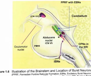
Figure 1.6: Illustration o f the Brainstem and Location o f Burst N eurons...40
Figure 1.7: An Illustration o f the Main Cortical Areas Involved with the Control and and Generation o f Saccadic Eye Movements ... ... ...43
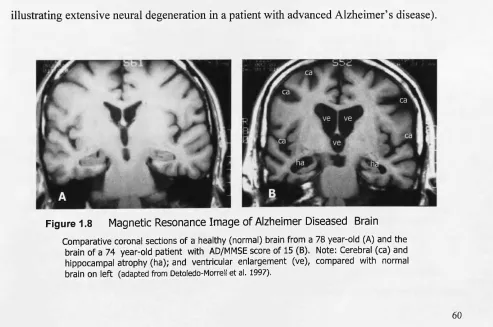
Figure 1.8: Magnetic Resonance Image o f Alzheimer Diseased Brain ...60
Chapter 2 Figure 2.1: Dementia Patients Participation over tim e ... ... 83
Figure 2.2: Control Participant Recruitment Response Rates for the Range of Promotional Methods ... ... ...85
Figure 2.3: Elderly Controls Participation over tim e ...86
Figure 2.4: Calculating the Visual Angle o f the Stimulus ...94
Figure 2.5: Experimental Conditions Prosaccade and Antisaccade Paradigms... 96
Figure 2.6: Experimental Conditions NO-GO and GO / NO-GO Paradigms... 99
Figure 2.7: The Express Eye H eadset ...102
Figure 2.8: Mechanical adjustment o f infrared emitter/sensor apparatus ... 103
Figure 2.9 : Recording o f saccadic eye movements ... 105
Figure 2.10: Antisaccade Gap Task: Correct Primary Saccade, Uncorrected Error and Corrected E rro r... 107
Figure 2.11: The Day/Night T e st... 119
Figure 3.1: An Illustrative Representation of Responses in the Antisaccade ‘ Gap’ Task Displaying Temporal and Spatial Characteristics of the
Visual Stim ulus... 132
Figure 3.2: An Illustration o f the Working Memory M o d e l... 134
Figure 3.3: Inhibitory Errors for Alzheimer’s Disease Compared with Dementia o f
other types and Elderly Controls in Voluntary Saccade T a sk s... 155
Figure 3.4: A Scatter Plot Illustrating the Relationship between Alzheimer’s
Disease Patients’ Inhibitory Error During the Antisaccade Gap Task
and Digit Span Reverse Test S c o re... 161
Figure 3.5: Stacked Bar Charts Illustrating the Proportions of Correct Saccades,
Corrected Errors and Uncorrected Errors by Sub-group for the
Antisaccade Gap T a sk ... 164
Figure 3.6: An Illustration using the Unitary Ratio to Display the Ratio for the
Proportion o f Correct Saccades to Inhibitory Errors Compared to the Proportion o f Correct Saccades+Corrected Error saccades to
uncorrected Errors in the Antisaccade Gap Task by Sub-group... 165
Figure 3.7: Graphs Displaying Correctness o f Performance for Sub-groups in the
Antisaccade Gap T a s k ...166
Chapter 4
Figure 4.1: The Forbes & Klein Model Illustrating the Functional Activity
Between Endogenous (ENDO) and Exogenous (EXO) Systems in the Control of Saccade (SAC) G eneration...192
Figure 4.2: Interaction Between Reflexive Fixation Offset and Group
(Dementia Patients and Elderly Controls)... 203
Figure 4.3: Interaction Between Reflexive Fixation Offset and Group
(Alzheimer’s disease, Elderly Controls and Other Dementia Types) 204
Figure 4.4: Histograms Displaying the Frequency o f Saccade Latency in the
Reflexive Gap and Overlap T a sk s... 205
Figure 4.5: A Line Graph Illustrating the Fixation Offset Effect Within Sub-groups
for the Antisaccade T a s k ...210
Figure 4.6: Histograms Displaying the Frequency o f Antisaccade Saccade Latency
in the Gap and Overlap T a s k ... 211
Figure 5.1: Illustration locating the Basal Ganglia in the Human B ra in ...224
Figure 5.2: Inhibitory Errors Across Voluntary Saccade T a sk s...237
Figure 5.3: Stacked Bar Charts Illustrating the Proportions o f Correct Saccades,
Corrected Errors and Uncorrected Errors Including Parkinson’s Disease Patients and Young C ontrols... 242
Figure 5.4: An Illustration using the Unitary Ratio to Display the Ratio for the
Proportion o f Correct Saccades to Inhibitory Errors Compared to the Proportion o f Correct Saccades + Corrected Error saccades
to uncorrected Errors in the Antisaccade Tasks by Sub-group... 243
Figure 5.5: Graphs Displaying Correctness o f Performance in the Antisaccade Gap
Task for Parkinson’s Disease Patients and Young C ontrols... 244
Figure 5.6: The Magnitude o f Fixation Offset Effect in the Reflexive Saccade
Paradigm for Young Controls and Parkinson’s Disease Patients
Compared with Elderly Controls and Dementia P atien ts...248
Figure 5.7: Histograms Displaying the Frequency of Saccade Latency in the
Reflexive Gap and Overlap T a sk s...250
Chapter 6 no figures
Chapter 7
Figure 7.1: Graph Displaying the Interaction Between SMMSE Test Session and
Group (Alzheimer’s Disease and Elderly Controls)...281
Figure 7.2: Graphical Representation of the Interaction Between EADAS cog Test
Session with Group (Alzheimer’s Disease and Elderly Controls)... 283
Figure 7.3: A Graph Displaying the Three-Way Interaction Between Digit Span Test
Session (baseline, 6 months, 12 months, 18 months), Digit Span Test
(Forward and Reverse) and Group (ADs and E C s)...286
Figure 7.4: Graphical Representation of the Interaction Between Verbal Fluency Test
Session and Group (Alzheimer’s Disease and Elderly Controls)...295
Figure 7.5: A Graphical Representation of the Three-Way Interaction Between Trail
Making Test Session (baseline, 6 months, 12 months, 18 months), Trail Making Test (Form A and Form B) and Group (ADs and E C s)... 299
Figure 7.6: A Graph Representing the Interaction Between National Adult Reading
Performance on the Antisaccade T a sk ... 312
Figure 7.8: A Graph to Display Longitudinal Antisaccade Uncorrected Error Rates
for Alzheimer’s Disease Patients and Elderly Controls... 313
Figure 7.9: A Graphical Representation of Reflexive Saccade Fixation Offset Effect
Over Time for the Alzheimer’s Disease and Elderly Control G roups... 316
Figure 7.10: Reflexive Saccade Fixation Offset Effect: Two-Way Interactions at each Test Session for the Alzheimer’s Disease and Elderly Control G roups... 317
Figure 7.11: A Bar Chart Displaying the Reflexive Saccade Fixation Offset Effect for Elderly Control Participants and Alzheimer’s Disease Patients
Over T im e ...334
Figure 7.12: A Graphical Representation o f Longitudinal Reflexive Saccade Overlap Task Latency with Projected Trend line to Estimate Future
Saccade L atency... 335
Chapter 8
Figure 8.1: Receiver Operating Characteristic Curves for Neuropsychological
Assessment and Eye Movement Models with Dementia P atien ts...358
Figure 8.2: Antisaccade Gap Task Error Correction R a te ...360
Chapter 9
Figure 9.1: The Components o f Working Memory and Inhibitory Control Co-vary
Depending on the Nature of a Given T a s k ...373
Figure 9.2: The Forbes & Klein Model Illustrating the Functional Activity
Between Endogenous (ENDO) and Exogenous (EXO) Systems in the Control o f Saccade (SAC) Generation. Modified to Highlight
the Theorised Neurodegenerative Links for Alzheimer’s D isease 378
Page Chapter 1
Table 1.1: The Classification o f Eye M ovem ents...29
Table 1.2: Brainstem Innervation o f Extraocular M uscles... 30
Chapter 2 Table 2.1: Clinical Investigations...82
Table 2.2: Composition o f Dementia Patient C andidates...84
Table 2.3: Generic M edications... 89
Table 2.4: CAPE Scoring System for the Gibson Spiral M a z e ...121
Chapter 3 Table 3.1: Prepotent Responses, Alternative Responses and Working Memory Demands for the Voluntary Saccade Tasks and Working Memory Tasks in the Study I, Following the Roberts, Hager and Heron F ram ew ork... 143
Table 3.2: Clinical Rating Scale and Neuropsychological Assessment S co res 149 Table 3.3: Descriptive Statistics for Error A nalysis... 153
Table 3.4: Pair-wise Within-Group Comparisons of Voluntary Saccade Task Inhibitory Errors Corresponding to Figure 3 .3 ... 158
Table 3.5: An Analysis o f Relationships Between Voluntary Saccade Task Inhibitory Error and Psychometric Test S co res... 160
Table 3.6: Correlations Between Inhibitory Errors and Psychometric Test Scores for Dementias o f Other T y p e ... 162
Table 3.7: The Inter-saccadic Interval for Corrected Errors in the Antisaccade Gap T a sk ... 172
Chapter 4 Table 4.1: Clinical Rating Scale S co res... 199
Table 4.4: Descriptive Statistics for Oculomotor Measures in the Antisaccade
Paradigm ... 209
Table 4.5: Attenuation o f the Fixation Offset Effect in the Antisaccade Paradigm ...212
Table 4.6: Descriptive Statistics for Inhibition Error Rates in the
Antisaccade Paradigm ... 214
Chapter 5
Table 5.1: Clinical Rating Scale and Neuropsychological Assessment Scores to
Include Parkinson’s Disease Patients and Young Control Participants...233
Table 5.2: Between-Groups Statistical Analyses for Clinical Rating Scale and
Neuropsychological Assessment S co res... 234
Table 5.3: Descriptive Statistics for Error Analysis with Parkinson’s Disease
Patients and Young C ontrols... 236
Table 5.4: Within-group t-tests Comparing Proportions o f Correct Primary
Saccades, Corrected Errors and Uncorrected E rro rs... 245
Table 5.5: Descriptive Statistics for Reflexive Saccade Latency with Parkinson’s
Disease Patients and Young Controls Added to the g ro u p s... 247
Table 5.6: Descriptive Statistics for Antisaccade Latency with Parkinson’s Disease
Patients and Young Controls Added to the g ro u p s...251
Chapter 6
Table 6.1: Education, Clinical Rating Scale and Neuropsychological Assessment
Scores for Medicated and Non-Medicated Dementia P atien ts... 263
Table 6.2: Descriptive Statistics for Error A nalysis...264
Table 6.3: Saccade Latency for Reflexive and Antisaccade Paradigms... 265
Chapter 7
Table 7.1: Descriptive Statistics for Longitudinal Clinical Rating Scale and
Neuropsychological Assessment Scores...278
Table 7.2: Longitudinal Statistical Analyses (ANOVA) Between-Groups for Clinical
Rating Scales and Neuropsychological Assessments...280
Table 7.3: Longitudinal Gibson Spiral Maze Credit Scores (CAPE Scoring System).. 290
305
325
.331
342
343
345
349
350
352
353
355
356
362 Longitudinal Statistical Analyses (ANOVA) Between-Groups for
Saccadic Eye Movement D a ta ...
Longitudinal Statistical Analyses (ANOVA) Between-Groups for Spatial Span Total Scores...
Inhibition Error Rate in Studies I and III compared with Baseline measurement in Study V ...
Descriptive Statistics for Saccadic and Neuropsychological Variables ...
Possible Positive and Negative Outcomes from the Logistic Regression A nalyses...
Correlations Between Clinical Rating Scales Scores, Saccadic
Variables and Neuropsychological Assessment Scores...
Accumulative Loss for the Logistic Regression Model with
Neuropsychological A ssessm ents...
Parameter Estimates for the Logistic Regression Model with
Neuropsychological A ssessm ents...
Accumulative Loss for the Logistic Regression Model with Saccadic Eye Movement V ariables...
Parameter Estimates for the Logistic Regression Model with Saccadic Eye Movement V ariables...
Accumulative Loss for the Logistic Regression Model with Saccadic Eye Movement and Neuropsychological Assessment V ariables...
Parameter Estimates for the Logistic Regression Model with Saccadic Eye Movement and Neuropsychological Assessment V ariables...
Chapter One
Introduction to the Study of Eye Movement
Research in Alzheimer’s Disease
1.1
Introduction to the Study of Eye Movements
Eye movement research offers the scientist (and clinician) a valuable tool with which to
gather important information regarding activity in oculomotor control systems, brain function
and the localisation o f disease. Using eye movements as a model system to study the
regulation o f neural activity provides the researcher with a number of benefits over other motor
systems. Leigh and Zee Leigh (1999) outline the following points: i). Choosing from a range
of oculographic technologies, it is possible to record accurate measurements of eye movement
activity as rotations of the eyes are limited to three planes; ii). Eye movements fall into a
number of different categories which correspond with visual activity, physiology and
neuroanatomical substrates; iii) As the mechanical load that the eye muscles move against is
constant, there is a lack o f monosynaptic stretch; iv) Eye movement abnormalities are often
characteristic of a particular pathophysiology, anatomical location or pharmacological
disturbance.
Eye movements have been used extensively in the study of psychiatric and neurological
illness taking advantage of neuropsychological insights, derived from versatile experimental
design. Detection o f the cortical structures involved in the control of saccadic eye movements
revealed by research employing various neuroimaging techniques, animal models and human
lesion investigations, has highlighted the crucial role of the prefrontal cortex and the parietal
lobes (Comelissen et al., 2002; Guitton, Buchtel & Douglas, 1985; Kimmig et al., 2001; Law,
Svarer, Rostrup & Paulson, 1998; Nieuwenhuis, Ridderinkhof, Blom, Band & Kok, 2001;
Paus, Petrides, Evan & Meyer, 1993; Pierrot-Deseilligny, 1991; Pierrot-Deseilligny, Milea &
Miiri, 2004; Pierrot-Deseilligny, Rivaud, Gaymard, Mtiri & Vermersch, 1995; Pierrot-
Deseilligny, Ploner, Miiri, Gaymard & Rivaud-Pechoux, 2002; Schall, 2004; Schlag & Schlag-
Rey, 1987; Sweeney et al., 1996). Thus, a profile of disturbance indicated by performance on
specific saccadic eye movement paradigms (saccadic eye movements and paradigms are
outlined in Section 1.3) can give a valuable insight o f brain dysfunction and oculomotor
control.
Neuropsychological research has been employed widely as a means of investigating
sensorimotor integration and executive function (see Section 1.1.1), yielding connections with
high-level cognition. Planned control of action and cognition is governed by the prefrontal
cortex (dorsolateral prefrontal cortex, the frontal eye fields, the supplementary eye fields, and
the anterior cingulate cortex), linked with sub-cortical areas of the brain via distinct neural
pathways (Section 1.4). Neuropsychological enquiry has thus utilized eye movement
methodology extensively to probe executive function. The field of eye movement research
benefits from a range o f accurate recording systems that has the potential to deliver a plethora
o f measurements and behavioural information.
Behavioural oculomotor paradigms have indicated selective impairments in
neurological patients, psychiatric patients and other groups such as dyslexics, highlighting the
potential o f eye movements to reveal abnormalities. Eye movement research on patients with
schizophrenia has revealed deficits in smooth pursuit (see section 1.2.1 gaze shifting) eye
movements (Broerse, Crawford & den Boer, 2001; Crawford & Broerse, 2001; Crawford et al.,
1998; Diefendorf & Dodge, 1908; Holzman, Proctor & Hughes, 1973) and with saccadic eye
movements where patterns o f cognitive dysfunction have been elucidated (Crawford &
Broerse, 2001), as identified by deficits of inhibitory control (McDowell & Clementz, 1997;
Sereno & Holzman, 1995), prolonged latency (Hutton & Kennard, 1998; Klein, Heinks,
Andresen, Berg & Moritz, 2000a) and saccadic accuracy (McDowell, Myles-Worsley, Coon,
paradigms has also revealed a number of abnormalities including dysmetric responses and a
characteristic multi-stepping pattern in the primary response, using a remembered target
location paradigm (Crawford, Henderson & Kennard, 1989b). Additionally, abnormalities
have been found for antisaccade latency and error rates (Briand, Strallow, Hening, Poizner &
Sereno, 1999) and the relationship between antisaccade latency and error rates and clinical
symptoms in Parkinson’s disease (Briand et al., 1999; Kitagawa, Fukushima & Tashiro, 1994).
A range o f saccadic abnormalities have been revealed in dyslexia from erratic saccadic
eye movements in visual tracking (Pavlidis, 1981) and reduced centre-of-gravity effect in a
double-spot paradigm (Crawford & Higham, 2001), to possible attentional deficits where
dyslexic participants produce high frequencies of express saccades (Fischer & Weber, 1990).
Additionally, dyslexic participants have been found to have poor fixation control, lower
vergence amplitudes and poor smooth pursuit compared with controls (Eden, Stein, Wood &
Wood, 1994). A study into patients suffering from human immunodeficiency virus (HIV)
discovered that abnormal saccadic accuracy (amplitude) was a sensitive measure between
patients and healthy control participants (Merrill, Paige, Abrams, Jacoby & Clifford, 1991).
Huntington’s disease (Lasker, Zee, Hain, Folstein & Singer, 1987, 1988) and
progressive supranuclear palsy (Pierrot-Deseilligny, Rivaud, Pillon, Fournier & Agid, 1989)
are two additional diseases where eye movement abnormalities have been revealed. A further
line o f enquiry in the study of eye movements has been to conduct research on patients with
dementia and o f particular importance for this thesis, the study eye movements in Alzheimer’s
disease (AD). A review of these studies can be found in Section 1.5.
Eye movement research provides a conduit by which researchers can thus understand
more thoroughly, the neurocognitive systems underlying oculomotor processes; for example,
inhibition of prepotent response and self-monitoring by evaluating error correction. The
relative ease by which eye movement data can be collected in the laboratory or clinical setting
demonstrates the neuropsychological utility of oculomotor methodology and its capacity to
study both reflexive and complex behaviour (Leigh & Kennard, 2004). This property is
particularly useful given the encumbrance of secondary behavioural characteristics that present
in certain diseases (e.g. Alzheimer’s disease), which can overshadow primary cognitive
dysfunction.
This thesis will focus on the investigation of primary horizontal saccadic eye
movements and explore saccadic error and correction, self-monitoring, attention and a variety
of temporal and spatial measurements in dementia patients o f the probable Alzheimer type.
The research employs a range of oculographic paradigms, utilising involuntary and voluntary
oculomotor methodology.
1.1.1 Executive Function and Cognitive Terminology
The present thesis uses some terminology that is often applied vaguely in the wider
literature, including the terms: executive function, working memory, visual attention, and
inhibitory control. Therefore, this section aims to clearly define these terms and show how
they are related in the context of the saccadic eye movement research described throughout the
chapters that follow.
Executive Function: The term executive function stems from traditional theories of
working memory (Baddeley, 1986) and is used in the present thesis to refer to higher-order
cognitive processing for purposeful action such as planning, self-regulation, monitoring,
volition and problem solving, i.e. the flexible control of cognition and action. The issue of
there being a central control mechanism (such as the central executive in Baddeley’s original
model o f working memory) that controls the various mechanisms o f cognitive control (e.g.
memory and attention) remains a source of debate. There is a substantial amount of evidence
to support the concept of a control mechanism that integrates the various cognitive functions
and motor control (see Sections 1.4.2.3 and 3.1). Whereas in the past the central executive
from Baddeley’s working memory model may have been considered for this purpose, in the
prefrontal cortex function’ (Miller & Cohen, 2001). In Miller and Cohen’s theory executive
function, i.e. cognitive control, is orchestrated by the prefrontal cortex through the “ ... active
maintenance of patterns of activity that represent goals and the means to achieve them” (p.
171) and via the resolution of competitive processes between weak task-relevant information
and stronger (automated) task-irrelevant information pathways, to achieve goal-directed
behaviour. Furthermore, Miller and Cohen’s theory corresponds with Massen’s hypothesis for
the parallel programming of exogenous (externally stimulated) and endogenous (internally
generated) components in volitional saccade tasks (Massen, 2004). Massen’s approach
exemplifies the notion of task-relevant information (e.g. goal = antisaccade) and task-irrelevant
information (e.g. antisaccade error = automated/reflexive saccade) and is therefore useful in
explaining the inhibitory mechanisms responsible for successful completion o f the antisaccade
task and how inhibition errors may occur (see Section 1.3, 1.3.1, 1.3.2 & 1.3.2.1). A more
detailed account of these theoretical constructs is discussed in Chapter 3, Section 3.1.
Working Memory: In the present thesis the term working memory is used to describe
an active store which can hold information for short periods of time (i.e. short-term memory)
for online processing and manipulation. Thus, working memory is part of executive function
where information can be integrated with long-term memory and other cognitive modules e.g.
prior to motor action, and can produce dynamic outcomes for example in arithmetic and
problem solving by the manipulation of task rules and goals. This definition o f working
memory is basically the same as in Baddeley’s model (1986), except that it is used here to
describe executive functioning from within the framework of Miller and Cohen’s integrative
theory o f prefrontal cortex function. Miller and Cohen’s theory is useful as a fundamental
theoretical construct for executive function, where the prefrontal cortex is viewed as key to the
active maintenance of task rules and goals. Allied to this theory the goal activation approach
o f Nieuwenhuis and colleagues applies the connectionist modelling of Miller and Cohen’s
theory (and others) in the context o f antisaccade task (Nieuwenhuis, Broerse, Nielen & de
Jong, 2004). Central to this approach and commensurate with Miller and Cohen’s theory, the
level by which a given task goal is activated is vital to the success o f volitional control.
Although task requirements may be fully understood by participants, goal activation failures
result in goal neglect, which Nieuwenhuis et al. consider to be a characteristic o f executive
dysfunction. Nieuwenhuis et al.’s goal activation approach directly supports Miller and
Cohen’s theory of prefrontal cortex function, and is a useful framework when attempting to
explain why failures to consistently focus attention on task requirements may occur (see
Chapter 3, Section 3.1). In summary, Miller and Cohen’s theory of prefrontal cortex function
provides a contemporary framework for understanding working memory function (i.e.
executive control) and the goal activation approach of Nieuwenhuis et al. can be
conceptualised as a function of working memory.
Visual Attention and Inhibitory Control: Visual attention can be externally/exogenously
stimulated or internally/endogenously generated. This can result in an overt shift o f attention
with an eye movement (to salient objects or events of interest) or in covert attention without an
eye movement (Humphreys & Bruce, 1995). Furthermore, attention can be broadly
categorised into i) selective attention, ii) sustained attention and iii) divided attention.
Selective attention is where attention is directed to a particular stimulus whilst ignoring other
irrelevant stimuli. Sustained attention is the ability to maintain an attentional focus for a
prolonged period e.g. in a visual fixation task. Divided attention can be defined as the ability
to share attention over more than one process at a time e.g. during dual task experiments
(Peny & Hodges, 1999).
The prefrontal cortex has long been associated with endogenous selective visual
attention and inhibitory control (Banich et al., 2000; Chao & Knight, 1997; Doricchi et al.,
1997; Fukuyama et al., 1997; Kimberg & Farah, 2000; Lecas, 1995; Mishkin, 1964; Posner &
Petersen, 1990). Therefore, in the present thesis visual attention and inhibitory control are
viewed from the perspective of Miller and Cohen’s integrative theory of prefrontal cortex
function (Miller & Cohen, 2001). The theory views attention and inhibition as a reflection of
behaviour stemming from a single underlying mechanism of cognitive control, following on
from competition between processing pathways that are responsible for task performance. This
corresponds with the biased competition model o f Desimone and Duncan in which attention is
the result o f biasing competition in support o f task-relevant information, and inhibition is the
consequence o f the attentional biasing against the irrelevant information (Desimone & Duncan,
1995) i.e. attending to a stimulus automatically results in the inhibition o f other stimuli.
Chapter 3, Section 3.1 explains (in the context o f the present thesis) how attentional processing
and working memory are closely related in endogenous tasks and how the success of
attentional processing relies on the extent to which a particular goal is activated in working
memory.
1.2
The Importance of Eye Movements for Foveation
The visual system in humans has evolved to elicit functionally specific, useful and
helpful information for the problem solving brain, thereby enhancing fitness and ultimately
facilitating continued existence in the natural world. Inextricably linked to the fully
operational healthy visual system, various types o f eye movements play a crucial role in
enabling the eyes to scan a scene, track a moving target and to locate objects o f interest
through a combination o f movements and fixations. Therefore, eye movements perform two
vital functions: firstly they serve to shift the direction o f gaze and secondly, to stabilise the
position o f gaze so that the image, perhaps first detected in peripheral vision, falls onto the
fovea (Figure 1.1). Thus, eye movements can actually facilitate foveation independently o f
head movements in foveate animals (Delgado-Garcia, 2000), but also serves to counter
movements o f the head that would otherwise disrupt visual processing due to sweeping visual
stimulation across the retina.
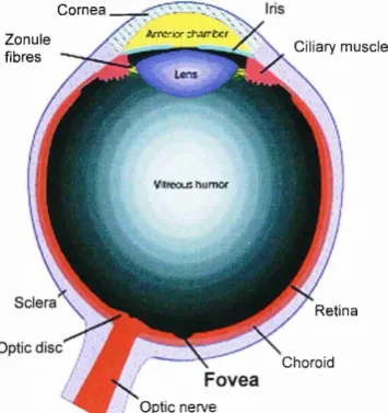
Figure 1.1 Diagram Illustrating a Cross-section of the Human Eye Highlighting the Location of the Fovea (sagittal section, adapted from Zigmond etal., 1999)
C o rn ea
A r r o x rtvurfccf Zon ule
fibres C iliary m uscle
V .tr e o u s h iin r v c f
R etina
Choroid
[image:28.528.260.438.594.783.2]This function is very important for the visual system as the fovea, which is
approximately centred on the visual axis serving 1° o f visual field (Hughes, 1975), is the point
o f highest resolution on the retina (Hess, Burgi & Bucher, 1946; Jacobs, 1979; Perry & Cowey,
1985).
The retina is a highly complex part o f the central nervous system (CNS), comprising a
multifaceted array o f photoreceptors (i.e. rods and cones) and three layers o f ganglion cells
(with five different cell types) that enable temporal, spatial and chromatic aspects o f visual
processing in the physical world (Figure 1.2). Vitally significant to the present topic, retinal
ganglion cells consist o f two major categories, M cells and P cells. M cells receive most o f
their input from rod photoreceptors, whereas P cell input is derived mainly from cone
photoreceptors. M cells and P cells form the basis o f two morphologically and physiologically
distinct visual channels. The channels project from the retina via the optic nerve, through the
optic chiasm and on to form the optic tracts. The optic tracts proceed to the dorsal lateral
geniculate nuclei (LGN), which are linked to the striate cortex (primary visual cortex) via the
optic radiations to form the magnocellular and parvocellular pathways (Leventhal, Rodieck &
Dreher, 1981; Perry & Cowey, 1981; Perry, Oehler & Cowey, 1984). Retinotopic mapping is
maintained at each level o f the retina - geniculate - striate pathway (Figure 1.3).
rod cone
pigmented layer {
i j - ,, | f
photoreceptors (rods & cones)
} outer plexdorm layer
} inner plexiform layer
axons ot retinal ganglion cells form the optic nerve
light light
Figure 1.2 A Diagram to Illustrate the Complex Layers of the Human Retina
intcrblob
visual co rtex
retin a
blobs
parvoccllular %,
layers y
T
LG N magnocellular %
layers
contralateral: ipsilatcral:
M-type M-typc
ganglion ganglion
cell cell
contralateral: P-type ganglion cell
ipsilatcral: P-typo ganglion cell
Figure 1.3 Illustration Representing the M and P Visual Pathways
(from Hall & Robinson, 1998)
The characteristics o f a third channel, the koniocellular pathway links with a third
category o f ganglion cell, W cells which are not relevant to the focus o f the present thesis.
There is a variation in the quantities o f M ganglion cells and P ganglion cells at retinal
eccentricity. The fovea and parafoveal areas o f the retina contain a higher density o f P
ganglion cells than M ganglion cells, whereas M ganglion cells are evenly distributed across
the retina. P ganglion cells are physiologically more sensitive to images o f high contrast and
low spatial frequencies and M ganglion cells more sensitive to low contrast and high spatial
frequencies (Derrington & Lennie, 1984). Thus, foveation enables visual perception via the
area o f the retina with highest visual acuity, where the P ganglion cells o f the parvocellular
system are most prolific. This physiology provides the visual system with mechanics for a
parallel dual-processing system, where the high resolution o f a foveated image enables detailed
analysis and focused attention. Superior sensitivity to high spatial frequencies, motion and low
contrast in peripheral areas o f the retina - afforded readily by the magnocellular system -
permits the visual system to easily detect movement and objects o f potential interest for
[image:30.529.90.406.60.303.2]subsequent eye movements (and head and limb movements). Objects or salient events entering
peripheral vision frequently trigger a reflexive ocular movement, known as the visual grasp
reflex (VGR; Hess et al., 1946; Ingle, 1973), orienting the eyes so as to foveate an image of the
object. The eye movement facilitates foveal fixation to within roughly 0.5° of midpoint on the
fovea (Leigh & Zee, 1999). Essentially, the combined physiological characteristics of foveal
and peripheral vision have evolved to provide an efficient system for survival in nature.
1.2.1 Stabilising and Shifting Gaze
Fundamentally important to the visual system, there are two versatile groups of eye
movements that facilitate efficient foveation, gaze-stabilising and gaze-shifting mechanisms.
By definition, gaze-stabilisation mechanisms serve to maintain a given visual input on the
fovea, whereas gaze-shifting mechanisms provide the capacity for conveying an image onto the
fovea (Leigh & Zee, 1999). There are numerous classes of gaze-stabilisation eye movements,
which include the vestibulo-ocular system, the optokinetic system, smooth pursuit, visual
fixation and vergence, where the eyes are able to binocularly converge or diverge
disconjugately, as a target moves towards or away from the eyes. Stabilisation of gaze is
activated automatically as a reflexive compensatory strategy (the vestibulo-ocular reflex;
VOR) during head movements and thus retains foveation. The labyrinthine semicircular canals
possess angular acceleration sensors that mediate the VOR. A combination of the VOR and
supplementary optokinetic system correction, achieves accurate stabilisation across a range of
head movements and postures (Robinson, 1977). The optokinetic system provides visually
mediated saccades, as a result of sustained rotation when the VOR signal declines.
Mechanisms for gaze-shifting include quick-phase nystagmus, which resets the eyes to
their normal working range so as to view objects and subsequent scenes during regular bodily
rotations. Part o f the vergence system also falls into this category of eye movement, enabling
range. A further type o f gaze-shifting mechanism is the saccadic system. Leigh and Zee
(1999) provide the a useful summary for the functional classification o f eye movements in
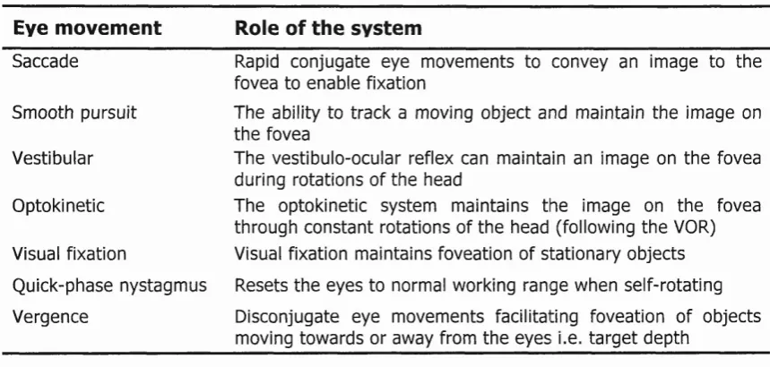
Table 1.1.
Table 1.1 The Classification o f Eye M ovem ents (adapted from Leigh & zee 1999)
Eye movem ent Role of the system
Saccade Rapid conjugate eye m ovem ents to convey an image to the fovea to enable fixation
Smooth pursuit The ability to track a moving object and maintain th e image on th e fovea
Vestibular The vestibulo-ocular reflex can maintain an image on th e fovea during rotations of th e head
Optokinetic The optokinetic system maintains th e image on th e fovea through co n stan t rotations of th e head (following th e VOR)
Visual fixation Visual fixation maintains foveation of stationary objects
Quick-phase nystagm us Resets th e eyes to normal working range when self-rotating
Vergence Disconjugate eye m ovem ents facilitating foveation of objects moving tow ards or away from th e eyes i.e. ta rg e t depth
1.2.1.1
Mo ving the Eye
The eye is positioned in the orbit, a socket-type recess in the front o f the skull. It is
held in position by three pairs o f extraocular muscles, which are able to move the eye with
synergistic action through horizontal, vertical and oblique directions (Figure 1.4).
S u p erio r rectus (turns eye upward
and inward) S u perior oblique (turns eye dow nw ard and
M edial rectus (turns eye inward)
Figure 1.4 The Extraocular Muscles
(adapted from Zigmond etal., 1999) 29
Inferior oblique (turns eye upward
and outw ard)
Inferior rectus (turns eye dow nw ard and
inward)
[image:32.527.40.470.162.367.2] [image:32.527.155.374.518.746.2]Horizontal eye movements (from side-to-side) are implemented by the lateral and
medial recti muscles. Vertical eye movements (up and down) are facilitated by the superior
and inferior recti muscles, whereas rotational eye movements are enabled by the superior and
inferior oblique muscles. Brainstem motor neurons innervate the extraocular muscles.
Specifically, this involves in the third {oculomotor), fourth {trochlear) and sixth {abducens)
cranial nerve nuclei (Sparks, 2002). The functions o f the cranial nerves involved in eye
movements are displayed in Table 1.2 below.
Table 1.2 Brainstem Innervation of Extraocular Muscles
Cranial Nerve Extraocular Muscles
Oculom otor (III) Ipsilateral medial and inferior rectus, contralateral superior rectus and inferior oblique Trochlear (IV) Contra-lateral superior oblique
A bducens (VI) Inpsilateral lateral rectus
The primary area of investigation for this thesis is the saccadic eye movement, which is
discussed in Section 1.3 below. Further discussion detailing the neurological control of
saccadic eye movements can be found in Section 1.3.1.
1.3
Saccadic Eye Movements
Fundamental to day-to-day vision saccadic eye movements are generated for example,
when we read text and thus serve to shift gaze direction and minimize drift of retinal image
between fixations. The word saccade can be defined as ‘jum p’ and saccades may occur as a
series o f rapid conjugate jerks of the eyes, which can be horizontal, vertical or oblique. When
a saccade is executed direction cannot be altered, thus, the saccade is a ballistic movement of
the eyes facilitating efficient foveation for a given fixation point. The saccadic system enables
the eyes to make rapid shifts of gaze from one point to another, with a peak velocity of up to
700V 1 for large amplitude saccades (Becker, 1991). There is a consistent saturating
duration and amplitude are also related linearly). As saccade size increases the faster the speed
o f the movement. The relationship between saccade velocity and amplitude is often termed the
main sequence (Bahill, Clark & Stark, 1975), and the main sequence is also found in
microsaccades and quick-phase nystagmus.
Saccadic behaviour manifests as two main categories, comprising involuntary and
voluntary eye movements. Classification of these two categories is discussed in the following
sections (1.3.1 & 1.3.2).
1.3.1 Involuntary Saccadic Eye Movements
Involuntary saccadic eye movements can be classified by a number of behavioural
characteristics. The most basic form is quick phases of vestibular nystagmus, resulting from
stimulation o f the vestibular or optokinetic system to realign the eyes as a consequence of drift
(Leigh & Zee, 1999). Involuntary saccades may appear spontaneously, without stimulation of
the visual system by internal or external cues. The rapid eye movement activity that takes
place whilst sleeping, is also involuntary. The end of Section 1.2 outlined the VGR, which is
a saccadic response that occurs as a result of the sudden appearance of an external stimulus
(visual, auditoiy or tactile). These saccades are frequently called reflexive saccades and
involve bottom-up processing (the term prosaccade is often used interchangeably with
reflexive saccade). However, the VGR is not a fully formed primary reflex as it can be
inhibited, for example, during the antisaccade task. Despite the fact that visually-guided
saccades involve an accurate motor system, they do not require response inhibition and
working memory. Therefore, the cognitive system is placed under a relatively low load, the
demand perhaps comparable with that required by visual attention (Broerse et al., 2001) where
there is focussed awareness by the visual system. Reflexive saccades {horizontal) are included
in the experimental design for the present study and a representation of the spatial
characteristics for this type o f saccade, are illustrated in Figure 1.5 (A) (a detailed account o f
the experimental paradigm can be found in Chapter 2, Section 2.3.3.1 & Figure 2.5(A)).
1.3.2 Voluntary Saccadic Eye Mo vements
Voluntary saccades are under volitional control and may be generated towards a known
activity. Volitional saccades can also be made to remembered locations. Additionally,
voluntary responses include anticipatory and predictive behaviour, for example, in searching
o f the target location or temporal characteristics. Thus, voluntary saccadic eye movements
high-level executive functions, that include planning, visual attention, anticipation, memory,
inhibition o f prepotent response (to the VGR) and sensory-motor integration. Voluntary
saccades are therefore a product o f top-down cognitive processing and can be considered as
concept-driven (see Section 1.4). One such volitional task is the antisaccade first used by
Hallett (1978).
stimulus location in a prosaccade fashion, in response to a command or during purposeful
for a target a saccade may be initiated ahead of appearance o f the stimulus, due to a prediction
invoke a higher load on the cognitive system, utilizing multiple cognitive centres including
Reflexive Saccade and Antisaccade Oculomotor Paradigms
A B
Reflexive Antisaccade
-4° 0‘ 4‘ O' 4‘
Figure 1.5 The diagram above illustrates the basic concept o f reflexive saccade (A) and antisaccade (B) paradigms. In the reflexive saccade paradigm a saccade is
produced directly towards the target. Conversely for the antisaccade paradigm a
[image:35.528.106.391.526.671.2]1.3.2.1
The Antisaccade Task
The antisaccade task is one of the most widely used paradigms in the study of voluntaiy
saccades and the inhibition of inappropriate action (Amador, Schlag-Rey & Schlag, 2004;
Dorris & Munoz, 1995; Everling, Dorris & Munoz, 1998a; Fischer, Gezeck & Hartnegg, 2000;
Mockler & Fischer, 1999; Roberts, Ralph, Hager & Heron, 1994), and places a high level of
demand on the cognitive system. During the task the eyes have to move to an equidistant
location in space, in the opposite hemifield (the mirror location) from where the target is
(Figure 1.5 (B) & 2.5 (B)). To achieve this the visual system must first avoid overt capture of
visual attention by the stimulus, which is presented randomly in the left or right visual field.
This is done by volitionally maintaining attention and visual fixation on the central fixation
point, thereby inhibiting the prepotent response created by the newly presented visual target
appearing in peripheral vision {note, the VGR would normally result in a prosaccade towards
the target). Concurrently, the top-down processing must also generate a representation of an
imaginary target location in the opposite hemifield from that of the actual target. This
endogenous process must to be initiated with sufficient time to spare so that the competing
saccade programme of the VGR can be overridden. Attending to the coordinates of the
imaginery target location, a volitional saccade must be generated immediately to the imagined
spatial location. Therefore, compared with involuntary reflexive saccades antisaccades incur
additional reprocessing time. This is due to the fact that attentional mechanisms inhibit
saccadic response and attention must shift to the opposite hemifield from that where the visual
stimulus is actually located (provided that attention was allocated in the first place).
1.3.2.2
Inhibition of Response Tasks
Alternative experimental paradigms can be designed to probe other aspects o f volitional
and inhibitory control. By manipulating the rules, saccadic inhibition tasks can be conducted
to exert higher levels of cognitive load thereby taxing the executive system. Due to the