http://www.scirp.org/journal/jct ISSN Online: 2151-1942
ISSN Print: 2151-1934
Molecular
Marker of Tumours
Marian Surma
Department of Physics, Adam Mickiewicz University, Poznań, Poland
Abstract
Molecular structure of the marker of tumour is determined by magneto-optical analysis of blood serum. The marker is the laevorotatory enantiomer of alanine. The cancer status of a subject is described by the number of molecules of the laevorotato-ry alanine enantiomer (−)ρ and the effectiveness of therapy is measured by the num-ber of molecules of the dextrorotatory alanine enantiomer (+)ρ. The values of (−)ρ and (+)ρ are determined separately for the patient before and after therapy.
Keywords
Cancer, Molecular Marker, Protein Markers
1. Introduction
Molecular structure of the marker of cancer status has not been known so far. The opt-ically active components of human blood are the free laevorotatory alanine enantiomer of the structure of an electric quadrupole and the dextrorotatory enantiomer of the structure of an electric dipole as well as some optically active protein structures. Protein markers provide information on the cancer disease status but with a large margin of error, the information is often unreliable for evaluation of the actual status of disease development and effectiveness of the therapy applied. As shown in paper [1], reliable information is provided by the magnetic field B2 induced angle of rotation bexp of the polarisation plane of light passing through the serum from the blood of the patient. The human blood serum in a magnetic field B2 becomes optically birefringent (Magneto- optical Circular Birefringence). This effect, MOCB, measured by the value of the angle of rotation bexp is an indicator informing about cancer status if the serum is optically laevorotatory (bexp < 0). The serum from the blood of patients after effective therapy and from healthy people is dextrorotatory (bexp > 0).
How to cite this paper: Surma, M. (2016) Molecular Marker of Tumours. Journal of Cancer Therapy, 7, 675-679.
http://dx.doi.org/10.4236/jct.2016.710070 Received: June 17, 2016
Accepted: September 19, 2016 Published: September 22, 2016
Copyright © 2016 by author and Scientific Research Publishing Inc. This work is licensed under the Creative Commons Attribution International License (CC BY 4.0).
http://creativecommons.org/licenses/by/4.0/
2. Experiment and Experimental Data
Protein markers have been dominant in use and in literature, however, it should be rea-lised that the procedures of serum denaturation destroys molecular structures of the optically active enantiomers which also provide information on the cancer status or ef-fectiveness of therapy or lack of cancer. The complexity of protein structures is illu-strated by the fact of the presence of 20 amino acids in a human cystatin C [2]. The presence of amino acids in different percent contributions in proteins is well known in literature [3]. Analysis of results of bexp parameter of MOCB marker for about 600 sam-ples of blood serum from subjects diagnosed with cancer at different degrees of the tu-mour development [4]-[6] permits making diagnosis on the basis of this parameter value. Although the structure of the marker is not known yet, on the basis of analysis of bexp < 0 it can be concluded that it contains laevorotatory alanine Ala(). This result means that in these serum samples the contribution of laevorotatory enantiomers do-minates over the content of dextrorotatory ones. The laevorotatory enantiomer of ala-nine is a molecular marker of tumour. This conclusion is supported by: MOCB data analysis [4]-[6], Follicle Receptor protein marker analysis [7] and literature data (tables 3-1 [3]).
According to literature, the alanine enantiomers make about 7.8% of amino acid re-sidues in the patient blood.
The authors of [7] report results of protein marker determination for typical tumours “selectively expressed on the surface of the blood vessels of a wide range of tumours lo-cated in the prostate, breast, colon, pancreas, urinary bladder, kidney, lung, liver, sto-mach, testis and ovary” to support the indications obtained by the protein marker Fol-licle Receptor. These data are compared with the independent method based on deter-mination of MOCB marker in blood serum from the patients diagnosed with the same tumours: prostate, breast, lung, ovary and kidney. The comparison between the results obtained by the protein marker and the MOCB marker indicates that the values of both markers are determined by the presence of the laevorotatory enantiomer of alanine Ala() in the blood serum of cancer patients. In the blood serum from healthy people and patients after successful therapy the dextrorotatory enantiomer Ala(+) is dominant. Results of MOCB marker for a few chemically pure enantiomers have been reported in [8].
The alanine density is 1.42 g/cm3, and its molecular mass is M
e = 89.09 g/mol. Analy-sis of magneto-optical measurements of blood serum samples implies that the presence of cancer and advancement of its development are correlated with the content of lae-vorotatory, the electric quadrupole structure, enantiomer of alanine Ala().
3. Results
Comparison of the results of MOCB molecular marker and tPSA protein marker are given in Tables 1-3.
Table 1. Prostate.
bexp < 0; (1,2,3,4,5): different cancer patients. ()ρ = 1.79 ×10²² (−bexp )
Mocb tPSA tPSA Mocb/tPSA
105bexp/degT−2mm1 1018()ρ/mm3 ng/mL 103 g/mm3 102 degT2g1
1 −1.36 0.24 6.70 6.70 −0.20
2 −5.93 1.06 5.45 5.45 −1.08
3 −9.49 1.69 5.14 5.14 −1.84
4 −15.43 2.76 6.09 6.09 −2.53
5 −18.07 3.23 3.40 3.40 −5.31
bexp > 0; (1a,2a,3a,4a,5a): different recovered patients after effective therapy. (+)ρ = 1.79 ×10²² (bexp )
Mocb tPSA tPSA Mocb/tPSA
105bexp/degT2mm1 108(+)ρ/mm3 ng/mL 103 g/mm3 102 degT2g1
1a 1.45 0.26 1.87 1.87 0.77
2a 6.64 1.18 7.00 7.00 0.94
3a 9.56 1.71 2.37 2.37 4.03
4a 14.58 2.61 1.53 1.53 9.52
5a 18.63 3.33 2.26 2.26 8.24
Table 2. Prostate cancer patients and after effective therapy.
1. Patient P (1); Medical diagnose date 12.05.2003. Mocb
105bexp/degT2mm1 1018()ρ/mm3 tPSA/ng/mL
−5.93 1.06 5.45
After effective therapy RP1 medical diagnosis dated 28.01.2004. Mocb
105bexp/degT2mm1 1018(+)ρ/mm3 tPSA/ng/mL
8.85 1.55 5.35
2. Patient P (2); Medical diagnose date 12.05.2003. Mocb
105bexp/degT2mm1 1018()ρ/mm3 tPSA/ng/mL
2.22 0.39 5.76
After effective therapy RP2 medical diagnosis dated 10.03.2004. Mocb
105bexp/degT2mm1 1018(+)ρ/mm3 tPSA/ng/mL
14.58 2.60 8.00
3. Patient P (3); Medical diagnose date 02.06.2003. Mocb
105bexp/degT2mm1 1018()ρ/mm3 tPSA/ng/mL
−1.36 0.24 6.70
Mocb
105bexp/degT2mm1 1018(+)ρ/mm3 tPSA/ng/mL
For patients (1, 2, 3, 4) the results obtained by the two methods MOCB and protein markers are fully consistent: bexp < 0, tPSA > 4 ng/mL.
Disagreement of Mocb with tPSA data: Table 1 (5), bexp < 0, tPSA < 4 ng/mL. The value of the ratio MOCB/tPSA decreases. It means that the content of Ala() enantio-mers residues in the patients (1, 2, 3, 4, 5) serum increases as their data (bexp < 0) de-creases.
Development of tumour is manifested in increase in the content of Ala() in the blood serum of cancer patient and a negative value of MOCB marker, bexp < 0. Effectiveness of therapy is manifested as an increase in the content of Ala(+) in the blood serum and an increase in bexp > 0, Table 2 Cancer patients: P (1), P (2), P (3); bexp < 0, tPSA > 4 ng/mL, agreement of MOCB with tPSA data.
Recovered patient RP3; bexp >0, tPSA < 4 ng/mL, agreement of MOCB with tPSA da-ta. Recovered patients: RP1, RP2; bexp >0, tPSA > 5 ng/mL, disagreement of MOCB with tPSA data.
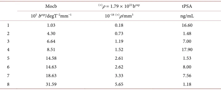
Agreement of MOCB with tPSA data: Table 3 (2,5,8), bexp > 0, tPSA < 4 ng/mL. Ta-ble 3 (1, 3, 4, 6, 7), bexp > 0. Marker Ala (+)dominant in the blood serum, (+)ρ ≠ 0 and in-crease as inin-crease bexp > 0.
4. Discussion
The authors of [7] analyze the presence of Follicle-Stimulating Hormone Receptor in tumour. All the above mentioned tumours contain the protein of the mass of Me ≈ 87,000 Dalton, which is indicated by the glycosylated FSH receptor. The results corres-pond to those based on MOCB marker, for example for prostate cancer Me ≈ 30,000 Dalton and on the data from [4] [5] monomers occur in tumours in contrast to dimers whose presence is natural in glycosylated FSH receptor. Their presence results, reported in table 3-3 [3], for the multimers forms of protein, are natural in glycosylated FSH re-ceptor. Their presence detected by the MOCB method suggests that the laevorotatory enantiomer of alanine Ala() present in the proteins ([3], tables 3-1), is a marker of tu-mour, irrespective of its localisation in the organism.
[image:4.595.194.553.561.708.2]The paper presents arguments supporting the use of alanine enantiomers Ala() and a molecular markers informing about the presence and state of development of cancer.
Table 3. Data for healthy subjects.
Mocb (+)ρ = 1.79 ×10²² bexp tPSA 105bexp/degT2mm1 1018(+)ρ/mm3 ng/mL
1 1.03 0.18 16.60
2 4.30 0.73 1.48
3 6.64 1.19 7.00
4 8.51 1.52 17.90
5 14.58 2.61 1.53
6 14.63 2.62 8.00
7 18.63 3.33 7.56
Alanine enantiomers Ala() and Ala(+) are present in the protein markers used in clinical treatment. The agreement and disagreements of MOCB marker and tPSA marker indi-cations are given in Tables 1-3.
Electric quadrupole moment and magnetic dipole moment were measured for opti-cally inactive structures of CH3Cl [9] and CO2[10]. Theoretical analysis of B2 field in-duced magneto-optical birefringence in optically active media is given in [11].
Analysis of the results presented in this paper indicates potential of a new method for diagnostics of cancer changes based on determination of the content of optically active alanine enantiomers Ala() and Ala(+) in the blood. The measurement of the content of the laevorotatory enantiomer Ala() provides reliable information on the presence of cancer changes. The effectiveness cancer therapy can be measured by the ratio of Ala()/ Ala(+).
References
[1] Surma, M. (2007) Magnetooptical Characterization of Human Blood Serum: Correlation between Neoplasmic Changes and Their Biomolecular Information Carriers. Physics and Chemistry of Liquids, 45, 271-279. http://dx.doi.org/10.1080/00319100600620912
[2] Jankowski, R., Kozak, M., Jankowska, E., Grzonka, Z., Grubb, A., Abrahamson, M. and Jaskulski, M. (2001) Human Cystatin C, An Amyloidogenic Protein, Dimerizes through Three-Dimensional Domain Swapping. Nature Structural Biology, 8, 316-320.
http://dx.doi.org/10.1038/86188
[3] Nelson, D. and Cox, M. (2005) Lehninger Principles of Biochemistry. 4th Edition,W.H. Freeman & Co, Table 3-1, 77, Table 3-3, 88.
[4] Surma, M. (2012) Cancer Status: MOCB and tPSA Prostate Cancer Markers. Journal of Cancer Therapy, 3, 1101-1103.http://dx.doi.org/10.4236/jct.2012.36144
[5] Surma, M. (2014) Enantiomers Present in Serum Carry Cancer and/or Recovery Status In-formation. Journal of Cancer Therapy, 5, 618-621.http://dx.doi.org/10.4236/jct.2014.56071
[6] Surma, M. (1998) Experimental Evidence of the B2 and B3 Dependent Circular Birefrin-gence of Chiral Molecules in High Magnetic Fields. Molecular Physics, 93, 271-278.
http://dx.doi.org/10.1080/00268979809482210
[7] Radu, A., Pichon, Ch., Antoine, M., Allorry, Y., Couvelard, A., Fromont, G., Hai, M.T.V. and Ghinea, N. (2010) Expression of Follicle-Stimulating Hormone Receptor in Tumor Blood Vessels. The New England Journal of Medicine, 363, 1621-1630.
http://dx.doi.org/10.1056/NEJMoa1001283
[8] Surma, M. (1999) Correlation between Quadratic Magnetic Field Induced Circular Bire-fringence and the Natural Optical Activity of Chiral Media. Molecular Physics, 96, 429-433.
http://dx.doi.org/10.1080/00268979909482976
[9] Buckingham, A.D. and Disch, R.L. (1963) The Quadrupole Moment of the Carbon Dioxide Molecule. Proceedings of the Royal Society, 273, 275-289.
http://dx.doi.org/10.1098/rspa.1963.0088
[10] Barron, L.D. and Vrbancich, J. (1984) Magneto-Chiral Birefringence and Dichroism. Mo-lecular Physics, 51, 715-730.http://dx.doi.org/10.1080/00268978400100481
Submit or recommend next manuscript to SCIRP and we will provide best service for you:
Accepting pre-submission inquiries through Email, Facebook, LinkedIn, Twitter, etc. A wide selection of journals (inclusive of 9 subjects, more than 200 journals)
Providing 24-hour high-quality service User-friendly online submission system Fair and swift peer-review system
Efficient typesetting and proofreading procedure
Display of the result of downloads and visits, as well as the number of cited articles Maximum dissemination of your research work