ScholarWorks @ Georgia State University
ScholarWorks @ Georgia State University
Biology Dissertations Department of Biology
Summer 6-18-2012
Tyrosine Phosphorylation of p68 RNA Helicase Promotes
Tyrosine Phosphorylation of p68 RNA Helicase Promotes
Metastasis in Colon Cancer Progression
Metastasis in Colon Cancer Progression
Chia Yi Liu
Georgia State University
Follow this and additional works at: https://scholarworks.gsu.edu/biology_diss
Recommended Citation Recommended Citation
Liu, Chia Yi, "Tyrosine Phosphorylation of p68 RNA Helicase Promotes Metastasis in Colon Cancer Progression." Dissertation, Georgia State University, 2012.
https://scholarworks.gsu.edu/biology_diss/117
IN COLON CANCER PROGRESSION
by
CHIA YI LIU
Under the Direction of Zhi-Ren Liu
ABSTRACT
The initiation of cancer metastasis usually requires Epithelial-Mesenchymal Transition (EMT),
by which tumor cells lose cell-cell interactions and gain the ability of migration and invasion.
Previous study demonstrated that p68 RNA helicase, a prototypical member of the DEAD-box
RNA helicases, functions as a mediator to promote platelet-derived growth factor
(PDGF)-induced EMT through facilitating nuclear translocation of β-catenin in colon cancer cells. In this
context, p68 RNA helicase was found to be phosphorylated at the tyrosine 593 residue (referred
as phosphor-p68) by c-Abl kinase, and this phosphorylation is required for the activation of
β-catenin signaling and the consequent EMT. The phosphor-p68 RNA helicase-mediated EMT was
characterized by the repression of an epithelial marker, E-cadherin, and the upregulation of a
membrane and thus inhibit its transcriptional activity. The functional loss of E-cadherin is the
fundamental event of EMT. Despite the role of phosphor-p68 RNA helicase in regulating nuclear
translocation of β-catenin, whether phosphor-p68 is involved in the regulation of E-cadherin
remains unknown. Here, our data indicated that phosphor-p68 RNA helicase initiated EMT by
transcriptional upregulation of Snail1, a master transcriptional repressor of E-cadherin. The data
suggest that phosphor-p68 RNA helicase displaced HDAC1 from the chromatin remodeling
MBD3:Mi-2/NuRD complex at the Snail1 promoter, thereby activating the transcription of
Snail1. In the xenograft tumor model, abolishing the phosphorylation of p68 RNA helicase by
the expression of Y593F mutant resulted in a significant reduction of metastatic potential in
human colon cancer cells. Analyses in the colon cancer tissues also revealed that the tyrosine 593
phosphorylation level of p68 RNA helicase is substantially enhanced in the tumor tissues
comparing to that in the corresponding normal counterparts, suggesting a correlation of
phosphor-p68 and tumor progression. In conclusion, we showed that tyrosine phosphorylation of
p68 RNA helicase positively correlated to the malignant status of colon cancer progression. The
molecular basis behind this correlation could be partly through the transcriptional regulation of
Snail1.
IN COLON CANCER PROGRESSION
by
CHIA YI LIU
A Dissertation Submitted in Partial Fulfillment of the Requirements for the Degree of
Doctor of Philosophy
in the College of Arts and Sciences
Georgia State University
Copyright by Chia Yi Liu
IN COLON CANCER PROGRESSION
by
CHIA YI LIU
Committee Chair: Zhi-Ren Liu
Committee: Ritu Aneja
Susanna F. Greer
Electronic Version Approved:
Office of Graduate Studies
College of Arts and Sciences
Georgia State University
ACKNOWLEDGEMENTS
I would like to thank Dr. Zhi-Ren Liu for accepting me into his laboratory and having the
patience to guide me throughout my graduate research. Thank you for your support and
encouragement. I would like to acknowledge my exceptional doctoral committee, Dr. Ritu Aneja
and Dr. Susanna Greer, for their continuous support and helpful suggestions on the improvement
of this dissertation work. I would like to especially thank Dr. Jenny Yang for always being there
for me when I needed help. Debby Walthall, thank you for your technical support and
willingness to help me when I needed it. LaTesha Warren, I could not have done this without
you, thank you for providing all the assistance in my graduate career. Special thanks to the
members in Dr. Liu’s lab. William and Yinwei, you have been good friends and thank you for all
the help in my graduate career. And thanks to all of you for making my time in the Liu lab.
I would like to especially thank Dr. Hui-Wen Lue for the advice in both my research and
life and for being a great friend. Thanks to Vaishali Pannu for giving me the advice on improving
the organization of my defense slides. Special thanks to all my friends who have supported me
along the way.
Finally, I would like to thank my family: Mom, brother, and sister in law, for their
encouragement throughout my graduate career. Thanks to my boyfriend, John, for giving me the
advice on my dissertation and supporting me to achieve my goal of becoming a molecular
TABLE OF CONTENTS
ACKNOWLEDGEMENTS ... iv
LIST OF TABLES ... ix
LIST OF FIGURES ... x
CHAPTER 1: GENERAL INTRODUCTION ... 1
1.1 Helicases and Nucleic Acid Translocases ... 1
1.2 RNA Remodeling Proteins/ RNA Helicases ... 2
1.3 DEAD Box RNA Helicases ... 2
1.4 DEAD Box Protein-p68 RNA Helicase ... 5
1.4.1 Gene and Structure of p68 RNA Helicase ... 5
1.4.2 Expression of p68 RNA Helicase ... 6
1.4.3 Posttranslational Modification of p68 RNA Helicase ... 7
1.5 Transcriptional Regulation ... 12
1.5.1 General Transcriptional Regulation ... 12
1.5.2 p68 RNA Helicase in Transcriptional Regulation ... 15
1.5.3 p68 RNA Helicase in Pre-mRNA Splicing ... 17
1.5.4 p68 RNA Helicase in Ribosome Biogenesis ... 20
1.6 p68 RNA Helicase in Cancer Development... 21
1.7 Cancer Metastasis ... 24
1.7.2 Downregulation of E-cadherin in Epithelial-Mesenchymal Transition ... 28
1.7.3 Signaling Pathways toward Epithelial-Mesenchymal Transition ... 31
1.7.4 Convergence of Signaling Pathways toward Snail1 ... 34
1.7.5 Snail1 Expression and Cancer Metastasis... 35
1.7.6 Regulation of Snail1 ... 36
1.8 Rationale and Aims of the Dissertation ... 40
1.9 References ... 43
CHAPTER 2: PHOSPHORYLATED p68 RNA HELICASE ACTIVATES SNAIL1 TRANSCRIPTION BY PROMOTING HDAC1 DISSOCIATION FROM THE SNAIL1 PROMOTER1 ... 72
2.1 Abstract ... 72
2.2 Introduction ... 72
2.3 Results ... 74
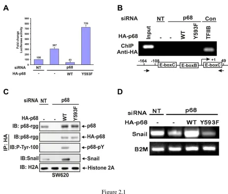
2.3.1 The Phosphor-p68 Repressed E-cadherin by Upregulating Transcription of Snail1 ... 74
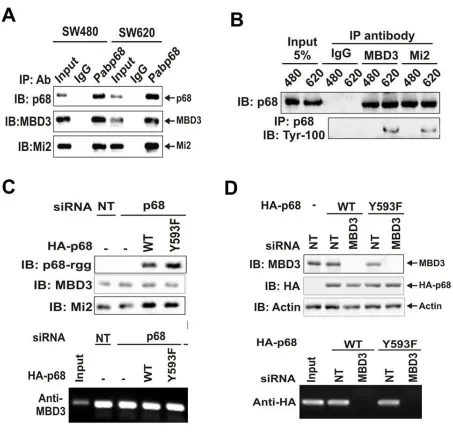
2.3.2 p68 Associates with the MBD3:Mi2/NuRD Complex ... 77
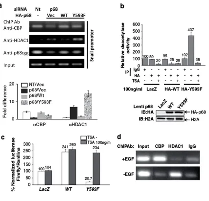
2.3.3 HDAC1 Dissociated from the Snail1 Promoter in the Presence of Phosphor- p68 ... 78
2.3.4 p68 Phosphorylation Correlates with Snail1 Expression in Metastatic Cancer Cells ... 80
2.4 Discussion ... 81
2.6 References ... 88
CHAPTER 3: PHOSPHORYLATION OF p68 RNA HELICASE PROMOTES COLON CANCER METASTASIS ... 102
3.1 Abstract ... 102
3.2 Introduction ... 102
3.3 Results ... 104
3.3.1 Characterization of Sublines with Endogenous p68 Knockdown and Exogenous p68 wt and Y593F Mutant Expressions ... 104
3.3.2 Knockdown of p68 and Expression of Y593F Mutant Significantly Reduced Cancer Metastasis ... 106
3.3.3 p68 is Phosphorylated at Y593 in the Colon Cancer Tissue Samples ... 107
3.4 Discussion ... 108
3.5 Materials and Methods ... 110
3.6 References ... 116
CHAPTER 4: MATERIALS AND METHODS ... 134
4.1 Materials ... 134
4.1.1 Chemicals ... 134
4.1.2 Kits ... 135
4.1.3 Laboratory Equipment ... 136
4.1.4 Enzymes and Recombinant Proteins ... 136
4.1.6 Vectors and siRNA/shRNA Sequence ... 137
4.1.7 Bacteria Stains ... 137
4.1.8 The Mammalian Cell Lines ... 137
4.1.9 Buffers... 138
4.2 General Techniques... 138
4.2.1 Bacterial Culture ... 138
4.2.2 Transformation ... 139
4.2.3 Deoxyribonucleic Acid Techniques ... 139
4.2.4 RNA Isolation ... 143
4.2.5 Protein Techniques... 144
4.2.6 Antibody Generation and Purification ... 150
4.2.7 Cell Proliferation Assay ... 151
4.3 References ... 152
CHAPTER 5: CONCLUSIONS ... 153
5.1 Transcriptional Regulation of Snail1 by p68 RNA Helicase ... 154
5.2 The Role of p68 RNA Helicase in Cancer Metastasis ... 160
LIST OF TABLES
LIST OF FIGURES
Figure 1.1 Classification of helicases and translocases ... 66
Figure 1.2 The conserved motifs of DEAD-box proteins and their interaction with ATP .... 67
Figure 1.3 DEAD-box posttranslational modifications and protein-protein interactions ... 68
Figure 1.4 The crystal structure of conserved helicase core of DEAD box RNA helicase ... 69
Figure 1.5 Epithelial-mesenchymal transition (EMT) ... 70
Figure 1.6 The molecular model describes the structural change of chromosome between transcription repression and activation. ... 71
Figure 2.1 The phosphor-p68 down-regulated E-cadherin by upregulation of transcription of Snail1. ... 92
Figure 2.2 p68 interacted with Snail1 promoter. ... 94
Figure 2.3 P68 interacted with the MBD3:Mi-2/NuRD complex. ... 96
Figure 2.4 Phosphor-p68 dissociated HDAC1 from the Snail1 promoter. ... 98
Figure 2.5 P68 phosphorylation correlates with Snail1 expression in metastatic and non -metastatic cancer cells ... 100
Figure 3.1 P68/HA-p68 levels in sublines of SW620 ... 118
Figure 3.2 Growth conditions of established SW620 sublines ... 120
Figure 3.3 Cell migration and epithelial/mesenchymal marker characterization in SW620 sublines ... 122
Figure 3.4 Morphology of SW620 sublines and SW480 cells ... 124
Figure 3.5 The effects of p68 mutations on metastasis of xenograft of SW620 tumors. .... 125
Figure 3.6 mRNA levels of p68 in SW620 tumors ... 127
Figure 3.7 Phosphorylation level of p68 at Y593 in colon cancer tissues ... 129
CHAPTER 1: GENERAL INTRODUCTION
1.1 Helicases and Nucleic Acid Translocases
Helicases are a highly diverse group of proteins that perform a variety of functions in
cells. These proteins are capable of unwinding deoxyribonucleic acid (DNA) or ribonucleic acid
(RNA) duplex substrates by hydrolyzing nucleotide triphosphate (NTP), mainly adenosine
triphosphate (ATP), to provide energy. The helicases exist in every living organism and virus,
and they play a role in almost every process that involves nucleic acids, including DNA
replication and repair, transcription, translation, ribosome synthesis, RNA maturation and
splicing, and nuclear export processes. Originally, the helicases were identified by a series of
conserved motifs that form a core domain to exert their ATP hydrolysis and unwinding activity
(Koonin 1993b, Koonin and Deutscher 1993). With accumulative evidence over years, it has
become clear that these motifs are actually characteristics of nucleic acid translocases that use
ATP hydrolysis to move directionally along nucleic acid strands. Therefore, helicases are now
classified as a subgroup of translocases (Berger 2008, Singleton et al 2007).
Based on the fundamental works by Gorbalenya, Koonin, and others, this large group of
helicases/translocases is classified into six superfamilies (Erzberger et al 2006, Gorbalenya et al
1989, Koonin 1993a, Koonin 1993b, Korolev et al 1998, Mahdi et al 2003, Pause and Sonenberg
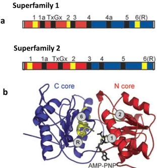
1992, Tanner et al 2003). For the largest two superfamilies (SF1 and SF2), their helicase/ATPase
core domain shares several common motifs, summarized in Fig.1.1a. The available structural
evidence suggests that the active interface (cleft) of the helicase/ATPase core domain in SF1 and
SF2 is shaped by the amino-terminal and carboxyl-terminal recombinase A (RecA)-like domains
shown in Fig.1.1b (Singleton et al 2007). The RecA-like domain is a universal structural feature
nucleotide binding and hydrolysis that provides energy in mechanical work, such as moving
polypeptides or nucleic acids (Ye et al 2004).
1.2 RNA Remodeling Proteins/ RNA Helicases
RNA helicases are ubiquitous, highly conserved enzymes involved in most aspects of
RNA metabolism, including transcription, pre-mRNA splicing, translation, mRNA decay, and
microRNA biogenesis (Aratani et al 2001, Hilliker et al 2011, Jia et al 2011, Moore et al 2011).
It is generally believed that RNA helicases modulate dynamic RNA structures and RNA-protein
complexes in an ATP-dependent manner, by which the hydrolysis of the ATP drives the
dissociation of RNA-RNA and/or RNA-protein interactions (Chen et al 2002, Jankowsky et al
2001, Linder et al 2001). With their important role in RNA metabolism, RNA helicases are
found in all kingdoms of life, and many viruses express one or more of these proteins to execute
their biological functions, for example, to replicate in the host (Jin and Peterson 1995, Wen et al
2009).
1.3 DEAD Box RNA Helicases
The family of DEAD box RNA helicases, one of the largest subdivisions of SF2
helicases, is characterized by the presence of an Asp-Glu-Ala-Asp (DEAD) motif and plays an
essential role in almost every cellular event, including cell survival, growth, proliferation, and
differentiation (Abdelhaleem 2005, Jankowsky 2011). Similar to all SF2 helicases, the DEAD
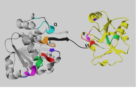
box proteins share nine conserved sequence motifs (Fig.1.2) that build the helicase core of two
nearly identical RecA-like domains. These sequence motifs encompass an approximate 300-400
amino acid region involved in ATP binding/hydrolysis and RNA binding/unwinding (Caruthers
The crystal structures of a number of DEAD-box proteins or domains have been solved,
including eIF4A (Benz et al 1999, Caruthers et al 2000, Johnson and McKay 1999); the
DEAD-box protein of Methanococcus jannaschii (Story et al 2001) and Bacillus stearothermophilus
(Carmel and Matthews 2004); Vasa in Drosophila melanogaster (Sengoku et al 2006); DDX3X,
the closely related form of the DDX3 protein (Hogbom et al 2007); yeast DEAD box protein
Mss116p (Del Campo and Lambowitz 2009); as well as some human DEAD box proteins
(Schutz et al 2010). According to the comparative structural analysis of DEAD-box RNA
helicases, motifs I, II (DEAD box), VI, and the Q-motif participate in nucleotide binding,
whereas motifs Ia, Ib, IV, and V contribute to the RNA binding process (Linder 2006, Schutz et
al 2010). Moreover, the helicase core of the DEAD box proteins is flanked by variable
N-terminal and C-N-terminal auxiliary domains, which are thought to be critical for the diverse
functions of these enzymes by posttranslational modification or interacting with cofactors, as
Fig.1.3 summarizes (Gustafson and Wessel 2010, Linder 2006).
The biological functions of DEAD box RNA helicases in eukaryotic cells are very
diverse and range from transcription to the degradation of RNA (Fuller-Pace 2006, Linder 2006).
How the single-folded enzymes perform such diverse functions in the cells remains elusive. In
vitro, the vast majority of RNA helicases do not display sequence or structural preferences
except for the polarity requirements of some enzymes for unwinding RNA duplexes. However,
in vivo studies have revealed that different RNA helicases, even with a high structural similarity,
perform distinct functions on various substrates (Fairman-Williams et al 2010, Franca et al 2007,
Strohmeier et al 2011). For example, the eukaryotic translation initiation factor 4A (eIF4A, also
known as DDX2A/B), a prototypic member of the DEAD box helicase family, has been shown
translation process (Hilbert et al 2011, Imataka and Sonenberg 1997). During translation
initiation, eIF4A is capable of unwinding the secondary structure in the 5’-untranslated region of
eukaryotic mRNAs, thus promoting ribosome binding (Hilbert et al 2011, Imataka and
Sonenberg 1997, Ozes et al 2011). However, eIF4AIII (DDX48), which is structurally similar to
eIF4A, has a distinct function in the exon junction complex (EJC). The EJC is a multi-protein
complex deposited roughly 20 nucleotides upstream of exon-exon junctions during pre-mRNA
splicing in higher eukaryotes (Le Hir and Andersen 2008, Tange et al 2004). Crosslinking and
antibody inhibition studies suggested that eIF4AIII constitutes at least part of the platform that
anchors other EJC components, such as nuclear-cytoplasmic shuttling proteins, Y14 and Magoh,
to the spliced mRNAs. In mammalian cells, it is also known that eIF4AIII is essential for
nonsense-mediated mRNA decay. Therefore, eIF4AIII may represent a new functional class of
DEAD box proteins that act as RNA clamps for the sequence-independent attachment of
additional factors to RNAs (Shibuya et al 2004).
Many DEAD box RNA helicases appear to participate solely in a single step of one
cellular process. A multi-step process, such as ribosome biogenesis, requires a large number of
DEAD box RNA helicases, suggesting a functional specificity for the proteins in each step
(Burger et al 2000, Daugeron and Linder 2001, Hilliker et al 2011). Interestingly, several DEAD
box proteins, including Ded1p (DDX3) and p68 (DDX5), were found to play a role in various
multi-component complexes, such as chromatin remodeling, transcription, and splicing
complexes (Carter et al 2010, Fuller-Pace and Moore 2011, Hilliker et al 2011, Mooney et al
2010a, Rosner and Rinkevich 2007). Therefore, understanding the role(s) of DEAD box RNA
helicases in the formation and modulation of these complexes will help to elucidate possible
1.4 DEAD Box Protein-p68 RNA Helicase
1.4.1 Gene and Structure of p68 RNA Helicase
Human p68 RNA helicase is encoded in DDX5 gene located at chromosome 17q21 (Iggo
et al 1989). The cDNA sequence of human p68 shows an open reading frame encoding a
polypeptide of 614 amino acids (Hloch et al 1990). The p68 RNA helicase was first identified
because of its immunological cross-reaction with the anti-SV40 large T monoclonal antibody
DL3C4 (PAb204) (Crawford et al 1982a, Lane and Hoeffler 1980a). Sequence analysis of human
p68 cDNA revealed the molecular basis for its cross-reaction with the SV40 large T antigen and
its extensive homology to the eIF4A protein, both of which have nucleic acid unwinding activity
(Pause and Sonenberg 1992, Scheffner et al 1989). Purified p68 protein has been shown to
exhibit ATP binding, RNA-dependent ATPase, and RNA helicases activities in vitro (Hirling et
al 1989, Iggo and Lane 1989b).
The p68 protein is evolutionarily conserved. The similarity of the amino acid sequence of
mouse and human p68 is as high as 98% (Lemaire and Heinlein 1993). The amino acid sequence
of the p68 protein contains multiple conserved motifs that place it in the family of DEAD box
RNA helicases. Although the crystal structure of p68 has not yet been solved, the structure of
eIF4A has been rendered (Benz et al 1999, Caruthers et al 2000, Johnson and McKay 1999). The
crystal structure of the eIF4A helicase core resembles a dumbbell-like shape that consists of two
RecA-like globular domains connected by a flexible linker (Fig.1.4) (Caruthers et al 2000). The
structure of the helicase core reveals that the motifs conserved in the DEAD box family form a
pocket within the two globular domains, providing the binding sites for ATP and RNA. The
latest identified Q motif has been reported to regulate ATP binding and hydrolysis (Tanner et al
which are essential for ATP binding/hydrolysis in the family of DEAD box RNA helicases
(Carmel and Matthews 2004, Lorsch and Herschlag 1998, Rocak et al 2005, Schneider and
Hunke 1998). Mutations in the motif I affect ATP binding, whereas mutations in the motif II
affect ATP hydrolysis that couples with the RNA helicase activity. Furthermore, the first residue
in the motif VI (HRxGRxGR) is involved in ATP hydrolysis, and mutations in the motif III
(SAT) abolish RNA unwinding while the binding of RNA and ATP is unaffected (Pause and
Sonenberg 1992). The motif V (RGID) has been shown to contribute to the RNA binding process
(de la Cruz et al 1999). Outside the helicase core, an IQ motif, identified in the C-terminal of the
p68 protein, has been shown to contain the conserved protein kinase C phosphorylation site and
the calmodulin binding site, both of which have negative regulatory effects on p68 ATPase
activity (Buelt et al 1994, Yang et al 2004). An RGG (Arg-Gly-Gly) repeat region following the
helicase core of the p68 protein has been suggested to regulate the protein function via
posttranslational modification (Yang and Liu 2004b, Yang et al 2005d).
1.4.2 Expression of p68 RNA Helicase
The expression of the p68 RNA helicase has been shown in a wide range of vertebrate
species and is essential for normal development and cell growth (Lane and Hoeffler 1980a,
Lemaire and Heinlein 1993). The chicken homologue of p68 was found to be tightly associated
with the 5-methylcytosine-DNA glycosylase complex in developing embryos. This glycosylase
complex is involved in the active de-methylation of DNA on the CpG repeats, which is vital for
the normal development of vertebrates (Jost et al 2001). In the Swiss 3T3 cells, the expression of
p68 is detectable in quiescent cells, but is enhanced greatly by serum stimulation, suggesting a
growth-related role of this protein (Stevenson et al 1998b). In adult rats and the human tissues,
types; however, the expression levels do not correlate completely with the proliferation status,
suggesting that the regulation of expression of this protein is more complicated in the tissue than
in the cultured cells (Rossler et al 2000, Stevenson et al 1998b).
In the mouse brain, it has been shown that the expression level of the p68 RNA helicase
is significantly lower in old males compared with adult males. Furthermore, the p68 RNA
helicase in the mouse brain was found to interact with the transactivation domain of the estrogen
receptor α (ERα), and this interaction is relatively lower in old mice of both sexes and even
lower in male mice of the same ages compared with their counterparts. Such age- and
sex-dependency of the p68-ERα interaction suggests a role for p68 in ERα-mediated brain function
during aging (Ghosh and Thakur 2009).
In the screening of the differential cDNA library, nitric oxide (NO) stimulation in HaCaT
human keratinocytes was found to induce the p68 RNA helicase. NO is known to pivotally drive
epithelial movement and gene expression during skin repair. Wound healing experiments on
mouse skin demonstrated that the p68 RNA helicase is essential for developing epithelium,
possibly through stimulating keratinocyte proliferation and the expression of the vascular
endothelial growth factor to accelerate the healing process (Kahlina et al 2004).
1.4.3 Posttranslational Modification of p68 RNA Helicase
Posttranslational modification is a process, in which proteins are added with other
biochemical functional groups, such as acetates, phosphates, lipids, carbohydrates, and small
polypeptides, to specific amino acid residue(s). The posttranslational modification changes the
In the cells, phosphorylation is accomplished by transferring the γ-phosphoryl group of
ATP to serine, threonine, or tyrosine residues of a protein, which is catalyzed by protein kinases
(Berg et al 2007). Phosphorylation modification grants the host protein two extra negative
charges, which are likely to induce a conformational change of the protein and thus alter its
enzymatic activity, binding affinity to interacting factors, and/or cellular localization (Berg et al
2007). Also, a protein can be phosphorylated simultaneously or sequentially at multiple residues
to perform its function precisely in a spatiotemporal manner. In addition, protein phosphorylation
can be reversed by phosphatases, which confers a more flexible regulatory mechanism onto the
cells in response to different stimulations. Therefore, kinases and phosphatases play a critical
role in most, if not all, signaling pathways that are vital for regulating cellular behaviors, such as
cell growth and survival. Imbalanced regulation of protein phosphorylation may cause severe
diseases, such as cancer (Brognard and Hunter 2011).
A previous study from our laboratory demonstrated that the recombinant p68 RNA
helicase purified from bacteria, Escherichia coli, has the phosphorylation modifications on
serine, threonine and tyrosine residues (Yang and Liu 2004b). Biochemical analysis showed that
the phosphorylation modification on the recombinant p68 RNA helicase affects its ATPase and
RNA binding/unwinding activities, which may suggest an important mechanism in controlling
the function of this protein at the posttranslational level (Yang and Liu 2004b). Further studies
showed that the recombinant C-terminal domain of p68 RNA helicase is responsible for the
binding of single stranded RNA, which is negatively regulated by protein kinase C (PKC).
Phosphorylation of the C-terminal domain of p68 RNA helicase by PKC abolishes its RNA
binding activity in vitro (Yang et al 2004). These data suggested that the C-terminal domain of
be regulated through phosphorylation by PKC in the cells. Another study also showed that the
phosphorylation on tyrosine residue(s) of p68 RNA helicase is enhanced in tumor cell lines
compared with their normal counterparts, suggesting a role of p68 in abnormal cell proliferation
and cancer development (Yang et al 2005c). Furthermore, phosphorylation of p68 at threonine or
tyrosine residues was found to respond differently to tumor necrosis factor α (TNF-α) treatment
in HeLa cells (Yang et al 2005d). In the absence of TNF-α, p68 RNA helicase is phosphorylated
on tyrosine but not on serine or threonine residue(s) in HeLa cells. In the presence of TNF-α,
tyrosine phosphorylation of p68 RNA helicase is first enhanced and then replaced by threonine
phosphorylation due to the activation of a serine/threonine kinase, p38 MAP kinase. The data
also revealed that the threonine phosphorylation of p68 is essential for its ATPase and RNA
unwinding activities, whereas the tyrosine phosphorylation attenuates its enzymatic activities and
the functional role in the pre-mRNA splicing, suggesting that phosphorylation pattern of p68
RNA helicase is associated with different cellular functions (Yang et al 2005d).
In glioblastoma cells, a site-directed mutagenesis study specified that double
phosphorylation of tyrosine 593 and 595 residues on p68 RNA helicase confers the cells with the
resistance to apoptosis upon the treatment of tumor necrosis factor-related apoptosis-inducing
ligand (TRAIL) (Yang et al 2007a). In colon cancer cells, p68 is phosphorylated at the tyrosine
593 residue by c-Abl kinase upon the stimulation of platelet-derived growth factor (PDGF)-BB.
The tyrosyl (593)-phosphorylated p68 protects a transcription activator, β-catenin, from
GSK3β-medicated protein degradation in cytoplasm, enriches the nuclear portion of β-catenin, and thus
promotes epithelial to mesenchymal transition (EMT) (Yang et al 2006). The tyrosine
phosphorylation of p68 also mediates the effects of PDGF on promoting cell proliferation by
phosphorylation of p68 RNA helicase on different residues may play an important role in
regulating various cellular functions.
Ubiquitination is a posttranslational modification in which a small conserved peptide,
ubiquitin or ubiquitin chains, are added to the target proteins, giving rise to mono or
polyubiquitination (Busch and Goldknopf 1981, Schlesinger and Goldstein 1975). The process of
ubiquitination involves a series of steps. First, an E1 ubiquitin-activating enzyme driven
by ATP hydrolysis activates an ubiquitin. Second, the ubiquitin is transferred from E1 to
the active cysteine of an E2 ubiquitin-conjugating enzyme. Finally, an isopeptide bond is created
between a lysine of the target protein and the C-terminal glycine of ubiquitin, which is catalyzed
by E3 ubiquitin ligase with substrate specificity (Scheffner et al 1995). The effect of the
ubiquitination depends on the number and the position of ubiquitin appended to the protein.
Polyubiquitination marks proteins for proteasome-mediated degradation to maintain the protein
homeostasis in the cells (Busch and Goldknopf 1981), whereas monoubiquitination regulates
proteins in multiple ways, including changing the cellular localization, stability, and activity. The
monoubiquitination modification of proteins affects many cellular functions, such as membrane
trafficking, chromatin remodeling, transcription regulation, DNA repair, and DNA replication
(Sigismund et al 2004).
Sumoylation is another posttranslational modification, in which a small ubiquitin-like
modifier (SUMO) is added to a protein (Hay 2005). Similar to ubiquitination, posttranslational
modification of proteins by the SUMO-1 regulates their cellular localization, stability, and
activity, with implications in many cellular processes, such as cell cycle control, DNA repair,
and transcription activation/repression (Boyer-Guittaut et al 2005, Gill 2003, Johnson and
It has been shown that the expression of p68 protein is correlated with the development
of colorectal adenocarcinomas (Causevic et al 2001b). Interestingly, the increased level of p68
protein is accompanied by polyubiquitination modification, suggesting a possible defect in
proteasome-mediated degradation and/or an unknown mechanism by which the
polyubiquitinated p68 regulates the cellular proliferation in tumorigenesis.
The diverse effects on the transcriptional regulators by SUMO modification are often
through altering their localization or interactions with other factors. The p68 RNA helicase has
been shown to function as a transcriptional coactivator or corepressor in a context-dependent
manner (Rossow and Janknecht 2003, Wilson et al 2004a). A recent review indicated that SUMO
modification is a key in switching the transcriptional regulatory role of p68 between activation
and repression (Fuller-Pace et al 2007). It has been shown in vivo that p68 is modified on its
lysine 53 by the small ubiquitin-like modifier-2 (SUMO-2). This SUMO-2 modification of p68,
catalyzed by the SUMO E3 ligase, PIAS1, enhances its transcriptional repression activity
through altering chromatin structure by the recruitment of histone deacetylase 1 (HDAC1)
(Jacobs et al 2007). The SUMO modification has been shown to increase protein stability by
reducing polyubiquitination and the consequent proteasome-mediated protein degradation
(Geiss-Friedlander and Melchior 2007). Interestingly, a bioinformatics study revealed that p68
contains one consensus sumoylation motif, ψKxE, where ψ is an aliphatic-branched amino acid
and x is any amino acid. The SUMO modification of p68 increases its stability, whereas
mutation of the consensus sumoylation site enhances its polyubiquitination and degradation
(Mooney et al 2010b). Dysfunctional regulation of both ER and p53 signaling pathways has been
correlated with breast cancer development (Pelosi et al 1994, Shirley et al 2009). A recent study
promoters containing ER response elements, whereas the p68 sumoylation status does not affect
p53-dependent MDM2 transcription (Mooney et al 2010b). Therefore, this study suggests that
the pleiotropic effects of SUMO modification of p68 may be a critical mechanism by which ER
and p53 activities are regulated in breast cancer cells.
According to previous studies, it is obvious that p68 plays multiple roles in cells through
different posttranslational modifications. Emerging evidence also suggests that the
posttranslational regulation gains more weight in maintaining the normal function of cells and is
associated with pathogenesis, such as tumorigenesis. Therefore, understanding the underlying
mechanisms and the effects of posttranslational modification may shed light on its therapeutic
purpose in cancer treatment.
1.5 Transcriptional Regulation
1.5.1 General Transcriptional Regulation
Transcriptional regulation is very important in living cells, as it responds to various
cellular signals, and it involves a large number of proteins, including sequence-specific DNA
binding factors, chromatin regulators, general transcription factors, RNA polymerase II (Pol II),
and the coactivators or repressors (Li et al 2007, Orphanides and Reinberg 2002, Pugh 2000,
Struhl et al 1998). The general view of transcriptional activation is initiated by the interaction of
sequence-specific regulators and their cognate DNA motifs. The sequence-specific regulators
further recruit coactivators to alter and expose the local chromatin structure, facilitate the
assembly of the pre-initiation complex (PIC) consisting of general transcription factors (GTFs)
and RNA polymerase II (Pol II), and eventually lead to gene transcription (Venters and Pugh
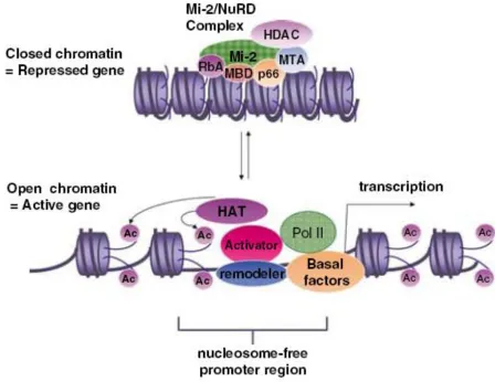
hindrance mechanism by which chromatin is compacted with histone proteins into a dense
nucleosome structure that limits its accessibility to transcription activators. Two groups of
enzymes with antagonistic activities, called histone acetyltransferases (HATs) and histone
deacetylases (HDACs), play a crucial role in activating or repressing gene transcription
(Johnsson et al 2009). HATs, such as CBP/p300, acetylate histone and make the chromatin more
accessible to transcription factors, whereas HDACs, such as HDAC1, function in a reverse way
to repress gene expression. Another group of enzymes, called DNA methylases, also plays a role
in transcriptional regulation, mainly in gene repression (Bhakat and Mitra 2003, Kang et al
1999). These regulatory enzymes can be recruited to the promoters by sequence-specific factors
to provide additional active or repressive effects on gene transcription (Lande-Diner et al 2007,
Li et al 2007).
Many sequence-specific regulators have been determined and granted with a crucial role
in regulating gene transcription. Growing evidence indicates that the sequence-specific
regulators orchestrate gene transcription in multiple aspects, including the recruitment of
chromatin remodeling complexes, general transcription factors, chromatin modifying complexes,
and RNA Pol II (Cosma et al 1999, Garbett et al 2007, Goldmark et al 2000, Green 2005,
Larschan and Winston 2001, Neely et al 2002, Nourani et al 2004, Ranish et al 1999, Yudkovsky
et al 1999).
It is evident that transcriptional activation involves the movement of nucleosome, which
is driven by ATP hydrolysis catalyzed in chromatin remodeling complexes (Li and Reese 2001,
Lohr 1997, Ravindra et al 1999). Based on sequence conservation, many identified chromatin
remodeling complexes are classified into four families, SWI/SNF, INO80/SWR1, ISWI, and
The SWI/SNF family of chromatin remodeling complexes generally functions as a
positive regulator through sliding or ejecting the nucleosomes, exposing the DNA biding sites to
transcription factors, and thus activating gene expression (Liu et al 2011). Unlike the SWI/SNF
family, the INO80/SWR1 family of chromatin remodeling complexes regulates transcription
positively by affecting the composition of nucleosomes rather than their position. For instance,
the SWR1 complex replaces the H2A with H2A.Z in an ATP-dependent manner in the promoter
region, where the substitute histone variant destabilizes the nucleosome structure and increases
its accessibility (Guillemette et al 2005, Raisner et al 2005, Zhang et al 2005). In contrast to the
positive regulatory roles of SWI/SNF and INO80/SWR1 families, the ISWI (Initiation SWItch)
family of chromatin remodeling complexes tends to repress gene transcription. Since the ISWI
chromatin remodeling complexes lack a proven DNA binding domain, it has been suggested that
they are recruited by sequence-specific repressors and may function in concert with the HDACs
to repress gene transcription (Fazzio et al 2001, Goldmark et al 2000). The intracellular role of
the CHD (Chromatin organization modifier, Helicase, and DNA-binding domains) family of
chromatin remodeling complexes is less understood. The Chd1 protein is a component of the
SAGA (Spt-Ada-Gcn5 Acetyltransferase) chromatin remodeling complex, and it interacts in
vitro with methylated H3K4, a gene activating marker, suggesting that the CHD family may
cooperate with other chromatin modifying complexes to regulate gene transcription (Pray-Grant
et al 2005). Taken together, chromatin remodeling and modifying complexes play important
roles in regulating gene transcription, and their functions rely largely on the sequence-specific
factors.
While chromatin remodeling complexes keep the promoter region dynamic, the
mediated by the mediator complex. The mediator complex is composed of a large number of
proteins involved in many aspects of transcription (Biddick and Young 2005). Step-wise
assembly of PIC indicates that TBP (TATA-Binding Protein) and TAFs (TBP-Associated
Factors) in the TFIID (Transcription Factor II D) complex first associate with the specific
promoter region (Meisterernst et al 1990). The TFIIA and TFIIB join afterward and recruit Pol
II, which is then stabilized by TFIIF (Chung et al 2003, Lagrange et al 1996). The further
association of TFIIE, TFIIH, and other regulators controls Pol II activity and mediates the
transition from transcription initiation to elongation, which is mainly through regulating the
phosphorylation state of a subunit of Pol II, CTD (Carboxyl-Terminal Domain). It is now evident
that Pol II CTD phosphorylation and isomerization regulate all stages in a transcription cycle,
including initiation, elongation, and termination (Egloff and Murphy 2008, Meinhart et al 2005,
Phatnani and Greenleaf 2006). As a result, controlling the activity and the configuration of Pol II
plays a critical role in efficient and accurate regulation of gene transcription.
1.5.2 p68 RNA Helicase in Transcriptional Regulation
The first evidence indicating that p68 RNA helicase acts as a transcription coactivator is
finding its interaction with nuclear estrogen receptor α (ERα) (Endoh et al 1999a). ERα is a
ligand-activated transcription factor that contains two transactivation domains, AF1 (activating
factor 1 containing N-terminal A/B domain) and AF2 (activating factor 2 containing C-terminal
E (ligand binding) domain). The activity of AF1 is ligand-independent, whereas the binding of
estrogen mediates AF2 activation (Nilsson et al 2001). It has been shown that p68 interacts with
AF1 but not AF2 of human ERα. Moreover, mitogen-activated protein kinase (MAPK)-induced
phosphorylation of ERα on Ser118 potentiates the interaction of p68 and AF1, which enhances
helicase activity of p68 is dispensable as a coactivator of ERα-AF1. Specificity tests also showed
that p68 has no effect on ERβ- AF1 and AF2. These findings suggest that p68 functions as a
coactivator that is specific for ERα-AF1, and MAPK signaling regulates the interaction between
p68 and ERα-AF1 (Endoh et al 1999a).
Other than ERα, p68 was found to interact with other ERα coactivators, such as steroid
receptor coactivator 1 (SRC1) and steroid receptor RNA activator (SRA), suggesting that p68
might serve as a bridge to facilitate the interaction between ERα and its coactivators (Watanabe
et al 2001). Indeed, some reports have strengthened this idea of p68 acting as a bridge. First, p68
was found to enhance CBP/p300-mediated transcriptional activation by interacting with
CBP/p300 and RNA Pol II (Rossow and Janknecht 2003). Second, it was found that p68
interacts with Smad3, a member of the Smad family of transcriptional activators, and facilitates
the formation of a transcriptionally active complex comprising smad3, p68, and CBP to enhance
TGF-β mediated cellular responses (Warner et al 2004). Third, p68 was shown to serve as an
adapter between the CCCTC-binding factor (CTCF), which is a DNA-binding protein, and its
interacting factors (Yao et al 2010). Therefore, these findings suggest that p68 may serve either
as a bona fide coactivator through its ATPase/helicase activities or as an adaptor to regulate gene
transcription in the cells.
A recent report demonstrated that p68 functions as a transcription coactivator of p53, a
tumor suppressor that transcriptionally activates certain genes involved in cell cycle arrest, DNA
repair, and apoptosis in response to stresses and DNA damage. p68 was found to be recruited to
the p53-responsive promoters to stimulate transcription in a p53-dependent manner. Depletion of
p68 inhibits the expression of p53 target genes, such as p21 (a cell cycle inhibitory protein), as
(Bates et al 2005a). The coactivator activity of p68 in p53 signaling echoes the finding for p68
and ERα in response to estrogen, suggesting that p68 may regulate transcription through the
recruitment of other transcription factors or coactivators, such as CBP, p300, SRC1, and RNA
Pol II (Endoh et al 1999a, Rossow and Janknecht 2003, Watanabe et al 2001).
In contrast to functioning as a transcription coactivator, p68 has been suggested as acting
as a transcription corepressor by interacting with HDAC1 (Wilson et al 2004a). A recent study
showed that Drosophila homolog of human p68 RNA helicase, Dmp68, plays a role in RNA
export and gene deactivation. Termination of gene transcription requires the maturation or
destruction of pre-existing transcripts and the return of the chromatin structure to an inactive
state. Mutation of Dmp68 revealed an accumulation of transcripts in the nucleus and a delayed
gene shutdown (Buszczak and Spradling 2006a). Therefore, these findings suggest that p68 may
regulate transcription at multiple stages, including early initiation and termination.
1.5.3 p68 RNA Helicase in Pre-mRNA Splicing
The precursor messenger RNAs (pre-mRNAs) in higher eukaryotes usually comprise
multiple non-coding intervening sequences, so-called introns, which must be removed by the
splicing process. The process of splicing pre-mRNAs involves the precise excision of the introns
and the ligation of the flanking exons. Alternative splicing is a mechanism by which the introns
in the pre-mRNAs are selectively excised to produce variants of mature mRNAs, thus increasing
the complexity of the entire proteome. In the pre-mRNA splicing process, RNAs and proteins
cooperate extensively as ribonucleoproteins (RNPs) to form a MegaDalton machine, called the
Extensive studies revealed that the spliceosome is composed of several major building
blocks, including U1, U2, U4/U6, and U5 small nuclear (sn)RNPs (Jurica et al 2002). In addition
to snRNPs, a core set of over 100 proteins with splicing activities is identified in this complex in
a stage-specific fashion (Agafonov et al 2011). Due to the spliceosome’s size and the dynamics
of its assembly, analyses of the splicing kinetics and the conformational rearrangements are
challenging. Recently, the basic molecular mechanism underlying the splicing process was
proposed. During the splicing process, the 5’splicing site (5’ss) in the intron is recognized by the
U1 snRNP complex that subsequently recruits the U2 snRNP to the 3’ splicing site (3’ss) and the
branching point sequence (BPS) in order to form a pre-spliceosome complex. The U4/U6 and U5
snRNPs are further recruited to the pre-spliceosome complex to form pre-catalytic spliceosome.
In order to activate the splicing activity of the spliceosome, the U1 and U4 snRNPs have to be
released from the complex with the assistance of some RNA-dependent ATPases/helicases to
unwind RNA-RNA and RNA-protein interactions. At the end of the splicing process, the intron
is removed from the pre-mRNA along with the rest of the snRNPs (Brow 2002, Wahl et al
2009). Within a single splicing cycle, numerous DEAD box RNA helicases have been identified
to catalyze RNA-RNA rearrangements and RNP remodeling events (Forch et al 2003, Tseng et
al 2011, Xu et al 2004).
The first clue linking the p68 RNA helicase to the pre-mRNA splicing process is
supported by the comprehensive analysis of mass spectrometry that showed the existence of p68
in the spliceosome complex (Neubauer et al 1998). Using methylene blue-mediated crosslinking
also showed that p68 interacts with the U1-5’ss duplex during spliceosome assembly. Depletion
of the p68 RNA helicase does not affect assembly of the pre-spliceosome complex; however, it
destabilizing the U1-5’ss interaction, which is essential in the transition from pre-spliceosome to
spliceosome (Liu 2002). Abolishing the ATPase/helicase activities of the p68 RNA helicase does
not affect the association of p68 to the splice sites, whereas it abrogates the dissociation of the
U1 snRNP from the 5’ss. Also, it was found that p68 participates in the addition of U4.U6/U5
snRNPs, and this does not require the ATPase and helicase activities (Lin et al 2005). These data
suggest that p68 may play multiple roles in the pre-mRNA splicing process and that some of
them may require the ATPase/helicase activities.
Human Ras (H-Ras) plays a crucial role in controlling proliferation, differentiation, and
apoptosis via coupling extracellular signals to intracellular networks. Alternative splicing has
been found to process H-Ras pre-mRNA to render two proteins, p19 and p21 H-Ras, by either
excluding or including the alternative intron D exon (IDX). This alternative splicing process is
regulated by some cis- and trans-acting factors (Camats et al 2008). An intronic silencer
sequence (rasISS1), acting along with IDX, has been characterized to regulate its upstream intron
negatively. The p68 RNA helicase was shown to associate with both IDX and rasISS1. Depletion
of p68 expression in HeLa cells results in an increase of IDX inclusion in the endogenous
mRNA, indicating that p68 plays a role in regulating H-Ras alternative splicing (Guil et al 2003).
Further studies revealed that the p68 RNA helicase promotes the exclusion of IDX from H-Ras
pre-mRNA by unwinding the IDX-rasISS1 structure, preventing the binding of hnRNP H to
IDX-rasISS1, and altering the dynamic localization of SC35, a splicing factor that facilitates IDX
inclusion (Camats et al 2008).
Another example of the p68 RNA helicase involving in alternative splicing is the
regulation of tau exon 10 splicing, which plays a crucial role in neurodegenerative diseases, such
splicing. During the splicing process, p68 induces a conformational change at the 5’ss, thereby
increasing the access of the U1snRNP to the 5’ss of tau exon 10. In concert with p68, RBM4, an
intronic splicing activator, interacts with the splicing region to enhance the spliceosome activity
(Kar et al 2011). Taken together, p68 was shown to play a role in the regulation of pre-mRNA
splicing, as well as alternative splicing, by interacting with different splicing regulators.
1.5.4 p68 RNA Helicase in Ribosome Biogenesis
Ribosome is an essential component for protein biogenesis in all living organisms. It has
been shown that some DEAD box RNA helicases are involved in the ribosome biogenesis
(Burger et al 2000, Daugeron and Linder 2001, Jalal et al 2007, Lavoie et al 1993, Saporita et al
2011). The p68 RNA helicase and its homologue, p72, play a role in ribosome biogenesis and
cell proliferation (Jalal et al 2007). In the case where the p68 gene is suppressed, p72
overexpression can rescue the cell proliferation, whereas co-silencing of both genes causes the
disorder of nucleolar structure and cell death. Furthermore, suppression of both p68 and p72
reduces the nucleolytic cleavage of 32S pre-rRNA, which causes failure of pre-rRNA maturation
and ribosome biogenesis. The p19ARF tumor suppressor has been shown to regulate ribosome
biogenesis negatively in response to hyper-proliferative signals by interfering nucleolar protein
or RNA polymerase I transcription factor, TTF-1 (Itahana et al 2003, Lessard et al 2010).
Recently, the p68 RNA helicase was shown to play a role in ARF-mediated inhibition of
ribosome biogenesis (Saporita et al 2011). This study demonstrated that ARF targets p68 and
prevents its interaction with nucleophosmin (NPM) protein, rDNA promoter, and nuclear
pre-ribosomes, ultimately reducing the ribosome output. Additionally, in ARF-deficient cells,
tumor growth in mice. Taken together, these findings indicate that p68 RNA helicase is a target
to regulate cell proliferation at least through participating in ribosome biogenesis.
1.6 p68 RNA Helicase in Cancer Development
Cancer, also known as a malignant neoplasm, involves unregulated cell growth. The
unrestricted growth observed in neoplasms is generally due to a stepwise acquisition of genetic
alterations, including the function gain of oncogenes and repression of tumor-suppressor genes
(Hanahan and Weinberg 2000). In cancer, cells divide uncontrollably to form malignant tumors,
which invade nearby tissues, and eventually spread throughout the body via the lymphatic
system or bloodstream (Hanahan and Weinberg 2000). Not all tumors are malignant. Benign
tumors do not grow unlimitedly, invade nearby tissues, or spread throughout the body.
Therefore, understanding the mechanisms causing uncontrolled cell division and growth, as well
as their dissemination ability, is a key to treating patients with cancers.
The p68 RNA helicase has been demonstrated to play an essential role in normal
development involving cell growth and differentiation (Stevenson et al 1998b). Recently, a
growing body of evidence links p68 to cancer development (Fuller-Pace and Moore 2011, Shin
et al 2007, Wang et al 2011, Yang et al 2005c). It has been shown that the tyrosine
phosphorylation of p68 is present in six different cancer cell lines and tumor tissues, but not in
the corresponding normal cells/tissues. Induction of the apoptosis in cancer cells by some
anticancer drugs, such as tumor necrosis factor α (TNF-α), tumor necrosis factor-related
apoptosis-inducer ligand (TRAIL), and STI-571, diminishes the tyrosine phosphorylation of p68,
suggesting that the tyrosine phosphorylation status of p68 is associated with abnormal cell
Another DEAD box RNA helicase, p72, highly related to p68, was discovered in 1996
(Lamm et al 1996). It was shown that p68 and p72 share a 90% identity across the conserved
helicase core and can form heterodimers in cells to function in mRNA processing. Moreover,
p68 and p72 can be found in a variety of protein complexes. The potential of p68 and/or p72 to
coexist with other factors in different complexes may imply a wide range of functions for p68
and p72 (Ogilvie et al 2003). A recent study reported that the expression of p68 and p72
increases during the transition of polyp-->adenoma-->adenocarcinoma in the progression of
colon cancer (Shin et al 2007). In this study, p68 and p72 form complexes with β-catenin to
activate the expression of some proto-oncogenes, including c-Myc, Cyclin D1, c-jun, and fra-1.
Moreover, double knockdown of p68 and p72 reduces the expression of these proto-oncogenes
and enhances the transcription of a cell cycle inhibitor, p21 (WAF1/CIP1), whose expression is
suppressed by c-Myc. Knockdown of p68 and p72 in colon cancer cells diminishes their ability
to form tumors in vivo. Thus, p68 and p72 may participate in colon cancer development by
activating proto-oncogenes directly and suppressing the cell cycle inhibitors indirectly.
The androgen receptor (AR) has been shown to play an important role in prostatic cancer
(PCa) development (Jenster 1999, Lonergan and Tindall 2011). Recently, p68 RNA helicase was
found to function as a coactivator in AR-mediated transcription activation in prostate cancer
(Clark et al 2008a). The p68 protein was identified to interact with AR in a yeast two-hybrid
screening, and coimmunoprecipitation in the PCa LNCaP cell line confirmed this interaction
further. Chromatin immunoprecipitation (ChIP) analysis showed that AR and p68 coexist in the
promoter region of the androgen responsive prostate-specific antigen (PSA) gene. Knockdown of
p68 reduces AR-mediated PSA expression. Luciferase reporter assay revealed that tyrosine
wherein the ATP-binding is dispensable. Tissue microarray screening also showed an increased
frequency and expression of p68 in prostate cancer compared with benign tissues. Therefore, the
study suggests that the p68 RNA helicase may control gene expression in an AR-dependent
manner to regulate prostate cancer development (Clark et al 2008b).
Although abundant evidence supports the role of the p68 RNA helicase in promoting
cancer progression, recent studies in breast cancer revealed an opposite role of p68 as a tumor
co-suppressor in a p53-dependent manner (Bates et al 2005a, Moore et al 2010). As mentioned
earlier, p68 has been shown to interact with p53 to activate the expression of cell cycle inhibitor,
p21, and other apoptosis-related genes in response to DNA damage (Bates et al 2005a). Also, a
recent study showed that the expression of p68 and the ∆133p53 isoform(s), negative regulators
of full-length p53, is inversely correlated in primary breast cancer (Moore et al 2010).
Consistently, depletion of p68 in culture cells results in an increase of the ∆133p53 isoform(s) in
response to DNA damage, and the increased ∆133p53α inhibits the p53-dependent transcription
of p21. The data suggest that p68, p53, and ∆133p53α may form part of a complex to regulate
p53-mediated transcription and may modulate the function of p53 in breast and other cancers that
harbor wild-type p53.
Novel mechanisms of microRNAs (miRNA)-mediated gene regulation at the
posttranscriptional level have emerged recently. MiRNAs regulate gene expression by targeting
mRNAs for translational repression or mRNA degradation (Chen 2005). Growing evidence
shows that many miRNAs are regulated in the normal development, and abnormal expression of
specific miRNAs may contribute to cancer progression (Chen 2005). Studies in cultured cells
and human tissues revealed that the expression of the p68 RNA helicase increases progressively
p68 was found to upregulate a subset of miRNAs, including miR-21 and miR-182, which
downregulate a tumor suppressor, PDCD4, and cytoskeleton regulators, cofilin and profilin,
respectively (Wang et al 2011). Depletion of p68 leads to cytoskeleton reorganization and
reduced cell proliferation (Wang et al 2011). These findings reveal a novel functional role of p68
via regulating microRNAs, and suggest a potential target, including p68 and its downstream
microRNAs in the treatment of breast cancer.
The compound (-)-epigallocatechin-3-gallate (EGCG), a major catechin found in green
tea, induces apoptosis and suppresses tumor growth by regulating a wide range of signaling
pathways (Nagle et al 2006, Singh et al 2011). It has been found that EGCG can bind covalently
to cysteine residues in proteins and thus regulate protein function. Recently, the p68 RNA
helicase was identified as a novel EGCG-binding target in AZ521 human gastric cancer cells
(Tanaka et al 2011). Treatment of AZ521 cells with EGCG reduces the protein level of p68 in
dose and time-dependent manners. Furthermore, EGCG inhibits AZ521 cell proliferation by
promoting protein degradation of β-catenin (Tanaka et al 2011), a transcription activator that was
shown to be upregulated by p68 in tumor progression (Yang et al 2006). Taken together, the p68
RNA helicase seems to play an essential role in the progression of various types of cancers,
which may strengthen the idea that targeting this protein could be an effective way to treat
cancers.
1.7 Cancer Metastasis
Cancer metastasis is the spreading of primary tumor cells through the lymphatic system
or bloodstream to distant organs. Metastasis causes most cancer deaths, yet the underlying
mechanism controlling it remains one of the most enigmatic aspects of this disease.
phase involves the escape of cancer cells from the original site to a distant organ. The second
phase, known as colonization, involves the adaptation of the cancer cells and the development of
micrometastasis at the new place. To start the metastatic cascade, cancer cells within the primary
tumors need to gain an invasive phenotype. The invasive cancer cells then invade the
surrounding tissues and migrate toward blood vessels, where they intravasate to enter the
circulation system. The cancer cells travel through the circulation and exhibit
anchorage-independent survival. Then, the circulating cancer cells leave the circulation via extravasation
and invade the foreign tissue. At the foreign site, the cancer cells have to escape the innate
immune response, adapt to the microenvironment, and initiate proliferation to form an active
macrometastatic colony (Chaffer and Weinberg 2011). Whether the acquisition of malignant
traits occurs as an inevitable consequence of primary tumor progression or as an accidental
outcome remains a question. Therefore, much work is needed to uncover the mechanisms behind
the entire metastatic cascade.
Critical thought from recent studies suggests that the cell population within individual
tumors is heterogeneous. The intra-tumoral heterogeneity is uncovered in many types of
carcinomas. The populations of cells within a tumor are arranged hierarchically like those in the
normal tissue, with the scheme of self-renewing stem cells (SCs), partially differentiated
progenitor cells, and fully differentiated end-stage cells (Ailles and Weissman 2007, Al-Hajj et al
2003). These cancer stem cells (CSCs), although not yet proved completely equal to the normal
tissue SCs, are defined with enhanced tumor-initiating potential relative to other cancer cells
within a tumor and should reveal the ability of self-renewal and producing non-CSC progeny
(Ailles and Weissman 2007). Many of the biological traits, such as motility, invasiveness, and
CSCs’ subpopulation in a tumor with larger populations of neoplastic cells. The traits that are
ascribed to CSCs, self-renewal and tumor-initiating ability, seem to be the inextricable trigger of
successful metastasis. Other traits in CSCs, such as notable motility, invasiveness, and the
resistance to apoptosis, also contribute to metastasis (Charafe-Jauffret et al 2009, Marcato et al
2011, Pang et al 2010). This implies a multi-faceted biological program that empowers cancer
cells within primary tumors to accomplish multiple steps of the metastasis process.
1.7.1 Epithelial-Mesenchymal Transition
Epithelial-mesenchymal transition (EMT) is a biological program characterized by the
loss of cell-cell adhesion, the loss of cell polarity, and the acquisition of migratory and invasive
properties. The well-organized epithelial cells attach to their neighbors and establish an
apical-basolateral polarity through various types of cell junctions, including tight junctions, adherens
junctions, desmosomes, and gap junctions. In contrast, mesenchymal cells are organized loosely
and have the abilities of migration and invasion (Alberts et al 2002). EMT and its reverse
program, MET, (mesenchymal-epithelial transition) are involved in many processes of
embryogenesis, including gastrulation, mesoderm formation, and neural crest maturation (Thiery
et al 2009). Recently, migratory tumor cells that lose E-cadherin-mediated cell-cell adhesion and
detach from the tumor mass and the surrounding stroma, have been observed, providing evidence
of EMT at the invasive front of tumors (Brabletz et al 2001, Prall 2007). The phenotype and
molecular changes during EMT are briefly illustrated in Fig.1.5.
Recent studies have shown that EMT stimulates non-CSCs to enter into a CSC-like state
(Mani et al 2008, Reiman et al 2010). Activation of EMT endows non-CSCs with the set of CSC
traits that empowers them to spread from primary tumors to distant sites (Thiery et al 2009).
property that is critical to survive the rigors of the journey from primary tumors to sites of
dissemination (Gal et al 2008). EMT may also contribute to the launch of new colonies during
metastasis.
How EMT is activated during tumorigenesis is not fully understood. Activation of EMT
during embryonic development and tumorigenesis requires signals from the neighboring stromal
cells (Yang and Weinberg 2008). It has been suggested that a variety of cell types, including
fibroblasts, myofibroblasts, granulocytes, macrophages, mesenchymal stem cells, and
lymphocytes, are recruited to the surrounding stroma of advanced primary tumors to create an
inflammatory microenvironment that releases EMT-inducing signals (Lopez-Novoa and Nieto
2009). The signals released from the stromal cells activate the expression of EMT-related
transcription factors (EMT-TFs) that orchestrate EMT programs within certain cancer cells
(Thiery et al 2009). Taken together, it suggests that two elements are required to activate the
EMT program. First, certain cancer cells have to undergo genetic and epigenetic alterations in
order to respond to EMT induction. Second, the reactive stroma has to release the inductive
signals to cause the expression of EMT-TFs and thus activate the EMT program within these
responsive carcinoma cells.
It seems that the EMT program is being induced in certain responsive carcinoma cells
during tumorigenesis. However, there is still a possibility that some carcinoma cells have
intrinsic metastatic ability (Chaffer and Weinberg 2011). Whether the metastatic ability is
intrinsic or induced through the EMT program within these carcinoma cells remains largely
unproved, due partly to the difficulties of capturing the transitory process in human cancer
patients or distinguishing the cancer cells after EMT from stromal cells or tumor-associated
to flip from one state into the other, in many tumors, epithelial carcinoma cells seem to advance
only partly toward the mesenchymal state, which yields cancer cells with concomitant expression
of epithelial and mesenchymal markers and thus brings more complexity to understand the EMT
program.
1.7.2 Downregulation of E-cadherin in Epithelial-Mesenchymal Transition
Loss of functional E-cadherin (encoded by CDH1 gene) is a key step in the transition of
epithelial to mesencymal phenotypes and is also considered a fundamental event in the
progression of adenoma to invasive carcinoma (Perl et al 1998). E-cadherin, a member of the
cadherin family of transmembrane proteins, serves as a major component in the formation of
adherens junctions (Takeichi 1993) that facilitate cell-cell adhesion and establish cell polarity in
epithelial cells (Shapiro et al 1995). The adherens junctions are stabilized by the interaction of
the intracellular domains of E-cadherin with α, β, and γ catenins, which link E-cadherin to the
actin filament cytoskeleton (Takeichi 1995). In epithelial cells, E-cadherin regulates not only
cell-cell adhesion, but also cell growth by interacting with β-catenin, a multifunctional
transcription factor that responds to Wnt signaling to promote cell proliferation (Nelson and
Nusse 2004).
The function of E-cadherin can be regulated in multiple aspects, including genetic,
epigenetic, transcriptional, and posttranslational levels (Guilford et al 1998). The loss-functional
mutation of CDH1 gene encoding E-cadherin protein is found less frequently in the majority of
E-cadherin-negative carcinomas. On the other hand, epigenetic silencing of the CDH1 promoter
via hypermethylation of CpG islands has been found in various types of cancers (Chang et al
2002, Nass et al 2000, Yeh et al 2002, Yoshiura et al 1995). Moreover, evidence in breast cancer