http://dx.doi.org/10.4236/health.2013.511259
Importance of neutralizing antibody positivity in
Turkish multiple sclerosis patients
Derya Kaya1*, Serap Tufan2, Serkan Özakbaş3, Hakki Bahar4, Emel Ada5, Egemen İdiman2,3 1Department of Neurology, Ordu Public Hospital, Ordu, Turkey; *Corresponding Author: [email protected]
2Neuroimmunology Laboratory, Faculty of Medicine, Dokuz Eylul University, Izmir, Turkey 3Department of Neurology, Faculty of Medicine, Dokuz Eylul University, Izmir, Turkey 4Department of Microbiology, Faculty of Medicine, Dokuz Eylul University, Izmir, Turkey 5Department of Radiology, Faculty of Medicine, Dokuz Eylul University, Izmir, Turkey
Received 1 September 2013; revised 8 October 2013; accepted 21 October 2013
Copyright © 2013 Derya Kaya et al. This is an open access article distributed under the Creative Commons Attribution License, which permits unrestricted use, distribution, and reproduction in any medium, provided the original work is properly cited.
ABSTRACT
The frequency and the consequences of binding and neutralizing antibodies (BAbs and NAbs) against Interferon beta (IFNbeta) in Turkish mul- tiple sclerosis (MS) patients have not been de- termined yet, which could differ in such a coun- try which is between Europa and Asia. The aim of the study is to assess the frequency of these antibodies, and to evaluate the impact of NAbs, from the clinical and radiologic aspects in Turk- ish patients with MS. One hundred and two MS patients were included. BAbs were screened us- ing capture enzyme-linked immunosorbent as- say (cELISA), and NAbs were detected via My- xovirus protein A (MxA) messenger RNA (mRNA) induction assay (real-time polymerase chain re- action-PCR) at the beginning and one year later. Relapse rate and expanded disability status scale (EDSS) were used to assess the clinical impact. Gadolinium enhanced lesions and T2 lesion volume were used as magnetic resonance imaging (MRI) parameters. Persistent NAb posi- tivity defines to be positive both at first and then one year later. NAbs were detected in 12.2% (6/49) of IFNbeta-1b treated patients, and in 7.5% (3/40) of IFNbeta-1a SC treated patients, but none of the IFNbeta-1a IM treated patients had detectable NAbs. It was found that the mean re- lapse rate difference was significantly higher in persistent NAb negative patients (p = 0.024). Persistent NAb positivity had no effect on T2 lesion volume and contrast enhancing lesions. 60% of the persistent NAb positive patients had at least one relapse during one-year of follow-up. On the other hand, 32% of persistent NAb nega-
tive patients were detected to have at least one relapse. Data from this study suggest that pa- tients may become unresponsive to IFNbeta therapy even when the frequency of NAbs does not prove to be as high as those in the literature. Nevertheless, one should keep in mind that disease activity is not always equal to NAb po- sitivity.
Keywords:
Interferon Beta; Multiple Sclerosis;
Neutralizing Antibody1. INTRODUCTION
Interferon beta (IFNbeta) is one of the first line of immune treatment options for multiple sclerosis (MS) for over 20 years. However, repeated IFNbeta injections may induce IFNbeta antibody production in some patients. Such antibodies are called binding antibodies (BAbs), which do not affect the biological activity of the mo- lecule, and neutralizing antibodies (NAbs), which are associated with a decrease in the efficacy of the treat- ment [1,2]. In fact, NAbs are a subset of BAbs which prevent the binding of the IFNβ to its receptor on the surface of cells. When BAbs are detectable it is likely that NAbs are also present [3].
tice, in patients doing poorly clinically, a switch to a non-IFNbeta therapy is initiated independent of NAb.
To our knowledge, this is the first study that assesses the frequency of the neutralizing antibodies and their effect on disease activity in Turkish MS patients. The fre- quency and the impact of NAbs could differ in our country which is located at the crossroads of Europe and
Asia. From another point of view, it is important to re-
evaluate the treatment of those whose disease activity is higher due to NAb.
In the present study, the frequency of BAbs and NAbs, and impact of these antibodies, from the clinical and radio- logic aspects in Turkish patients with MS were assessed.
3. METHODS
3.1. ParticipantsWe included 274 consecutive MS patients between May 2008 and October 2009. All patients were routinely attending the Neurology Department of Dokuz Eylul University Hospital, Izmir. Patients were receiving one of three IFN-beta preparations: interferon b-1B (“Beta- feron”, Bayer) or interferon b-1A (“Rebif 44 mg”, both Merck-Serono, or “Avonex”, Biogen). Inclusion criteria required: a) a neurologist-confirmed diagnosis of de- finite relapsing-remitting MS according to the McDonald criteria [5]; b) age of 18 - 55; c) to be already treated only with IFNbeta for at least 18 months; d) to be re- lapse-free for at least 30 days prior to testing. Exclusion criteria were: a) to receive immunosuppressive treat- ment within the preceding year; b) to receive intra-
venous immunoglobulin (IVIG) or plasmapheresis wi-
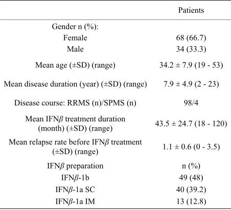
thin the last six months; c) to have primary progressive MS; d) to have upper respiratory tract infection within three weeks; e) to receive corticosteroid treatment due to an attack within three months. The local University Hospital Medical Ethics Committee approved the re- search proposals for the study. Written informed consent was obtained from patients who participated in this study. A total of 102 patients were eligible for the study. Clinical characteristics of the 102 patients are shown in Table 1.
3.2. Binding Antibody Analysis
2.5 ml venous blood was drawn in serum tubes for BAb analysis, and samples were screened using capture enzyme-linked immunosorbent assay (cELISA) first at the time of study and later, the following year. Persistent BAb-positivity indicates patients who were BAb positive both at their first and final assessments.
3.3. Neutralizing Antibody Analysis
Blood samples were collected in PAXgene tubes (Pre Analytix GmbH, Hombrechticon, CH) 12 - 14 hours af-
ter an injection of IFN, and NAbs were detected via Myxovirus protein A (MxA) messenger RNA (mRNA) induction assay (real-time polymerase chain reaction- PCR), first at the time of study and later, the following year.
RNA extraction and cDNA synthesis were done using commercially available kits (Preanalytix by Qiagen, and Superscript II Reverse Transcriptase, Invitrogen, Carls- bad, CA). PCR was performed on an ABI 7500 Fast Real Time PCR System (Applied Biosystems) using a com- mercially available TaqMan Universal PCR Master Mix and primer/probe kits. Gene expression in each sample of the target mRNA relative to GAPDH (ΔCt) was com- pared to a calibrator consisting of pooled cDNA from healthy controls. A normalization ratio (NR) was cal- culated using the formula NR = 2−ΔΔCt, where ΔΔCt =
ΔCt(sample) − ΔCt(pool). NR reflects fold induction of gene expression as compared to expression in the control pool. Samples were run in duplicate.
Persistent NAb-positivity indicates patients who were NAb positive both at their first and final assess- ments. Both binding and neutralizing antibody analy- ses were performed in the neuroimmunology labora- tory of Faculty of Medicine of Dokuz Eylul University, Izmir.
[image:2.595.310.536.503.708.2]3.4. Clinical and Radiologic Evaluation A relapse was recorded only if the physician described new findings consistent with the patient’s reported symptoms, and had excluded the possibility of a pseu- dorelapse. Relapse rate and Expanded Disability Status Scale (EDSS) [6] were used to assess the clinical impact.
Table 1. Demographical and clinical features of the patients.
Patients
Gender n (%): Female
Male
68 (66.7) 34 (33.3)
Mean age (±SD) (range) 34.2 ± 7.9 (19 - 53)
Mean disease duration (year) (±SD) (range) 7.9 ± 4.9 (2 - 23)
Disease course: RRMS (n)/SPMS (n) 98/4
Mean IFNβ treatment duration
(month) (±SD) (range) 43.5 ± 24.7 (18 - 120) Mean relapse rate before IFNβ treatment
(±SD) (range) 1.1 ± 0.6 (0 - 3.5) IFNβ preparation
IFNβ-1b IFNβ-1a SC IFNβ-1a IM
n (%) 49 (48) 40 (39.2) 13 (12.8)
EDSS scoring was performed at baseline and one year later. Also, number of relapses was recorded within one year of study.
Magnetic resonance imaging (MRI) scans were per- formed after blood sampling at the beginning and than one year later. Hyperintense lesions on T1-weighted post- gadolinium sequences were counted. T2 lesion total vol- ume was calculated within the proton density/T2 weight- ed images semi-automatically using “Lesion Annotation and Volume Assessment (LAVA) software, Medical Im- age Mining Laboratories (New York)” in Windows XP operating system on personal computers. All radiologic images were assessed by a blind radiologist.
3.5. Statistical Analysis
SPSS 15.0 for Windows was used for the statistical analysis. Descriptive features for continuous variables were implied as mean and standard deviation (SD), and for discontinuous variables as number and percent. The comparison of continuous variables between two in- dependent groups was done with Mann-Whitney U test, and when more than two groups with Kruskal-Wallis test if the normal distrubition could not be fulfilled. Wil- coxon test was used for the time effect in between de- pendent groups due to lack of normal distrubition of the difference between continous variables. The significancy of <0.05 had been accepted.
4. RESULTS
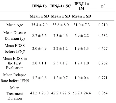
Demographic and clinical characteristics were similar in three different treatment groups (Table 2).
4.1. The First Evaluation
BAbswere detected in 26.5% (27/102) of the patients. Of 49 patients treated with IFNβ-1b, 40.8% were BAb positive at the beginning of our study, whereas of 40 pa- tients treated with IFNβ-1a SC, 15% were BAb positive; and of 13 patients treated with IFNβ-1a IM, 7.7% were BAb positive. BAb positivity was higher in IFNβ-1b treated group than IFNβ-1a treated group, and in IFNβ- 1a preparation, the patients treated with SC form had higher BAb positivity than IM form (p = 0.006).
NAbs were detected in 8.8% (9/102) of all the pa- tients. NAbs were present in 12.2% (6/49) of IFNβ-1b treated patients, and in 7.5% (3/40) of IFNβ-1a SC treated patients, but none of the IFNβ-1a IM (0/13) treated patients had detectable NAbs. The treatment duration was longer in NAb negative patients than in NAb positive patients, but it was not statistically sig- nificant (respectively 45.28 months; p = 0.055) (Table 3).
[image:3.595.310.540.112.316.2]98.7% of BAb negative patients were NAb negative, and 29.6% of BAb positive patients were NAb positive.
Table 2. Demographic and clinical characteristics of the three
different treatment groups.
IFNβ-1b IFNβ-1a SC IFNβ-1a
IM p
*
Mean ± SD Mean ± SD Mean ± SD
Mean Age 35.4 ± 7.9 33.8 ± 8.0 31.0 ± 7.3 0.210
Mean Disease
Duration (y) 8.7 ± 5.6 7.3 ± 4.6 6.9 ± 2.2 0.532 Mean EDSS
before IFNβ 2.0 ± 0.9 2.2 ± 1.2 1.9 ± 1.3 0.627 Mean EDSS in
the First Evaluation
2.0 ± 1.1 2.5 ± 1.7 1.7 ± 1.0 0.262
Mean Relapse
Rate before IFNβ 1.2 ± 0.6 1.2 ± 0.7 1.0 ± 0.4 0.771 Mean
Treatment Duration
41.2 ± 26.0 42.2 ± 22.6 56.2 ± 24.4 0.054
y: year; IFNβ: interferon beta; m: month; *Kruskal Wallis test.
There was only one NAb positive patient who was found to be BAb negative.
4.2. The Second Evaluation
Six of 102 patients dropped out one year after the first sampling time. 38.5% (n = 37/96) of patients were BAb positive. The increase in BAb positivity during one year follow-up was statistically significant only in IFNβ-1b treated patients (respectively 41%, 56%; p = 0.039). Per- sistent BAb positivity was found 25% (24/96).
In the second evaluation (one year later), NAb was present in 7.3 (7/96) of all the patients. Similar to the first evaluation, none of the IFNβ-1a IM treated patients had detectable NAbs. NAb had disappeared in three of the IFNβ-1b treated NAb positive patients and in one of the IFNβ-1a SC treated NAb positive patient, whereas NAb had appeared in two of the IFNβ-1b SC treated NAb negative patients. Persistent NAb positivity was found 5.2% (5/96).
4.3. Clinical Evaluation
Neither BAb positivity in the first evaluation nor per- sistent BAb positivity had any effect on relapse rate and progression of disability in terms of EDSS scoring.
The mean relapse rate difference was significantly higher in persistent NAb negative patients than in persis- tent NAb positive patients (p = 0.024) (Table 4). Mean
Table 3. Features of patients regarding to NAb status.
NAb
negative positive p
Mean Age ( ±SD) 34.4 ± 7.9 32.9 ± 6.1 0.816
Mean Disease Duration
(y) (±SD) 8.3 ± 5.1 4.6 ± 2.0 0.050 Mean IFNβ Duration
(m) (±SD) 44.8 ± 25.0 28.1 ± 10.1 0.055 Mean EDSS in the First
Evaluation (±SD) 2.1 ± 1.4 1.9 ± 0.8 0.835 Mean Relapse Rate
before IFNβ (±SD) 1.1 ± 0.6 0.9 ± 0.5 0.205
y: year; IFNβ: interferon beta; m: month.
of follow-up (p = 0.330).
4.4. Radiologic Evaluation
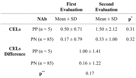
It was shown that persistent NAb positivity had no ef- fect on T2 lesion volume (Table 5) and contrast enhanc-
ing lesions in MRI (Table 6).
5. DISCUSSION
In the literature, it was reported that 28% - 47% of IFNβ-1b treated patients, 12% - 28% of IFNβ-1a SC treated patients, and 2% - 6% of IFNβ-1a IM treated pa- tients had developed NAbs [7-13]. In our study, NAbs were present in 12.2% of IFNβ-1b treated patients, and in 7.5% of IFNβ-1a SC treated patients, but none of the IFNβ-1a IM treated patients had detectable NAbs. The frequency of persistent NAb positive patients was also lower when compared to other similar studies [14-16], which might be due to genetic features of Turkish MS patients. Another possible explanation for the low pro- portion of persistent NAb positive patients could be about mean IFNbeta treatment duration. It was roughly 3.6 years in the present study. Several studies showed that 40% of patients treated with IFNb-1b reverted to NAb negative status within 4 years [17]. So that, some of our NAb positive patients could have reverted to NAb negative before the study. Three NAb positive patients receiving IFNbeta-1b and one NAb positive patient re- ceiving IFNbeta-1a reverted to NAb negative status even in one year time in our study. Consistent with the results to other studies, IFNβ-1b was found more immunogenic than IFNβ-1a SC, and IFNβ-1a SC was more immuno- genic than IFNβ-1a IM [9,18].
[image:4.595.310.536.111.242.2]Patients whose IFNbeta treatmet duration was at least 18 months were included in the present study. The rea- sons for that were, firstly, NAb appear between 3 to 18 months after the treatment [19], and secondly, determin- ing persistence of NAbs does not seem to be reliable
Table 4. The mean relapse rate difference between persistent
NAb positive and negative patients.
Before IFNβ Therapy
After IFNβ
Therapy
NAb Mean ± SD Mean ± SD p* Relapse
Rate PP (n = 5) 1.00 ± 0.35 0.60 ± 0.55 0.34
PN (n = 85) 1.57 ± 0.80 0.28 ± 0.40 <0.001
Difference in Relapse
Rate
PP (n = 5) PN (n = 85)
−0.40 ± 0.82
−1.37 ± 0.85
p** 0.024
IFNβ: interferon beta; Nab: neutralizing antibodies; PP: persistent positive; PN: persistent negative; *Wilcoxson Test; **Mann Whitney U Test.
Table 5. Mean difference in T2 lesion total volume between
persistent NAb positive and negative patients.
First Evaluation Second Evaluation
NAb Mean ± SD Mean ± SD p*
T2 Lesion
Volume PP (n = 5) 3.67 ± 0.51 2.83 ± 0.66 0.18
PN (n = 85) 8.59 ± 11.04 9.41 ± 11.91 0.02
T2 Lesion Volume Difference
PP (n = 5) −0.83 ± 0.15
PN (n = 85) 0.82 ± 2.24
p** 0.15
NAb: neutralizing antibodies; PP: persistent positive; PN: persistent nega- tive; SD: standard deviation; *Wilcoxson Test; ** Mann Whitney U Test.
Table 6. Mean difference in CELs between persistent NAb po-
sitive and negative patients.
First Evaluation
Second Evaluation
NAb Mean ± SD Mean ± SD p*
CELs PP (n = 5) 0.50 ± 0.71 1.50 ± 2.12 0.31
PN (n = 85) 0.17 ± 0.79 0.33 ± 1.00 0.32
CELs
Difference PP (n = 5) 1.00 ± 1.41
PN (n = 85) 0.16 ± 1.22
p** 0.17
CELs: contrast enhancing lesions; Nab: neutralizing antibodies; PP: persis- tent positive; PN: persistent negative; SD: standard deviation; *Wilcoxson
Test; **Mann Whitney U Test.
[image:4.595.309.537.300.442.2] [image:4.595.309.537.503.634.2]There are a number of methods that have been devel- oped to detect NAbs. Antiviral assays (AVA) in which IFN-β inhibits viral replication have been commonly used [17]. Another approach for measuring NAbs is the myxovirus resistance protein A (MxA) induction assay, which measures the expression of the IFN-inducible GTPase MxA in cultured cells [23]. A reporter gene as- say has been developed [24,25]. Recently, a new non- cell-based NAb assay has been described [26]. Besides, there is still ongoing researches for the development of a reliable, quick, standard and cost-effective NAb assay. In our study, NAbs are not detected using functional assays of interferon-responsive cell lines in culture, beacuse cell cultures are not feasible for our laboratory condition. Alternatively, NAbs were detected via MxA mRNA in- duction assay. It was shown that treatment of patients with IFNbeta leads to maximal MxA increase at about 12 h post injection [27]. Following treatment of the patient with IFNbeta at 12 - 14 h, we collected blood samples into special tubes designed to lyse the cells and stabilize the mRNA. The mRNA is extracted, converted to cDNA by reverse transcription and analyzed by real-time PCR. If the patient has NAbs to IFNβ the amount of MxA pro- duced is reduced or, in the case of particularly high NAb titres, abolished [27-29].
It was shown that BAb positivity precedes the devel- opment of NAbs and BAb positivity appears to be a pre- dictor of subsequent NAb development [30,31]. Regard- ing this, we found that 98.7% of BAb negative patients were NAb negative. As studies focused on the standardi- zation and detecting techniques of NAb analyses [16,28, 32-35], importance of BAb anaysis, which is cheaper and less time consuming, has been neglected. We think that if there are patients doing poorly clinically that are consid- ered due to NAbs, BAb analysis using ELISA could be used as a first step. When BAb is negative, it is most likely that the reason is not NAb. In relation to this, a recent paper also suggest to use ELISA measurements of BAbs to identify patients with high titres of NAbs, and in patients with low titres, they suggest to supplement ELISA with measurement of MX1 mRNA to establish whether the bioavailability of IFN-b is preserved [36].
During one-year of follow up, 60% of the persistent NAb positive patients had at least one relapse; this find- ing can be significant even for a clinically stable NAb positive patient and she/he should be followed up more closely. On the other hand, 32% of persistent NAb nega- tive patients were detected to have relapses which may be due to mechanisms by non-NAb mediated molecules [31]. This finding could also reinforce that NAbs account for only a minor part of breakthrough disease [37].
The only parameter that NAb was shown to be associ- ated with a decline in therapeutic effects was relapse rate. We found that NAb positive patients had a higher relapse
rate. Our study failed to demonstrate significant impact of NAbs on progression of disability and MRI measures of disease activity. This lack of effect on MRI parameters may be due to small number of persistent NAb positive patients.
The main limitation of this study relates to the number of patients in the study. It was mostly due to our study population included those who were treated only with IFNbeta for at least 18 months from one center. There- fore, a confirmation prospective multicenter study with a large number of MS patients would help to prove the real power of NAbs. Moreover, our study is a short-term one. The evaluation of the impact of NAbs on MS disease progression requires long-term studies in large cohorts of IFNb-treated MS patients.
6. CONCLUSION
From a clinical point of view, reliably identifying NAb positive and NAb negative patients is crucial for the de-velopment of a new treatment algorithm for the patient. The present study observation reinforces the importance of NAb on relapse rate and shows that patients may be-come unresponsive to IFNbeta therapy even when the frequency of NAbs does not prove to be as high as those in the literature. Nevertheless, further studies with a lar-ger number of Turkish MS patients should examine to confirm our clinical results.
REFERENCES
[1] Chou Bellomi, F., Scagnolari, C., Tomassini, V., Gas- perini, C., Paolillo, A. and Pozzilli, C. (2003) Fate of neutralizing and binding antibodies to IFN beta in MS patients treated with IFN beta for 6 years. Journal of Neurological Sciences, 215, 3-8.
[2] Deisenhammer, F., Schellekens, H. and Bertolotto, A. (2004) Measurement of neutralizing antibodies to inter- feron beta in patients with multiple sclerosis. Journal of Neurology, 215, 1131-1139.
[3] Bendtzen, K. (2003) Anti-IFN BAb and NAb antibodies: A minireview. Neurology, 61, 6-10.
[4] Sato, D.K., Nakashima, I., Fukazawa, T., Shimizu, Y., Tomizawa, Y. and Yokoyama, K. (2012) Neutralizing an- tibodies are associated with a reduction of interferon-β
efficacy during the treatment of Japanese multiple sclero- sis patients. The Tohoku Journal of Experimental Medi- cine, 228, 85-92 [5] McDonald, W.I., Compston, A., Edan, G., Goodkin, D.,
Hartung, H.P. and Lublin, F.D. (2001) Recommended di- agnostic criteria for MS: Guidelines from the interna- tional panel on the diagnosis of MS. Annuals of Neurol- ogy, 50, 121-127
multiple sclerosis: An expanded status scale (EDSS).
Neurology, 33, 1444-1452.
[7] Polman, C.H., Kappos, L., White, R., Dahlke, F., Beck- mann, K. and Pozzilli, C. (2003) European study group in interferon beta-1b in secondary progressive MS: Neutral- izing antibodies during treatment of secondary progres- sive MS with interferon beta-1b. Neurology, 60,37-43.
[8] Kappos, L., Clanet, M., Sandberg-Wollheim, M., Radue, E.W., Hartung, H.P. and Hohlfeld, R. (2005) Neutralizing antibodies and efficacy of interferon β-1a: A 4-year con- trolled study. Neurology, 65,40-47.
[9] Perini, P., Clabrese, M., Biasi, G. and Gallo, P. (2004) The clinical impact of interferon beta antibodies in relaps- ing-remitting MS. Journal of Neurology, 251, 305-309.
[10] Kivisakk, P., Alm, G.V., Tian, W.Z., Matusevicius, D., Fredrikson, S. and Link, H. (1997) Neutralizing and bind- ing anti-interferon-β-1b (IFN-β-1b) antibodies during IFN-
β-1b treatment of multiple sclerosis. Multiple Sclerosis, 3,
[11] Sorensen, P.S., Ross, C., Clemmesen, K.M., Bendtzen, K., Frederiksen, J.L. and Jensen, K. (2003) Clinical impor- tance of neutralising antibodies against interferon beta in patients with relapsing-remitting multiple sclerosis. Lan- cet, 362, 1184-1191.
[12] PRISMS (Prevention of Relapses and Disability by Inter- feron β-1a Subcutaneously in Multiple Sclerosis) Study Group (1998) Randomised double-blind plasebo con- trolled study of interferon β-1a in relapsing/remitting multiple sclerosis. Lancet, 352, 1498-1504.
[13] IFNβ Multiple Sclerosis Study Group, University of Brit- ish Colombia MS/MRI Analysis Group (1996) Neutraliz- ing antibodies during treatment of multiple sclerosis with interferon beta-1b: Experience during the first three years.
Neurology, 47, 889-894.
[14] Pachner, A.R., Cadavid, D., Wolansky, L. and Skurnick, J. (2009) Effect of anti-IFNβ antibodies on MRI lesions of MS patients in the BECOME study. Neurology, 73, 1485-
[15] Pachner, A.R., Dail, D., Pak, E. and Narayan, K. (2005) The importance of measuring IFNbeta bioactivity: Moni- toring in MS patients and the effect of anti-IFNbeta anti- bodies. Journal of Neuroimmunology, 166, 180-188.
[16] Aarskog, N.K., Marøy, T., Myhr, K.M. and Vedeler, C.A. (2009) Antibodies against interferon-beta in multiple sclerosis. Journal of Neuroimmunology, 212, 148-150.
[17] Sørensen, P.S., Koch-Henriksen, N., Ross, C., Clem- mesen, K.M. and Bendtzen K. (2005) Appearance and disappearance of neutralizing antibodies during inter- feron-beta therapy. Neurology, 65, 33-39.
[18] Malucchi, S., Sala, A. and Gilli, F. (2004) Neutralizing antibodies reduce the efficacy of IFN during treatment of multiple sclerosis. Neurology, 62, 2031-2037.
[19] Bertolotto, A., Malucchi, S., Sala, A., Orefice, G., Carrieri, P.B. and Capobianco, M. (2002) Differential effects of three interferon betas on neutralising antibodies in pa- tients with multiple sclerosis. A follow up study in an in- dependent laboratory. Journal of Neurology Neurosurgery Psychiatry, 73, 148-153.
[20] Hartung, H.P., Munschauer, F. and Schellekens, H. (2005) Significance of neutralizing antibodies to interferon beta during treatment of multiple sclerosis: Expert opinions based on the proceedings of an international consensus conference. European Journal of Neurology, 12, 588-601.
[21] Polman, C.H., Bertolotto, A., Deisenhammer, F., Gio- vannoni, G., Hartung, H.P. and Hemmer, B. (2010) Rec- ommendations for clinical use of data on neutralising an- tibodies to interferon-beta therapy in multiple sclerosis.
Lancet Neurology, 9, 740-750.
[22] Sørensen, P.S., Koch-Henriksen, N. and Bendtzen, K. (2007) Are ex vivo neutralising antibodies against IFN-beta al- ways detrimental to therapeutic efficacy in multiple scle- rosis? Multiple Sclerosis, 13, 616-621.
[23] Pungor Jr., E., Files, J.G., Gabe, J.D., Do, L.T., Foley, W.P. and Gray, J.L. (1998) A novel bioassay for the determina- tion of neutralizing antibodies to IFN-β1b. Journal of In- terferon Cytokine Research, 18, 1025-1030.
[24] Farrell, R., Kapoor, R., Leary, S., Rudge, P., Thompson, A. and Miller, D. (2008) Neutralizing anti-interferon beta antibodies are associated with reduced side effects and delayed impact on efficacy of Interferon-beta. Multiple Sclerosis, 14, 212-218.
[25] Lam, R., Farrell, R., Aziz, T., Gibbs, E., Giovannoni, G. and Grossberg, S. (2008) Validating parameters of a luci- ferase reporter gene assay to measure neutralizing anti- bodies to IFNbeta in multiple sclerosis patients. Journal of Immunological Methods, 336, 113-118.
[26] Cludts, I., Meager, A., Thorpe, R. and Wadhwa, M. (2013) Development and characterization of a non-cell-based assay to assess the presence of neutralizing antibodies to interferon-beta in clinical samples. Journal of Immu- nological Methods, 395, 37-44.
[27] Bertolotto, A., Gilli, F., Sala, A., Capobianco, M., Mal- ucchi, S. and Milano, E. (2003) Persistent neutralizing antibodies abolish the interferon β bioavailability in MS patients. Neurology, 60, 634-639.
377.
[29] Malucchi, S., Gilli, F., Caldano, M., Sala, A., Capobianco, M. and Di Sapio, A. (2011) One-year evaluation of fac- tors affecting the biological activity of interferon beta in multiple sclerosis patients. Journal of Neurology, 258,
[30] Pachner, A.R., Brady, J., Steiner, I. and Narayan, K. (2008) Management of neutralizing antibodies against beta-IFN in beta-IFN-treated multiple sclerosis patients.
Journal of Neurology, 255, 1815-1817.
[31] Gilli, F., Marnetto, F., Caldano, M., Valentino, P., Granieri, L. and Di Sapio, A. (2007). Anti-interferon-beta neutral- ising activity is not entirely mediated by antibodies.
Journal of Neuroimmunology, 192, 198-205.
[32] Pachner, A.R., Bertolotto, A. and Deisenhammer, F. (2003) Measurement of MxA mRNA or protein as a biomarker of IFN[beta] bioactivity: Detection of antibody-mediated decreased bioactivity (ADB). Neurology, 61, 24-26.
[33] Bertolotto, A., Gilli, F. and Sala, A. (2001) Evaluation of bioavailability of three types of IFNb in multiple sclerosis patients by a new quantitative-competitive-PCR method for MxA quantification. Journal of Immunological Me-
thods, 256, 141-152.
[34] Bertolotto, A., Sala, A., Caldano, M., Capobianco, M., Malucchi, S. and Marnetto, F. (2007) Development and validation of a real time PCR-based bioassay for quanti- fication of neutralizing antibodies against human inter- feron-beta. Journal of Immunological Methods, 321, 19-
[35] Gilli, F., Marnetto, F., Stefanuto, G., Rinaldi, V., Fari- nazzo, F. and Malucchi, S. (2004) Comparison of three PCR assays for the evaluation of interferon-beta biologi- cal activity in patients with multiple sclerosis. Journal of Molecular Diagnosis, 8, 185-194.
[36] Jensen, P.E.H., Sellebjerg, F., Søndergaard, H.B. and Sø- rensen, P.S. (2012) Correlation between anti-interferon-b binding and neutralizing antibodies in interferon-b-treat- ed multiple sclerosis patients. European Journal of Neu-rolology, 19, 1311-1317.
[37] Sørensen, P.S. (2012) Effects of neutralizing antibodies to interferon beta in multiple sclerosis: A logical paradox.
Multiple Sclerosis, 18, 131-132.