R E S E A R C H
Open Access
Routine chest x-rays in intensive care units:
a systematic review and meta-analysis
Anusoumya Ganapathy
1, Neill KJ Adhikari
2*, Jamie Spiegelman
3and Damon C Scales
2Abstract
Introduction:Chest x-rays (CXRs) are the most frequent radiological tests performed in the intensive care unit (ICU). However, the utility of performing daily routine CXRs is unclear.
Methods:We searched Medline and Embase (1948 to March 2011) for randomized and quasi-randomized controlled trials (RCTs) and before-after observational studies comparing a strategy of routine CXRs to a more restrictive approach with CXRs performed to investigate clinical changes among critically ill adults or children. In duplicate, we extracted data on the CXR strategy, study quality and clinical outcomes (ICU and hospital mortality; duration of mechanical ventilation and ICU and hospital stay).
Results:Nine studies (39,358 CXRs; 9,611 patients) were included in the meta-analysis. Three trials (N = 870) of moderate to good quality provided information on the safety of a restrictive routine CXR strategy; only one trial systematically assessed for missed findings. Pooled data from trials showed no evidence of effect of a restrictive approach on ICU mortality (risk ratio [RR] 1.04, 95% confidence interval [CI] 0.84 to 1.28,P= 0.72; two trials, N = 776), hospital mortality (RR 0.98, 95% CI 0.68 to 1.41,P= 0.91; two trials, N = 259), ICU length of stay (weighted mean difference [WMD] -0.86 days, 95% CI -2.38 to 0.66 days,P= 0.27; three trials, N = 870), hospital length of stay (WMD -2.50 days, 95% CI -6.62 to 1.61 days,P= 0.23; two trials, N = 259), or duration of mechanical ventilation (WMD -0.30 days, 95% CI -1.48 to 0.89 days,P= 0.62; three trials, N = 705). Adding data from six observational studies, one of which systematically screened for missed findings, gave similar results.
Conclusions:This meta-analysis did not detect any harm associated with a restrictive chest radiograph strategy. However, confidence intervals were wide and harm was not rigorously assessed. Therefore, the safety of abandoning routine CXRs in patients admitted to the ICU remains uncertain.
Introduction
Physicians often order routine daily antero-posterior chest x-rays (CXRs) for patients in intensive care units (ICUs) due to concerns about the severity of cardiopulmonary ill-ness and complexity of medical interventions [1] and for detection of complications associated with indwelling devices, such as endotracheal tubes and central venous catheters. The frequency of complications, such as device malpositioning or pneumothoraces, has led some guide-lines to recommend routine CXRs for all patients with acute cardiopulmonary problems or receiving mechanical
ventilation [2]. Advantages of routine CXRs may include prompt detection and thus earlier treatment of clinically unsuspected abnormalities, documentation of disease pro-gression or response to therapy, and educational value for trainees [3,4]. In contrast, a restrictive strategy limits CXRs to specific clinical indications, such as a change in clinical status or following certain procedures. Arguments for adopting a restrictive approach include variable inter-pretation of CXRs depending on clinician and patient factors, low incidence of clinically unsuspected abnormal-ities, potential harm arising from unnecessary treatment of minor or false positive findings, cost, radiation exposure and adverse events arising from repositioning of the patient to obtain the CXR [5,6].
Our objective was to systematically review the avail-able literature evaluating the effect on clinical outcomes * Correspondence: [email protected]
2
Department of Critical Care Medicine and Sunnybrook Research Institute, Sunnybrook Health Sciences Centre; Interdepartmental Division of Critical Care, University of Toronto; 2075 Bayview Avenue, Toronto ON M4N 3M5, Canada
Full list of author information is available at the end of the article
of abandoning routine CXRs in favor of a more restric-tive approach.
Materials and methods
We conducted our study following recommendations from the Preferred Reporting Items for Systematic Reviews and Meta-Analysis (PRISMA) statement [7] and the Meta-analysis of Observational Studies in Epidemiol-ogy (MOOSE) Group [8].
Search Strategy
We searched MEDLINE and EMBASE for relevant arti-cles in any language published between 1948 and March 2011 and limited the search to humans. The MEDLINE search strategy was‘radiography, thoracic’AND ‘inten-sive care units or critical care or critical illness’; the EMBASE strategy was‘radiography, thorax’AND ‘inten-sive care unit or inten‘inten-sive care or critical illness’AND ‘daily or day or routine’. We reviewed bibliographies of review articles and all included studies to identify addi-tional articles.
Study Selection
Three authors (AG, JS, DS) independently selected studies for inclusion if they were randomized or quasi-randomized (for example, alternate allocation or by medical record number) controlled trials or before-after observational stu-dies of restrictive versus routine CXR ordering in patients admitted to adult or pediatric medical or surgical ICUs. To be eligible for inclusion, studies had to report our pri-mary outcome of ICU mortality or one of our secondary outcomes (hospital mortality, ICU length of stay, hospital length of stay, duration of mechanical ventilation). In the case of duplicate data publication (several studies with overlapping samples), we included only the most recent study. We defined the‘routine CXR’ strategy as daily CXRs and the‘restrictive CXR’strategy to mandate CXRs only when a problem was clinically suspected or following certain procedures.
Data Extraction
Three authors (AG, JS, DS) independently extracted the following data using a standardized spreadsheet: year of study, location of study, type of ICU (adult medical, sur-gical, or mixed; pediatric), patient selection criteria and patient outcomes (as listed above) in the restrictive and routine CXR strategies. A fourth author (NA) verified outcomes data. Disagreements between reviewers were resolved by consensus.
Study Quality
Three authors (AG, DS, NA) assessed the randomized controlled trials (RCTs) and quasi-RCTs for quality based on the following factors: method of allocation,
allocation concealment, blinded outcomes assessment and losses to follow-up. We adapted the Newcastle-Ottawa Quality Assessment Scale to evaluate the before-after observational studies [9].
Data Analysis
Our primary outcome was ICU mortality; secondary out-comes were hospital mortality, ICU and hospital length of stay, and duration of mechanical ventilation. Our main analyses included data from RCTs and quasi-RCTs. We also conducted sensitivity analyses of all outcomes com-bining results of RCTs, quasi-RCTs, and before-after observational studies.
Meta-analyses were performed using Review Manager 5.1 software (Cochrane Collaboration, Oxford, UK) using random-effects models [10], which generally provide more conservative estimates of treatment effect in the presence of heterogeneity [10]. Risk ratios (RR) were cal-culated for binary outcomes and weighted mean differ-ences (WMD) for continuous outcomes, along with 95% confidence intervals (CI). All statistical tests were two-sided, withP≤0.05 interpreted as statistically significant. We measured heterogeneity asI2, the percentage of total variation across studies owing to between-study hetero-geneity rather than chance [11], and used suggested thresholds for low (I2= 25% to 49%), moderate (I2= 50% to 74%) and high (I2≥75%) [12] values. Given the small number of studies included in the meta-analysis, explora-tion of variaexplora-tion in treatment effects using meta-regres-sion was not possible.
One study reported a cluster RCT of 21 ICUs in 18 hospitals that mandated cross-over between the restric-tive and routine CXR strategies [13]. We adopted the approach recommended by the Cochrane Collaboration [14] to incorporate the results of this trial by adjusting the variance of the outcome measure for the design effect due to clustering. The design effect was calculated as 1 + ((m - 1) × ICC), where m is the number of observations per cluster and ICC is the intracluster correlation coeffi-cient. The ICC is a measure of the correlated (non-inde-pendent) nature of observations made within a cluster due to the similarity of individuals in a cluster [14]. It is calculated as between-cluster variance divided by total variance, where total variance = within-cluster variance + between-cluster variance. ICC may take on any value from 0 to 1. A very small value of ICC implies that indivi-duals within a cluster behave very dissimilarly; in other words, the variance within the clusters is much greater than the variance between the clusters. When ICC = 0, the sample size requires no adjustment because the indi-viduals within each cluster have zero similarity to each other. As ICC approaches 1, the effective sample size is reduced progressively. When ICC = 1, all individuals in a Ganapathyet al.Critical Care2012,16:R68
http://ccforum.com/content/16/2/R68
given cluster behave identically, and the effective sample size equals the number of clusters.
For the cluster RCT, we estimated a cluster size of 40 from Figure 1 of the publication [13] (that is, approxi-mately 20 patients per cross-over period per ICU) and an ICC of 0.01 (actual ICC not reported), which gives a design effect of 1.39. For binary outcomes, the sample size and number of events were divided by the design effect. For continuous outcomes, the sample size was divided by the design effect, but the mean and standard deviation were unchanged. We used the adjusted sample size for this trial when reporting the number of patients
in each meta-analysis. Sensitivity analyses using values of 0.05 or 0 for the ICC did not qualitatively change any results (Additional file 1). We were unable to account for possible treatment-order effects arising from the cross-over aspect of this trial’s design because of limita-tions of the data provided.
Results
Characteristics of included studies
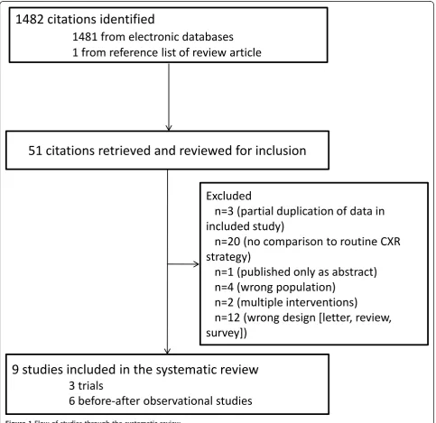
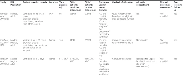
Our search identified 1,482 citations, of which we retrieved 51 citations for more detailed evaluation (Figure 1). Two RCTs [13,15], one quasi-randomized trial [16] (Table 1)
1482 citations identified
1481 from electronic databases
1
from
reference
list
of
review
article
51 citations retrieved and reviewed for inclusion
9 studies included in the systematic review
3 trials
6
before-after
observational
studies
Excluded
n=3 (partial duplication of data in
included study)
n=20 (no comparison to routine CXR
strategy)
n=1 (published only as abstract)
n=4 (wrong population)
n=2 (multiple interventions)
[image:3.595.57.541.252.721.2]n=12 (wrong design [letter, review,
survey])
Table 1 Characteristics and methodological quality of randomized and quasi-randomized controlled trials of restrictive versus routine CXR strategies.
Study ICU Population
Patient selection criteria Location Total patients (n)
CXRs/ patients, restrictive group (n/n)
CXRs/ patients, routine group (n/n)
Outcomes used in meta-analysis
Method of allocation Allocation concealment
Blinded outcomes assessmenta
Zero losses to follow up
Krivopal et al., 2003 [16]
Medical, Adult
Ventilated for 48 to 72 hours
Exclusion criteria: reintubated, transferred from other centers
USA 94 226/51 293/43 ICU and
hospital mortality ICU and hospital length of stay Duration of mechanical ventilation
Quasi-randomization based on last digit of medical record number
No Not
specified Yes
Clec’het al., 2007b
[15]
Medical-surgical, Adult
Ventilated for≥48 hours Exclusion criteria: reintubated, tracheostomy, or withdrawal of life support
France 165 94/81 885/84 ICU and
hospital mortality ICU and hospital length of stay
Computer-generated random number table
Not reported Not
specified Yes
Hejblum et al., 2009 [13]
Medical-surgical, Adult
Ventilated for≥2 days France 611, 849c 3,148/306,
425c
4,607/305, 424c
ICU mortality ICU length of stay Duration of mechanical ventilation
Computer-generated Not reported (’ open-label with respect to allocation
concealment’)
Not specified
Yes
a
Radiologists and clinicians were not blinded to the group when interpreting films;b
In this trial all patients had daily CXRs, but results in the restrictive group were concealed from clinicians, who were free to order CXRs as needed.c
The first sample size is adjusted for the design effect due to clustering [14], assuming intracluster correlation 0.01 (design effect 1.39); the second is the unadjusted sample size as reported in the study. CXR, chest X-ray; n, number.
Ganapathy
et
al
.
Critical
Care
2012,
16
:R68
http://ccfo
rum.com/con
tent/16/2/R6
8
Page
4
of
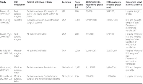
and six before-after observational studies [17-22] (Table 2) met selection criteria. We excluded 42 studies because of partial duplication of data included in the review [23-25]; lack of comparison to routine CXR strategy [26-45]; publi-cation as abstract only [46]; multiple interventions [47,48]; wrong population [49-52] and wrong design [letter, review, survey] [1,53-63].
The nine studies included a total of 39,358 CXRs done on 9,611 patients from the United States, Canada, France, The Netherlands and Germany. One study [18] was con-ducted in a pediatric ICU; the rest were concon-ducted in adult medical, surgical or combined medical-surgical ICUs. One study [13] was a cluster RCT including 849 patients, with an effective sample size of 611 after adjust-ing for clusteradjust-ing, assumadjust-ing an ICC of 0.01.
Overall study quality was moderate to good for trials (Table 1) but poor to moderate for before-after observa-tional studies (Table 3). Observaobserva-tional studies were often at risk for selection bias (that is, enrollment of non-conse-cutive patients) and secular trends in outcomes unrelated to the intervention. Both trials and observational studies generally did not blind radiologists to the CXR strategy in
place at the time the individual CXR was obtained; clini-cians and assessors of other outcomes were similarly unblinded. Only two studies (one RCT [15] and one before-after observational study [22]) mandated routine CXRs (with results concealed from clinicians) to screen for missed findings in patients in the restrictive CXR group.
Mortality outcomes
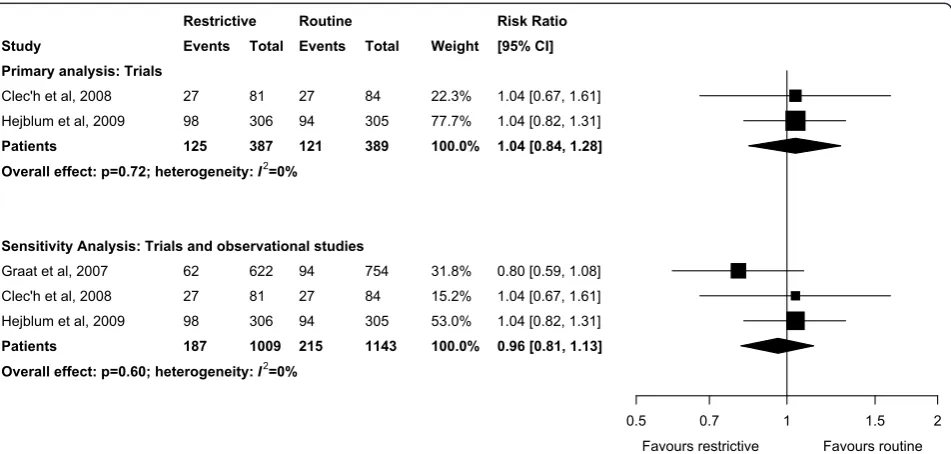
Pooled data showed that the primary outcome of ICU mortality (RR 1.04, 95% CI 0.84 to 1.27, P= 0.78; two trials, N = 776; Figure 2) was similar in the restrictive and routine CXR groups. Results were similar for hospi-tal morhospi-tality (RR 0.98, 95% CI 0.68 to 1.41, P = 0.91; two trials, N = 259; Figure 3).
Other secondary outcomes
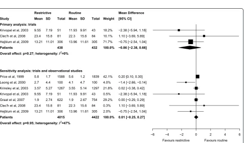
[image:5.595.56.541.386.672.2]Meta-analyses showed that ICU length of stay (WMD -0.86 days, 95% CI -2.38 to 0.66 days,P= 0.27; three trials, N = 870; Figure 4), hospital length of stay (WMD -2.50 days, 95% CI -6.62 to 1.61 days,P= 0.23; two trials, N = 259; Figure 5), and duration of mechanical ventilation
Table 2 Characteristics of included observational (before-after) studies of restrictive versus routine CXR strategies.
Study ICU Population
Patient selection criteria Location Total patients (n)
CXRs/patients, restrictive group (n/n)
CXRs/patients, routine group (n/n)
Outcomes used in meta-analysis
Raoet al., 1997 [17]a
Post cardiac surgery
Exclusion criteria: ICU length of stay > 36 hours, death within 36 hours
UK 200 36/100 304/100 Hospital length
of stay
Priceet al., 1999 [18]b
Pediatric Exclusion criterion: Cardiothoracic surgical patients
USA 3,427 5,939/1,588 10,585/1,839 ICU and hospital
length of stayc Duration of mechanical ventilationc Leonget al.,
2000 [19]d Postcardiac surgery
All patients included USA 200 334/100 520/100 Hospital mortality
ICU and hospital length of stay Duration of mechanical ventilation Krinsleyet
al., 2003 [20] Medical-surgical, Adult
All patients included USA 2,564 2,298/1,267 3,093/1,297 Hospital mortality
ICU length of stay Duration of mechanical ventilation Graatet al.,
2007 [21]
Medical-surgical, Adult
Exclusion criteria: Readmissions Netherlands 1,376 1,115/622 3,194/754 ICU and hospital mortality
Hendriskeet al., 2007 [22]
Medical-surgical, Adult
Exclusion criteria: Cardiothoracic surgical and neurosurgical patients
Netherlands 736 907/250 1,780/486 Hospital mortality
a
The study included one group with routine CXRs, a second group with less frequent routine CXRs, and a third group with CXRs ordered only as clinically
indicated. We abstracted data from the first and third groups;b
This study included a control phase with routine CXRs, an evaluative phase with a small convenience sample of patients with routine CXRs in which the investigators studied the impact of routine CXRs on patient management, and a post
intervention phase with no routine CXRs. We abstracted data from the first and third phases;c
Outcomes data were abstracted from Figure 5;d
Table 3 Newcastle-Ottawa quality assessment scale for before-after observational studies.
STUDY SELECTIONa COMPARABILITYb OUTCOMEc
Representativeness of the exposed cohort
Selection of the non-exposed cohort
Ascertainment of exposure
Outcome of interest not present at the start of study
Cohorts comparable on the basis of design or analysis
Assessment of outcome
Adequacy of duration of follow-up
Adequacy of completeness of follow-up
Raoet al., 1997 [17]
* * * *
Priceet al., 1999 [18]
* * * * * *
Leonget al., 2000 [19]
* * * * *
Krinsleyet al., 2003 [20]
* * * * * *
Graatet al., 2007 [21]
* * * * *
Hendrikse
et al., 2007 [22]
* * * * * *
Refer to reference [9] for a description of Newcastle-Ottawa Quality Assessment Scale for cohort studies. In general, more stars denote higher quality. A study can
be awarded a maximum of one star for each item within the‘Selection’and‘Outcome’categories.a
A maximum of four stars can be given for‘Selection’.
‘Representativeness’is awarded a star if the cohort is truly or somewhat representative of the population of interest. For selection of the non-exposed cohort, a
star is awarded if it is drawn from the same population as the exposed cohort. Exposure is satisfactorily ascertained if data are collected from a secure record.b
A
maximum of two stars can be given for‘Comparability’, one each for controlling of two important confounders in either the design or analysis phase.c
A
maximum of three stars can be given for‘Outcome’.‘Assessment of outcome’is awarded a star if the outcomes were assessed by independent blind assessment
or record linkage. The duration of follow-up was considered adequate if it was long enough for the outcomes to occur. Completeness of follow-up was considered adequate if all patients were accounted for or if the number lost to follow-up was sufficiently low to be unlikely to introduce bias.
0.5 0.7 1 1.5 2
Study
Primary analysis: Trials
Clec'h et al, 2008 Hejblum et al, 2009
Patients
Sensitivity Analysis: Trials and observational studies
Graat et al, 2007 Clec'h et al, 2008 Hejblum et al, 2009
Patients
Overall effect: p=0.60; heterogeneity: I2=0% Overall effect: p=0.72; heterogeneity: I2=0%
Favours restrictive Favours routine
Restrictive Events
27 98
125
62 27 98
187
Total
81 306
387
622 81 306
1009
Routine Events
27 94
121
94 27 94
215
Total
84 305
389
754 84 305
1143
Weight
22.3% 77.7%
100.0%
31.8% 15.2% 53.0%
100.0%
[image:6.595.62.538.431.658.2]Risk Ratio [95% CI]
1.04 [0.67, 1.61] 1.04 [0.82, 1.31]
1.04 [0.84, 1.28]
0.80 [0.59, 1.08] 1.04 [0.67, 1.61] 1.04 [0.82, 1.31]
0.96 [0.81, 1.13]
Figure 2Effect of a restrictive versus routine chest x-ray strategy on intensive care unit mortality among trials (primary analysis) and trials and observational studies (sensitivity analysis). The number of events and sample size of Hejblumet al.[13] have been adjusted for clustering (see Methods for details). Weight is the relative contribution of each study to the overall estimate of treatment effect on a log scale using a random effects model.
Ganapathyet al.Critical Care2012,16:R68 http://ccforum.com/content/16/2/R68
0.5 0.7 1 1.5 2
Study
Primary analysis: trials
Krivopal et al, 2003 Clec'h et al, 2008
Total
Sensitivity analysis: trials and observational studies
Leong et al, 2000 Krinsley et al, 2003 Krivopal et al, 2003 Graat et al, 2007 Hendrikse et al, 2007 Clec'h et al, 2008
Total
Overall effect: p=0.68; heterogeneity: I2=0% Overall effect: p=0.91; heterogeneity: I2=0%
Favours restrictive Favours routine
[image:7.595.58.540.85.355.2] [image:7.595.63.540.406.685.2]Restrictive Events 12 28 40 3 256 12 104 38 28 441 Total 51 81 132 100 1267 51 622 250 81 2371 Routine Events 11 29 40 2 265 11 132 79 29 518 Total 43 84 127 100 1297 43 754 486 84 2764 Weight 25.9% 74.1% 100.0% 0.4% 55.5% 2.6% 23.8% 10.3% 7.4% 100.0% Risk Ratio [95% CI]
0.92 [0.45, 1.87] 1.00 [0.66, 1.52]
0.98 [0.68, 1.41]
1.50 [0.26, 8.79] 0.99 [0.85, 1.15] 0.92 [0.45, 1.87] 0.96 [0.76, 1.21] 0.94 [0.66, 1.33] 1.00 [0.66, 1.52]
0.98 [0.87, 1.09]
Figure 3Effect of a restrictive versus routine chest x-ray strategy on hospital mortality among trials (primary analysis) and trials and observational studies (sensitivity analysis). Weight is the relative contribution of each study to the overall estimate of treatment effect on a log scale using a random effects model.
−6 −4 −2 0 2 4 6
Study
Primary analysis: trials
Krivopal et al, 2003 Clec'h et al, 2008 Hejblum et al, 2009
Patients
Sensitivity analysis: trials and observational studies
Price et al, 1999 Leong et al, 2000 Krinsley et al, 2003 Krivopal et al, 2003 Graat et al, 2007 Clec'h et al, 2008 Hejblum et al, 2009
Patients
Overall effect: p=0.95; heterogeneity: I2=47% Overall effect: p=0.27; heterogeneity: I2=0%
Favours restrictive Favours routine
Restrictive Mean 9.55 23.4 13.21 5.8 2.7 3.57 9.55 1.9 23.4 13.21 SD 7.19 15.8 11.01 1.7 4.4 5.27 7.19 2.74 15.8 11.01 Total 51 81 306 438 1588 100 1267 51 622 81 306 4015 Routine Mean 11.93 22.3 13.96 5.6 4.1 3.55 11.93 1.9 22.3 13.96 SD 9.91 15.6 11.61 1.2 4.7 5.14 9.91 2.67 15.6 11.61 Total 43 84 305 432 1839 100 1297 43 754 84 305 4422 Weight 18.2% 10.1% 71.7% 100.0% 42.1% 4.0% 21.8% 0.5% 29.2% 0.3% 2.0% 100.0% Mean Difference [95% CI]
−2.38 [−5.94, 1.18] 1.10 [−3.69, 5.89]
−0.75 [−2.54, 1.04]
−0.86 [−2.38, 0.66]
0.20 [0.10, 0.30]
−1.4 [−2.66, −0.14] 0.02 [−0.38, 0.42]
−2.38 [−5.94, 1.18] 0.00 [−0.29, 0.29] 1.10 [−3.69, 5.89]
−0.75 [−2.54, 1.04]
0.01 [−0.25, 0.27]
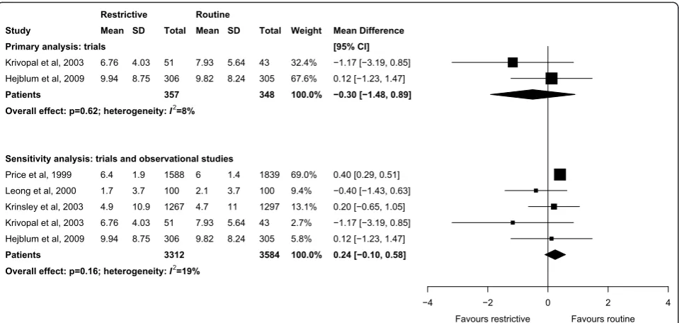
(WMD -0.30 days, 95% CI -1.48 to 0.89 days,P = 0.62; three trials, N = 870; Figure 6) were not significantly dif-ferent between groups. There was little heterogeneity in all analyses restricted to trials (I2≤8%).
Sensitivity analyses
Sensitivity analyses combining results of trials and observational studies showed no difference between groups for ICU mortality (RR 0.96, 95% CI 0.81 to 1.13,
−8 −6 −4 −2 0 2 4 6
Study
Primary analysis: trials
Krivopal et al, 2003 Clec'h et al, 2008
Patients
Sensitivity analysis: trials and observational studies
Rao et al, 1997 Price et al, 1999 Leong et al, 2000 Krivopal et al, 2003 Clec'h et al, 2008
Patients
Overall effect: p=0.48; heterogeneity: I2=87% Overall effect: p=0.23; heterogeneity: I2=0%
Favours restrictive Favours routine
[image:8.595.56.538.87.318.2] [image:8.595.59.539.448.676.2]Restrictive Mean 16.45 39.5 5.9 12.1 7.6 16.45 39.5 SD 11.87 29.4 1.5 2.8 6.6 11.87 29.4 Total 51 81 132 100 1588 100 51 81 1920 Routine Mean 19.34 40.3 6.1 11.3 11.1 19.34 40.3 SD 10.65 33.3 1.6 3.1 10.3 10.65 33.3 Total 43 84 127 100 1839 100 43 84 2166 Weight 81.6% 18.4% 100.0% 39.0% 40.9% 13.8% 5.0% 1.3% 100.0% Mean Difference [95% CI]
−2.89 [−7.44, 1.66]
−0.80 [−10.38, 8.78]
−2.50 [−6.62, 1.61]
−0.20 [−0.63, 0.23] 0.80 [0.60, 1.00]
−3.50 [−5.90, −1.10]
−2.89 [−7.44, 1.66]
−0.80 [−10.38, 8.78]
−0.39 [−1.48, 0.70]
Figure 5Effect of a restrictive versus routine chest x-ray strategy on hospital length of stay in days among trials (primary analysis) and trials and observational studies (sensitivity analysis). Weight is the relative contribution of each study to the overall estimate of treatment effect using a random effects model.
−4 −2 0 2 4
Study
Primary analysis: trials
Krivopal et al, 2003 Hejblum et al, 2009
Patients
Sensitivity analysis: trials and observational studies
Price et al, 1999 Leong et al, 2000 Krinsley et al, 2003 Krivopal et al, 2003 Hejblum et al, 2009
Patients
Overall effect: p=0.16; heterogeneity: I2=19% Overall effect: p=0.62; heterogeneity: I2=8%
Favours restrictive Favours routine
Restrictive Mean 6.76 9.94 6.4 1.7 4.9 6.76 9.94 SD 4.03 8.75 1.9 3.7 10.9 4.03 8.75 Total 51 306 357 1588 100 1267 51 306 3312 Routine Mean 7.93 9.82 6 2.1 4.7 7.93 9.82 SD 5.64 8.24 1.4 3.7 11 5.64 8.24 Total 43 305 348 1839 100 1297 43 305 3584 Weight 32.4% 67.6% 100.0% 69.0% 9.4% 13.1% 2.7% 5.8% 100.0% Mean Difference [95% CI]
−1.17 [−3.19, 0.85] 0.12 [−1.23, 1.47]
−0.30 [−1.48, 0.89]
0.40 [0.29, 0.51]
−0.40 [−1.43, 0.63] 0.20 [−0.65, 1.05]
−1.17 [−3.19, 0.85] 0.12 [−1.23, 1.47]
0.24 [−0.10, 0.58]
Figure 6Effect of a restrictive versus routine chest x-ray strategy on duration of mechanical ventilation in days among trials (primary analysis) and trials and observational studies (sensitivity analysis). The sample size of Hejblumet al.[13] has been adjusted for clustering (see Methods for details). Weight is the relative contribution of each study to the overall estimate of treatment effect using a random effects model.
Ganapathyet al.Critical Care2012,16:R68 http://ccforum.com/content/16/2/R68
P = 0.60; three studies, N = 2,152; Figure 2), hospital mortality (RR 0.98, 95% CI 0.87 to 1.09, P = 0.68; six studies, N = 5,135; Figure 3), ICU length of stay (WMD 0.01 days, 95% CI -0.25 to 0.27 days,P = 0.95; seven studies, N = 8,437; Figure 4), hospital length of stay (WMD -0.39 days, 95% CI -1.48 to 0.70, P= 0.48; five studies, N = 4,086; Figure 5) and duration of mechanical ventilation (WMD 0.24 days, 95% CI -0.10 to 0.58 days, P= 0.16; five studies, N = 6,896; Figure 6). In the sensi-tivity analyses, there was little heterogeneity for ICU or hospital mortality (I2 = 0% for both) or duration of mechanical ventilation (I2= 19%), but there was moder-ate heterogeneity for ICU length of stay (I2 = 47%) and substantial heterogeneity for hospital length of stay (I2= 87%). Additional sensitivity analyses using values of 0.05 and 0 for the ICC of the cluster RCT [13] did not quali-tatively change any results (see Additional file 1).
Discussion
We identified only three trials that evaluated the effects on clinical outcomes in critically ill adults and children of a restrictive strategy of obtaining CXRs only when abnormalities are clinically suspected compared to rou-tine daily CXRs. We found no differences in mortality when results of trials were combined, or when results of an additional six before-after observational studies were also considered. Similarly, duration of mechanical venti-lation and ICU and hospital lengths of stay were similar for both CXR strategies.
Strengths of our systematic review include a compre-hensive search strategy and broad inclusion criteria, with consideration of both RCTs and quasi-RCTs and before-after observational studies. Although we searched elec-tronic biobliographic databases with no restriction on publication date, studies included in the systematic review were all published in the last 15 years and, there-fore, are likely representative of current ICU practices. Our study is limited by small sample size, moderate methodological quality and heterogeneity of included studies. Only three trials evaluated the safety of a restric-tive versus routine CXR strategy; the combined sample size of these trials would only have sufficient power to detect an implausibly large difference in our primary out-come. Despite the absence of statistical heterogeneity in our analyses restricted to trials, all of which excluded patients mechanically ventilated for < 48 hours, there is likely substantial remaining clinical heterogeneity related to severity of illness and surgical status, for example.
Several studies have evaluated a restrictive CXR strat-egy with hypotheses that costs and radiation exposure would be reduced. The restrictive strategy can substan-tially reduce the number of CXRs and thus may reduce costs [18,22] but the anticipated cost-savings have varied greatly [29,31,42]. With respect to radiation exposure,
levels measured at ICU nursing stations have been shown to be below the maximum permissible for non-occupa-tional workers [64,65]. Although exposure to patients from a single CXR is felt to pose minimum risk, the cumulative exposure is thought to be significant due to the high frequency of this test [66]. In contrast, the main short-term risk of a restrictive CXR strategy in critically ill patients is the potential for a missed or delayed diag-nosis, leading to delayed treatment and thus additional morbidity and mortality. We were unable to estimate the number of missed diagnoses or delays in management of complications in the restrictive CXR group because only two studies [15,22] had a surveillance strategy for these patients.
The major design challenge for studies of different CXR ordering strategies is to overcome ascertainment bias, which may lead to more complications (including clinically non-apparent complications) being detected in the routine CXR group if surveillance CXRs are not obtained in the restrictive group. Complications may thus be underestimated in the restrictive CXR group. These study challenges can be overcome by obtaining surveillance CXRs (results blinded to clinicians) in the restrictive group to determine the delay between radio-graphically apparent and clinically detected complica-tions. Other design challenges include ensuring blinding of those adjudicating radiological and clinical outcomes subject to ascertainment bias (for example, pneumonia) and standardization of definitions of complications. Furthermore, establishing non-inferiority for clinical end-points in a RCT comparing CXR strategies requires a large sample size and modifications to the usual approach to hypothesis-testing [67]. An alternative approach would be to design a non-inferiority trial powered on the surro-gate outcome of delayed or missed diagnoses. The het-erogeneity of patients studied to date underscores the importance of a non-inferiority trial that tests the hypothesis that the restrictive CXR strategy does not increase harm and that is adequately powered to examine pre-specified subgroup effects. Finally, even if trials demonstrate the non-inferiority of the restrictive CXR strategy, ICUs implementing this strategy should have immediate access to clinicians who can reliably identify patients requiring urgent CXRs, interpret images that are promptly obtained and processed, and act rapidly on any findings requiring intervention [68].
mechanical ventilation for acute respiratory distress syn-drome or status asthmaticus. There was also consensus that CXRs were unnecessary in patients with an existing nasogastric tube for enteral feeding or with an existing central venous catheter previously documented to be cor-rectly positioned in the superior vena cava. However, there was no consensus on the need for routine CXRs in intu-bated patients. The clinical impact and safety of imple-menting these consensus recommendations have not been evaluated, but the findings of our review suggest no glar-ing safety concerns with a restrictive CXR strategy in criti-cally ill patients while underscoring the need for additional large randomized trials to establish definitively the non-inferiority of this approach.
Conclusions
Pooling the results of three trials alone or combined with six before-after observational studies that evaluated the safety of abandoning routine CXRs in favor of a more restrictive approach did not detect harm. How-ever, given the small overall sample size and infrequent systematic evaluation of missed findings in the restric-tive strategy group, the safety of abandoning routine CXRs in critically ill patients remains uncertain and mandates further investigation.
Key messages
•Few trials have compared restrictive versus routine CXRs in the ICU.
• Combined results from three trials did not show harm from a restrictive CXR strategy in the ICU; results were similar when six before-after observa-tional studies were also considered.
• However, given the small overall sample size and limited data on the effects of missed findings on routine CXRs, the safety of abandoning routine CXRs in critically ill patients remains uncertain.
Additional material
Additional file 1: Sensitivity analyses for the effect of a restrictive versus routine chest x-ray strategy on patient outcomes, assuming different values of ICC for the cluster randomized trial. The file contains two tables with meta-analytic data from sensitivity analyses that assume an intracluster correlation coefficient of 0 or 0.05 for the cluster RCT [13], instead of 0.01 as assumed in the main analyses.
Abbreviations
CI: confidence interval; CXR: chest x-ray; ICC: intracluster correlation coefficient; ICU: intensive care unit; RCT: randomized controlled trial; RR: risk ratio; WMD: weighted mean difference.
Acknolwedgments
We thank Dr Ruxandra Pinto for assistance with the statistical analysis and with the figures. This study received no funding. DS is supported by a New
Investigator Award from the Canadian Institutes of Health Research, which had no role in the design or conduct of the study or decision to submit for publication. Preliminary results have been presented in abstract form (Spiegelman J, Pinto R, Adhikari N, Scales D. Should routine daily chest radiography in Intensive Care Units (ICUs) be abandoned? A systematic review [abstract].Am J Respir Crit Care Med2009,179:A1618).
Author details
1
Department of Critical Care Medicine, Sunnybrook Health Sciences Centre; Interdepartmental Division of Critical Care, University of Toronto; 2075 Bayview Avenue, Toronto ON M4N 3M5, Canada.2Department of Critical Care Medicine and Sunnybrook Research Institute, Sunnybrook Health Sciences Centre; Interdepartmental Division of Critical Care, University of Toronto; 2075 Bayview Avenue, Toronto ON M4N 3M5, Canada.3Department of Medicine, Division of Critical Care Medicine, Humber River Regional Hospital, 2111 Finch Avenue West, Toronto ON M3N 1N1, Canada.
Authors’contributions
AG screened studies for inclusion, abstracted data, analyzed data, and drafted the manuscript. JS screened studies for inclusion, abstracted data, analyzed data, and drafted the manuscript. NA verified outcomes data, assessed study quality, analyzed data, and helped to draft the manuscript. DS screened studies for inclusion, abstracted data, analyzed data, and helped to draft the manuscript. AG, NA, and DS critically revised the manuscript for important intellectual content. All authors read and approved the final manuscript.
Competing interests
The authors declare that they have no competing interests.
Received: 8 November 2011 Revised: 14 February 2012 Accepted: 27 April 2012 Published: 27 April 2012
References
1. Rubinowitz AN, Siegel MD, Tocino I:Thoracic imaging in the ICU.Crit Care Clin2007,23:539-573.
2. American College of Radiology appropriateness criteria for routine chest radiographs in ICU patients.[http://www.acr.org/
SecondaryMainMenuCategories/quality_safety/app_criteria.aspx]. 3. Henschke CI, Yankelevitz DF, Wand A, Davis SD, Shiau M:Chest
radiography in the ICU.Clin Imaging1997,21:90-103.
4. Henschke CI, Yankelevitz DF, Wand A, Davis SD, Shiau M:Accuracy and efficacy of chest radiography in the intensive care unit.Radiol Clin North Am1996,34:21-31.
5. Leppek R, Bertrams SS, Holtermann W, Klose KJ:Radiation exposure during thoracic radiography at the intensive care unit. Dose accumulation and risk of radiation-induced cancer in long-term therapy.Radiologe1998, 38:730-736.
6. Meade MO, Cook RJ, Guyatt GH, Groll R, Kachura JR, Bedard M, Cook DJ, Slutsky AS, Stewart TE:Interobserver variation in interpreting chest radiographs for the diagnosis of acute respiratory distress syndrome.Am J Respir Crit Care Med2000,161:85-90.
7. Moher D, Liberati A, Tetzlaff J, Altman DG, PRISMA Group:Preferred reporting items for systematic reviews and meta-analyses: the PRISMA statement.Ann Intern Med2009,151:264-269, W64.
8. Stroup DF, Berlin JA, Morton SC, Olkin I, Williamson GD, Rennie D, Moher D, Becker BJ, Sipe TA, Thacker SB:Meta-analysis of observational studies in epidemiology: a proposal for reporting. Meta-analysis Of Observational Studies in Epidemiology (MOOSE) group.JAMA2000,283:2008-2012. 9. The Newcastle-Ottawa Scale (NOS) for assessing the quality of
nonrandomised studies in meta-analyses.[http://www.ohri.ca/programs/ clinical_epidemiology/oxford.asp].
10. DerSimonian R, Laird N:Meta-analysis in clinical trials.Control Clin Trials
1986,7:177-188.
11. Perera R, Heneghan C:Interpreting meta-analysis in systematic reviews.
Evid Based Med2008,13:67-69.
12. Higgins JP, Thompson SG, Deeks JJ, Altman DG:Measuring inconsistency in meta-analyses.BMJ2003,327:557-560.
13. Hejblum G, Chalumeau-Lemoine L, Ioos V, Boelle PY, Salomon L, Simon T, Vibert JF, Guidet B:Comparison of routine and on-demand prescription of chest radiographs in mechanically ventilated adults: a multicentre, Ganapathyet al.Critical Care2012,16:R68
http://ccforum.com/content/16/2/R68
cluster-randomised, two-period crossover study.Lancet2009, 374:1687-1693.
14. The Cochrane Collaboration. 2011 [http://www.cochrane-handbook.org]. 15. Clec’h C, Simon P, Hamdi A, Hamza L, Karoubi P, Fosse JP, Gonzalez F,
Vincent F, Cohen Y:Are daily routine chest radiographs useful in critically ill, mechanically ventilated patients? A randomized study.Intensive Care Med2008,34:264-270.
16. Krivopal M, Shlobin OA, Schwartzstein RM:Utility of daily routine portable chest radiographs in mechanically ventilated patients in the medical ICU.Chest2003,123:1607-1614.
17. Rao PS, Abid Q, Khan KJ, Meikle RJ, Natarajan KM, Morritt GN, Wallis J, Kendall SW:Evaluation of routine postoperative chest X-rays in the management of the cardiac surgical patient.Eur J Cardiothorac Surg1997, 12:724-729.
18. Price MB, Grant MJ, Welkie K:Financial impact of elimination of routine chest radiographs in a pediatric intensive care unit.Crit Care Med1999, 27:1588-1593.
19. Leong CS, Cascade PN, Kazerooni EA, Bolling SF, Deeb GM:Bedside chest radiography as part of a postcardiac surgery critical care pathway: a means of decreasing utilization without adverse clinical impact.Crit Care Med2000,28:383-388.
20. Krinsley JS:Test-ordering strategy in the intensive care unit.J Intensive Care Med2003,18:330-339.
21. Graat ME, Kroner A, Spronk PE, Korevaar JC, Stoker J, Vroom MB, Schultz MJ: Elimination of daily routine chest radiographs in a mixed medical-surgical intensive care unit.Intensive Care Med2007,33:639-644. 22. Hendrikse KA, Gratama JW, Hove W, Rommes JH, Schultz MJ, Spronk PE:
Low value of routine chest radiographs in a mixed medical-surgical ICU.
Chest2007,132:823-828.
23. Graat ME, Choi G, Wolthuis EK, Korevaar JC, Spronk PE, Stoker J, Vroom MB, Schultz MJ:The clinical value of daily routine chest radiographs in a mixed medical-surgical intensive care unit is low.Crit Care2006,10:R11. 24. Kroner A, Binnekade JM, Graat ME, Vroom MB, Stoker J, Spronk PE,
Schultz MJ:On-demand rather than daily-routine chest radiography prescription may change neither the number nor the impact of chest computed tomography and ultrasound studies in a multidisciplinary intensive care unit.Anesthesiology2008,108:40-45.
25. Mets O, Spronk PE, Binnekade J, Stoker J, de Mol BA, Schultz MJ: Elimination of daily routine chest radiographs does not change on-demand radiography practice in post-cardiothoracic surgery patients.J Thorac Cardiovasc Surg2007,134:139-144.
26. Bekemeyer WB, Crapo RO, Calhoon S, Cannon CY, Clayton PD:Efficacy of chest radiography in a respiratory intensive care unit. A prospective study.Chest1985,88:691-696.
27. Beyermann K, Kussmann J, Gartenschlager M, Herrmann A, Busch H, Stein K: Daily“routine chest x-ray”in ventilated surgical intensive care patients.
Chirurg1993,64:1032-1035.
28. Bhagwanjee S, Muckart DJ:Routine daily chest radiography is not indicated for ventilated patients in a surgical ICU.Intensive Care Med
1996,22:1335-1338.
29. Brainsky A, Fletcher RH, Glick HA, Lanken PN, Williams SV, Kundel HL: Routine portable chest radiographs in the medical intensive care unit: effects and costs.Crit Care Med1997,25:801-805.
30. Chahine-Malus N, Stewart T, Lapinsky SE, Marras T, Dancey D, Leung R, Mehta S:Utility of routine chest radiographs in a medical-surgical intensive care unit: a quality assurance survey.Crit Care2001,5:271-275. 31. Fong Y, Whalen GF, Hariri RJ, Barie PS:Utility of routine chest radiographs
in the surgical intensive care unit. A prospective study.Arch Surg1995, 130:764-768.
32. Gartenschlager M, Busch H, Kussmann J, Nafe B, Beyermann K, Klose KJ: Radiological thorax monitoring in ventilated intensive-care patients.Rofo
1996,164:95-101.
33. Greenbaum DM, Marschall KE:The value of routine daily chest x-rays in intubated patients in the medical intensive care unit.Crit Care Med1982, 10:29-30.
34. Hall JB, White SR, Karrison T:Efficacy of daily routine chest radiographs in intubated, mechanically ventilated patients.Crit Care Med1991, 19:689-693.
35. Hauser GJ, Pollack MM, Sivit CJ, Taylor GA, Bulas DI, Guion CJ:Routine chest radiographs in pediatric intensive care: a prospective study.
Pediatrics1989,83:465-470.
36. Henschke CI, Pasternack GS, Schroeder S, Hart KK, Herman PG:Bedside chest radiography: diagnostic efficacy.Radiology1983,149:23-26. 37. Horst HM, Fagan B, Beute GH:Chest radiographs in surgical intensive care
patients: a valuable“routine”.Henry Ford Hosp Med J1986,34:84-86. 38. Janower ML, Jennas-Nocera Z, Mukai J:Utility and efficacy of portable
chest radiographs.AJR Am J Roentgenol1984,142:265-267.
39. Lai MW, Yong SC, Boo NY:The effect of a user-guided request form for chest radiographs in a neonatal intensive care unit.Med J Malaysia2004, 59:598-603.
40. Marik PE, Janower ML:The impact of routine chest radiography on ICU management decisions: an observational study.Am J Crit Care1997, 6:95-98.
41. Quasney MW, Goodman DM, Billow M, Chiu H, Easterling L, Frankel L, Habib D, Heitschmidt M, Kurachek S, Moler F, Montgomery V, Moss M, Murman S, Rice T, Richman B, Tilden S:Routine chest radiographs in pediatric intensive care units.Pediatrics2001,107:241-248.
42. Silverstein DS, Livingston DH, Elcavage J, Kovar L, Kelly KM:The utility of routine daily chest radiography in the surgical intensive care unit.J Trauma1993,35:643-646.
43. Sivit CJ, Taylor GA, Hauser GJ, Pollack MM, Bulas DI, Guion CJ, Fearon T: Efficacy of chest radiography in pediatric intensive care.AJR Am J Roentgenol1989,152:575-577.
44. Strain DS, Kinasewitz GT, Vereen LE, George RB:Value of routine daily chest x-rays in the medical intensive care unit.Crit Care Med1985, 13:534-536.
45. Valk JW, Plotz FB, Schuerman FA, van Vught H, Kramer PP, Beek EJ:The value of routine chest radiographs in a paediatric intensive care unit: a prospective study.Pediatr Radiol2001,31:343-347.
46. Brivet FG, Jacobs FM, Maitre S, Rehel M, Prat D, Grigoriu BD, Musset D:A clinically based chest x-ray strategy reduces health care costs without adverse consequences [abstract].Intensive Care Med2006,32(Suppl 1): S136.
47. Prat G, Lefevre M, Nowak E, Tonnelier JM, Renault A, L’Her E, Boles JM: Impact of clinical guidelines to improve appropriateness of laboratory tests and chest radiographs.Intensive Care Med2009,35:1047-1053. 48. Roberts DE, Bell DD, Ostryzniuk T, Dobson K, Oppenheimer L, Martens D,
Honcharik N, Cramp H, Loewen E, Bodnar S:Eliminating needless testing in intensive care–an information-based team management approach.
Crit Care Med1993,21:1452-1458.
49. Sussman E, Goodwin P, Rosen H:Administrative change and diagnostic test use. The effect of eliminating standing orders.Med Care1984, 22:569-572.
50. Amshel CE, Palesty JA, Dudrick SJ:Are chest X-rays mandatory following central venous recatheterization over a wire?Am Surg1998,64:499-501, discussion 501-502.
51. Frassinelli P, Pasquale MD, Cipolle MD, Rhodes M:Utility of chest radiographs after guidewire exchanges of central venous catheters.Crit Care Med1998,26:611-615.
52. Vezzani A, Brusasco C, Palermo S, Launo C, Mergoni M, Corradi F: Ultrasound localization of central vein catheter and detection of postprocedural pneumothorax: an alternative to chest radiography.Crit Care Med2010,38:533-538.
53. Bedrick AD:To x-ray or not to x-ray? That is the question.Clin Pediatr (Phila)1993,32:520.
54. Graat ME, Stoker J, Vroom MB, Schultz MJ:Can we abandon daily routine chest radiography in intensive care patients?J Intensive Care Med2005, 20:238-246.
55. Hazelzet J:Where is the chest radiograph?Pediatr Crit Care Med2003, 4:119-120.
56. Iberti TJ:Daily chest radiographs: the jury is still out.Crit Care Med1991, 19:597.
57. Janower ML:Daily routine chest radiographs.Chest2004,125:1168, author reply 1168-1169.
58. Oba Y, Zaza T:Abandoning daily routine chest radiography in the intensive care unit: meta-analysis.Radiology2010,255:386-395. 59. Plotz FB, Valk JW:Routine chest radiographs in pediatric intensive care
units.Pediatrics2002,110:421, author reply 421.
60. Schultz MJ:Daily routine chest radiographs.Chest2004,125:1167, author reply 1168-1169.
62. Wosornu D, Pitt M, Beattie JM, Murray RG:Routine chest radiographs in admissions to coronary care.Lancet1995,345:62.
63. Graat ME, Hendrikse KA, Spronk PE, Korevaar JC, Stoker J, Schultz MJ:Chest radiography practice in critically ill patients: a postal survey in the Netherlands.BMC Med Imaging2006,6:8.
64. Boles JM, Boussert F, Manens JP, Le Cam B, Bellet M, Garre M:
Measurement of irradiation doses secondary to bedside radiographs in a medical intensive care unit.Intensive Care Med1987,13:60-63. 65. Herman MW, Patrick J, Tabrisky J:A comparative study of scattered
radiation levels from 80-kVp and 240-kVp x rays in the surgical intensive care unit.Radiology1980,137:552-553.
66. Veldkamp WJ, Kroft LJ, Geleijns J:Dose and perceived image quality in chest radiography.Eur J Radiol2009,72:209-217.
67. Piaggio G, Elbourne DR, Altman DG, Pocock SJ, Evans SJ, CONSORT Group: Reporting of noninferiority and equivalence randomized trials: an extension of the CONSORT statement.JAMA2006,295:1152-1160. 68. Siegel MD, Rubinowitz AN:Routine daily vs on-demand chest
radiographs in intensive care.Lancet2009,374:1656-1658.
69. Hejblum G, Ioos V, Vibert JF, Boelle PY, Chalumeau-Lemoine L, Chouaid C, Valleron AJ, Guidet B:A web-based Delphi study on the indications of chest radiographs for patients in ICUs.Chest2008,133:1107-1112.
doi:10.1186/cc11321
Cite this article as:Ganapathyet al.:Routine chest x-rays in intensive
care units: a systematic review and meta-analysis.Critical Care201216:
R68.
Submit your next manuscript to BioMed Central and take full advantage of:
• Convenient online submission
• Thorough peer review
• No space constraints or color figure charges
• Immediate publication on acceptance
• Inclusion in PubMed, CAS, Scopus and Google Scholar
• Research which is freely available for redistribution
Submit your manuscript at www.biomedcentral.com/submit
Ganapathyet al.Critical Care2012,16:R68 http://ccforum.com/content/16/2/R68