Detlef Schuppan, Yong Ook Kim
J Clin Invest.
2013;
123(5)
:1887-1901.
https://doi.org/10.1172/JCI66028
.
Fibrosis is an intrinsic response to chronic injury, maintaining organ integrity when
extensive necrosis or apoptosis occurs. With protracted damage, fibrosis can progress
toward excessive scarring and organ failure, as in liver cirrhosis. To date, antifibrotic
treatment of fibrosis represents an unconquered area for drug development, with enormous
potential but also high risks. Preclinical research has yielded numerous targets for
antifibrotic agents, some of which have entered early-phase clinical studies, but progress
has been hampered due to the relative lack of sensitive and specific biomarkers to measure
fibrosis progression or reversal. Here we focus on antifibrotic approaches for liver that
address specific cell types and functional units that orchestrate fibrotic wound healing
responses and have a sound preclinical database or antifibrotic activity in early clinical
trials. We also touch upon relevant clinical study endpoints, optimal study design, and
developments in fibrosis imaging and biomarkers.
Review Series
Find the latest version:
Evolving therapies for liver fibrosis
Detlef Schuppan1,2 and Yong Ook Kim1
1Institute of Molecular and Translational Medicine and Department of Medicine I, University Medical Center, Johannes Gutenberg University Mainz, Mainz, Germany. 2Beth Israel Deaconess Medical Center, Harvard Medical School, Boston, Massachusetts, USA.
Fibrosis is an intrinsic response to chronic injury, maintaining organ integrity when extensive necrosis or apoptosis
occurs. With protracted damage, fibrosis can progress toward excessive scarring and organ failure, as in liver
cir-rhosis. To date, antifibrotic treatment of fibrosis represents an unconquered area for drug development, with
enor-mous potential but also high risks. Preclinical research has yielded numerous targets for antifibrotic agents, some of
which have entered early-phase clinical studies, but progress has been hampered due to the relative lack of sensitive
and specific biomarkers to measure fibrosis progression or reversal. Here we focus on antifibrotic approaches for
liver that address specific cell types and functional units that orchestrate fibrotic wound healing responses and have
a sound preclinical database or antifibrotic activity in early clinical trials. We also touch upon relevant clinical study
endpoints, optimal study design, and developments in fibrosis imaging and biomarkers.
The clinical problem
Fibrosis is the excess accumulation of ECM, which results from chronic, nonresolving inflammation. This inflammation triggers a wound-healing process that mitigates inflammatory tissue destruc-tion but also leads to scar tissue formadestruc-tion. In the liver, fibrosis is mainly due to chronic viral hepatitis B or C, autoimmune and biliary diseases, alcoholic steatohepatitis (ASH) and, increasingly, nonalcoholic steatohepatitis (NASH) (1–5). While mild fibrosis remains largely asymptomatic, its progression toward cirrhosis, i.e., replacement of functional parenchyma by scar tissue accom-panied by severe architectural and vascular distortion, is the major cause of liver-related morbidity and mortality. Clinical sequelae of cirrhosis are (a) liver synthetic (functional) failure, including failing hemostatic, nitrogen handling, and detoxification systems; (b) por-tal hypertension with consequent formation of ascites and bleeding esophageal or gastric varices; (c) a high susceptibility to infection; and (d) a high risk to develop hepatocellular carcinoma (HCC) (2). Preventive measures, such as antiviral regimens for hepatitis B or C, are already decreasing the burden of viral cirrhosis and HCC, but other causes, such as NASH (which is linked to obesity and type 2 diabetes) are taking center stage. Moreover, numerous patients present initially in the clinic with advanced fibrosis or cirrhosis, which are largely irreversible. Therefore, antifibrotics that prevent progression toward cirrhosis or induce regression of advanced fibrosis and cirrhosis are urgently needed (6–9).
Liver fibrosis progression and reversal
Research has delineated key mechanisms and cells that determine fibrosis progression (fibrogenesis) and regression (fibrolysis) (1–19). Notably, liver fibrosis has much in common with fibrosis of other organs, such as lungs and kidneys, leading to a cross-fertilization of research across organ boundaries. The structural components of the fibrotic ECM, the growth factors, cytokines, chemokines, and proteases, as well as central signaling cascades implicated in fibrogenesis and fibrolysis, are nearly identical in these different tissues (18, 20–22). Importantly, fibrosis is no lon-ger considered static, but the result of a continuous remodeling process. Nonetheless, in contrast to kidneys and lungs, the liver has an extraordinary capacity to regenerate, even in advanced fibrosis.
Fibrosis is intimately linked to wound healing, serving to pre-vent tissues from disassembly during inflammation, apoptosis, necrosis, and release of lytic enzymes. Fibrosis usually reverses within days to a few weeks following the resolution of tissue dam-age, as demonstrated in less advanced rodent and human liver fibrosis (2, 8, 9, 23–25). However, the longer the damage persists, often at a low level, the more ECM is deposited. This chronic dam-age results in increasingly acellular scar tissue and a steep decline of potential reversibility, even after elimination of causative trig-gers (26, 27). Inefficient fibrolysis is due to several factors: (a) lack of cues for ordered cell repopulation and regeneration due to an atypical ECM and the loss of appropriate cellular context, (b) advanced vascular remodeling with architectural distortion, (c) extensive crosslinking of ECM components such as fibrillar collagen that make proteolytic removal difficult, and (d) the dis-appearance of cellular elements that digest the scar tissue. Here we discuss the cellular and molecular pathways that promote fibrosis progression and highlight current clinical trials as well as improved methods of monitoring fibrosis.
Cellular targets and multicellular fibrogenic units
Activated myofibroblasts, representing a spectrum of similar ECM-producing cells that mainly derive from hepatic stellate cells and portal fibroblasts, are the major producers of the fibrotic ECM and the most downstream cellular effectors of liver fibrosis (Figure 1). Very few hepatic myofibroblasts in fibrosis stem from BM-derived fibrocytes (12). Moreover, complete epithelial-mesen-chymal transition (EMT) of hepatocytes and bile duct epithelia to myofibroblasts may be a rare event — while an “incomplete” EMT of these cells with acquisition of a fibrogenic phenotype is common (28). Myofibroblasts and their products are primary tar-gets for antifibrotic therapies, which in principle would address all types of fibrosis, including advanced fibrosis.
Importantly, additional cellular elements that are either upstream of the myofibroblasts or tightly linked to fibrogenic activation within cellular units may provide a basis for comple-mentary and more disease-specific antifibrotic approaches. A combination therapy approach may be more effective, given that crosstalk between different cell types generally underlies fibro-genic activation. Conceptually, three major multicellular func-tional units can be defined according to their constituent cell types: (a) perisinusoidal/vascular — pericytes, i.e., hepatic stellate
Conflict of interest: The authors have declared that no conflict of interest exists.
cells, liver sinusoidal endothelial cells (LSECs), macrophages/ Kupffer cells, and hepatocytes; (b) stromal inflammatory — myofibroblasts, T cells, and macrophages; and (c) portal/peri-portal — cholangiocytes/ductular cells, portal/peri-portal fibroblasts, and various inflammatory cells (ref. 8 and Figure 2, A–C). Altered interactions within these functional units give rise to the major multicellular fibrogenic pathways.
Fibrogenic effectors
Activated myofibroblasts. Myofibroblasts that derive from both acti-vated hepatic stellate cells and portal fibroblasts are the primary producers of scar tissue (1, 2, 6–22, 29). Notably, myofibroblasts are essential for organ integrity, and their elimination promotes tissue necrosis and inflammation (30). Moreover, myofibroblasts can also contribute to fibrosis regression via release of
ECM-Figure 1
Myofibroblasts and their fibrogenic activation. Cells and major factors upstream of quiescent portal fibroblasts and hepatic stellate cells that induce transformation to fibrogenic myofibroblasts. This schematic highlights several major targets to treat liver fibrosis. Notably, the ECM itself can serve as modulator of fibrogenesis and fibrolysis. Thus collagen fibrils become crosslinked by LOXL2, which contributes to the reduced
reversibility of advanced fibrosis, and collagen-binding ECM receptors (especially the integrins α1β1, α2β1, and α11β1) confer signals of stress
[image:3.585.94.502.82.530.2]degrading proteases, when confronted with favorable (e.g., ECM-derived and integrin receptor–mediated) stimuli, in a process called stress relaxation. Stress relaxation is the basis for limiting ECM deposition once the wound is closed: the activated myo-fibroblasts contract on the accumulated loose collagen matrix, which triggers release of ECM-degrading proteases, mainly MMPs (31, 32). Consequently, treatment strategies should not eliminate myofibroblasts, but rather dampen their fibrogenic activation, confer signals of stress relaxation, and induce fibrolytic enzymes. Accordingly, two rodent studies demonstrated that approximate-ly 50% of activated hepatic stellate cells/myofibroblasts undergo apoptosis during fibrosis reversal, whereas the rest revert to a qui-escent phenotype (33, 34). Quiescence can be induced by inhibi-tion of certain fibroblast integrins, cellular receptors that confer mechanical cues in response to ECM attachment (20) with the
potential of converting activated to fibrolytic (myo-)fibroblasts (refs. 31, 32, 35, 36 and Figure 1). Specific integrin inhibitors have been developed for cancer therapy, but need better validation for treatment of fibrosis (37, 38). Myofibroblast stress relaxation and resultant amelioration of both fibrogenesis and portal hyperten-sion has been shown in rats by inhibition of Rho kinase, which is downstream of integrin signaling (39).
[image:4.585.48.538.80.477.2]Several agents that block fibrogenic activation and ECM produc-tion by myofibroblasts work well in culture and in some rodent models of liver fibrosis but carry a high risk of unwanted side effects in patients due to a lack of specificity for myofibroblasts. Three major strategies are currently in preclinical development to specifically target the pathogenic function of activated myofibro-blasts. First, therapies may address fibrosis-relevant pathways that are upregulated in these myofibroblasts, such as procollagen type I
Figure 2
Multicellular context of fibrogenesis and fibrolysis. Shown are the postulated major cellular functional units and secreted factors that should be
addressed in their complexity when designing effective antifibrotic strategies. (A) Vascular unit. (B) Biliary unit. (C) Inflammatory unit. (D) Cells
and factors that affect macrophage polarization. Macrophages (and monocytes as macrophage precursors) are major modulators of inflammation and tissue remodeling. Cells and factors that induce either M1 or M2 polarization are also linked to the generation of fibrogenic Th17 cells and
neutrophil recruitment. See text for details. B and C highlight factors not shown in A and B, respectively. Baso, basophil; EO, eosinophil; Mast,
or other key structural components of the ECM, or block cellular receptors for ECM components and growth factors/chemokines that are upregulated upon fibrogenic activation. Current blockers of collagen synthesis have unwanted off-target effects, but inhi-bition of upstream fibrogenic signaling, e.g., PDGFRβ, a strong myofibroblast mitogen, with the tyrosine kinase inhibitor ima-tinib or a more specific PDGFRβ-blocking antibody retarded early but not advanced liver fibrogenesis (40, 41).
A second approach to targeting activated myofibroblasts is to employ refined siRNA delivery techniques, such as liposomal for-mulations that intrinsically accumulate in liver due to their size, shape, and surface charge, and that deliver cargo to myofibroblasts as well as other liver cell types (42, 43). For example, biliary and parenchymal liver fibrosis was significantly mitigated in mice treated with liposomes loaded with procollagen α1(I) siRNA (44). Finally, the use of ligands specific to receptors on activated myofi-broblasts can target drugs or siRNA, thus increasing efficacy and minimizing detrimental off-target effects. Examples supporting this approach in vivo include delivery of IFN via a cyclic PDGFRβ
-binding peptide, of a PDGFRβ-specific kinase inhibitor via man-nose-6-phosphate (which addresses the IGF-II receptor), and of Hsp47 (which is involved in collagen processing) via vitamin A– coupled liposomes (45–48). Although these therapies would largely need to be given parenterally, such application can be justified in situations in which treatment is likely to be highly effective, e.g., for reversing advanced fibrosis. Moreover, modifications of deliv-ery systems such as pegylation (49) can be used to increase half-lives, permitting once-weekly or once-monthly dosing.
[image:5.585.63.519.77.427.2]Damaged hepatocytes. Ongoing hepatocyte apoptosis or necrop-tosis, as occurs predominantly in liver diseases characterized by enhanced oxidative and endoplasmic reticulum stress, lyso-somal activation, and mitochondrial damage (ASH, NASH), is a strong trigger of fibrogenesis (16, 50). Phagocytosis of apoptotic hepatocytes by myofibroblasts triggers their fibrogenic activation via NADPH oxidase 2 (NOX2) (51) and the JAK/STAT and PI3K/ Akt pathways (52). Notably, inhibition of hepatocyte apoptosis by a pan-caspase inhibitor or an antagonist of cathepsin B (a lyso-somal trigger of apoptosis) ameliorated (biliary) fibrosis in mice
Figure 3
(53, 54). On the other hand, as mentioned below, engulfment of apoptotic hepatocytes and biliary cells by macrophages can induce their fibrolytic activation.
Biliary progenitors. The hallmark of biliary fibrosis is the prolifera-tion of biliary progenitor cells (activated cholangiocytes) that tend to form small clusters or usually nonfunctional bile ductular struc-tures, termed ductular reaction. These cells replicate early devel-opmental programs of ductal plate formation, which includes secretion of several factors that attract and activate hepatic stel-late cells/myofibroblasts to proliferate and deposit ECM. This biliary progenitor response is amplified by the surrounding myo-fibroblasts, but also by inflammatory cells that release molecules that sustain ductular cell viability and proliferation (Figure 3). With the exception of infant fibrosis (biliary atresia, Caroli’s dis-ease, congenital hepatic fibrosis) and adult primary biliary cir-rhosis (PBC), primary sclerosing cholangitis (PSC), and secondary biliary fibrosis, all liver diseases of other etiologies, once advanced, develop into a portal fibrosis with proliferation of biliary progeni-tors, especially when excessive hepatocyte apoptosis forces the stem cell niche to produce biliary progenitors. These biliary pro-genitors are more resistant to enhanced oxidative stress and hepa-tocyte death, such as induced by ASH, NASH, or severe post-trans-plant hepatitis C (55–60). Drugs aimed at the biliary fibrogenic progenitors are effective antifibrotic agents in rodent biliary and advanced non-biliary fibrosis. Examples are antagonists to the bili-ary progenitor-specific integrin αvβ6 (a receptor for fibronectin and tenascin-C, and an activator of latent TGF-β1) (61–63) or inhi-bition of the hedgehog pathway, which is primarily upregulated in biliary fibrogenesis and in carcinogenesis (56–59, 64, 65). Nota-bly, inhibition of hedgehog signaling suppressed biliary fibrosis and even reversed hepatocellular cancer in phospholipid flippase (Mdr2) knockout mice (65).
LSECs. Hepatic (neo-)vascularization with LSEC activation and proliferation is tightly associated with perisinusoidal fibrosis (cap-illarization of the sinusoids) (Figure 1 and Figure 2A). During peri-sinusoidal fibrosis, activated LSECs contribute to ECM production (including basement membrane components, fibronectin, and interstitial collagen type I), produce cytokines (e.g., TGF-β1 and PDGF-BB) that activate hepatic stellate cells, and secrete factors (e.g., endothelin-1) that contribute to intrahepatic vasoconstric-tion, which exacerbates portal hypertension in cirrhosis. Converse-ly, myofibroblasts activate LSEC via secretion of angiogenic factors such as VEGF and angiopoietin-1 (66). Antiangiogenic therapies have mitigated experimental liver fibrosis, mostly in models with a prominent sinusoidal component. However, antifibrotic effects were evident with polykinase inhibitors such as sunitinib and sorafenib that, apart from angiogenic VEGF or FGF receptors on LSECs, also target numerous other cells and kinases involved in proliferation, ECM turnover, and immune regulation (67, 68). This lack of specificity may explain the finding that treatment with anti-VEGF antibody and an antagonist to integrin αvβ3, therapies that inhibit LSEC proliferation (but also affect the proliferation of endothelia of larger vessels) may worsen advanced biliary, peri-sinusoidal, and interstitial kidney fibrosis (69–71). Moreover, spe-cific inhibition of VEGF mitigates biliary fibrosis progression but retards fibrosis reversal after jejunoileal anastomosis (72). There-fore, as with many other therapies, the antifibrotic efficacy of anti-angiogenic therapies is highly context dependent.
T cells. CD4+ T cells with a Th2 polarization, which are prevalent
in allergies, asthma, or parasite infections, promote fibrogenesis in
the liver, lungs, or kidneys (18, 73–75). Th2 cells produce IL-4 and IL-13, which stimulate the differentiation of potentially fibrogenic myeloid cells and (alternatively) activated (M2) macrophages (refs. 73, 76, and Figure 2D). Thus rodents with Th2-dominant T cell infiltration (e.g., in experimental schistosomiasis or in experimen-tal models skewed toward Th2; ref. 77) display rapid fibrosis pro-gression, whereas CD4+ Th1 cells have an antifibrotic effect (78).
Accordingly, patients dually infected with HCV and Schistosoma show a 6-fold faster liver fibrosis progression than matched HCV-monoinfected patients (79).
Th17 cells are clear drivers of fibrosis in multiple tissues (80, 81). Th17 cells are induced by a special inflammatory environment, including the cytokines TGF-β1 and IL-6. Th17 cells secrete IL-17A, which drives fibrogenesis directly in myofibroblasts and indirectly via stimulation of TGF-β1 release from inflammatory cells (80, 82).
Regulatory T cells appear to either favor or inhibit fibrogenesis, again in a context-dependent manner. Subsets produce various amounts of the immunosuppressive cytokines IL-10 (potentially antifibrotic) and TGF-β1 (profibrotic). In most settings of chronic inflammation, TGF-β1 prevails.
NK and NKT cells are enriched in the liver and belong to the innate (NK) immune system or the interface between the innate and adaptive (NKT) immune system. In rodent models of liver fibrosis, NK cells repress fibrosis in two ways: (a) by killing early-activated or senescent hepatic stellate cells/myofibroblasts that express NK cell ligands and (b) via production of (antifibrotic) IFN (83). In rodent studies, the effect of invariant NKT (iNKT) cells on liver fibrosis is controversial and modest. At best, iNKTs attenuate early but not late toxin-induced fibrogenesis (84), whereas (vari-able) NKTs worsened fibrosis in the methionine- and choline-defi-cient diet NASH model (59). Similar to NK cells, beneficial activity may be explained by killing of hepatic stellate cells/myofibroblasts and IFN secretion, but subsets of iNKT cells can also produce pro-fibrotic IL-13. Notably, iNKT cells protected against diet-induced obesity, insulin resistance, and NASH (85), making them a poten-tial therapeutic target for this common cause of liver fibrosis.
Monocytes. Monocytes, which play a key role in inflammation and fibrosis, are also precursors of fibrocytes, macrophages, and dendritic cells and share characteristics with myeloid suppressor cells (86, 87). At the interface of innate and adaptive immunity, monocytes help orchestrate adaptive immune responses, with proinflammatory monocytes (Ly6C+Gr1+ in mice; CD14+CD16+
in humans) promoting fibrogenesis (88, 89). Chemokines and their receptors are important in monocyte recruitment and acti-vation, representing attractive targets for fibrosis modulation (16, 90). CCL2 and its receptor CCR2 are central to monocyte recruit-ment to the inflammatory lesion, and their inhibition ameliorates fibrosis progression in rodent models but retards fibrosis reversal (86). Conversely, the chemokine CXCL9 (and CXCL10) prevents pathological angiogenesis and fibrogenesis via activation of their receptor, CX3CR (91–93). Monocytes are also the precursors of cir-culating fibrocytes, cells that differentiate into collagen-producing fibroblasts and are related to BM mesenchymal stem cells (12). On the other hand, monocytes are the source of fibrolytic CD133+
cells that home to liver to induce fibrosis reversal after BM trans-plantation (12, 17). Chemokines and their receptors are important in monocyte recruitment and activation, representing attractive targets for fibrosis modulation.
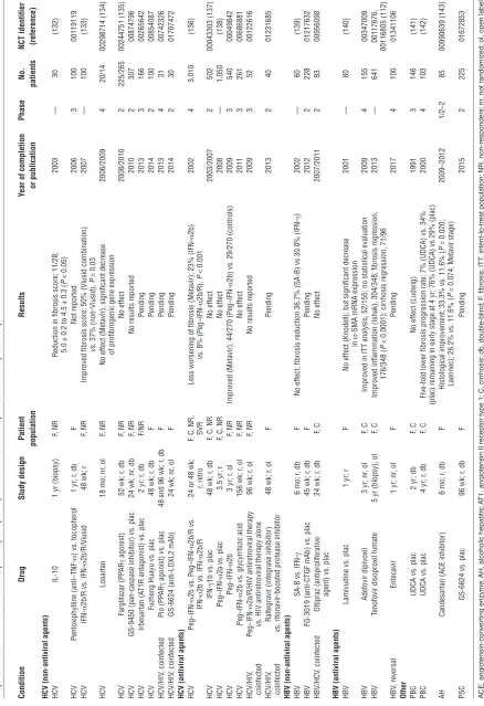
Tab
le 1
Major studies with liver fibrosis as primar
y endpoint (viral and alcoholic hepatitis and biliar
y fibrosis) Condition Drug Study design Patient Results
Year of completion
Phase No. NCT identifier population or publication patients (reference)
HCV (non-antiviral agents) HCV
IL-10
1 yr (biopsy)
F, NR
Reduction in fibrosis score; 11/28;
2003 — 30 (132) 5.0
± 0.2 to 4.5
± 0.3 (
P
< 0.05)
HCV
Pentoxiphylline
(anti–TNF-α
) vs. tocopherol
1 yr; r
, db F Not reported 2006 3 100 00119119 HCV IFN-α
2b/R vs.
IFN-α
2b/R/Viusid
48 wk; r
F, NR
Improved fibrosis score; 50% (Viusid combination)
2007
—
100
(133)
vs. 37% (non-Viusid),
P
= 0.03
HCV
Losartan
18 mo; nr
, ol
F, NR
No effect (Metavir), significant decrease
2006/2009
4
20/14
00298714 (134)
of profibrogenic gene expression
HCV
Farglitazar (PP
AR
γ
agonist)
52 wk; r
, db F, NR No effect 2008/2010 2 225/265 00244751 (135) HCV
GS-9450 (pan-caspase inhibitor) vs. plac
24 wk; nr
, db
F, NR
No results reported
2010 2 307 00874796 HCV Irbesartan (A
T1R antagonist) vs. plac
2 yr; r
, db F/NR Pending 2013 3 166 00265642 HCV
Fuzheng Huayu vs. plac
48 wk; r
, db F Pending 2014 2 100 00854087 HCV/HIV , coinfected Pio (PP AR γ
agonist) vs. plac
48 and 96 wk; r
, db F Pending 2013 4 31 00742326 HCV/HIV , coinfected
GS-6624 (anti-LOXL2 mAb)
24 wk; nr
, ol F Pending 2014 2 30 01707472
HCV (antiviral agents) HCV
Peg–IFN-α
2b vs.
Peg–IFN-α
2b/R vs.
24 or 48 wk;
F, C, NR,
Less worsening of fibrosis (Metavir); 23%
(IFN-α 2b) 2002 4 3,010 (136) IFN-α
2b vs.
IFN-α
2b/R
r, retro
SVR
vs. 8%
(Peg–IFN-α 2b/R), P < 0.001 HCV IFN-γ
1b vs. plac
48 wk; r
, db
F, C, NR
No effect 2003/2007 2 502 00043303 (137) HCV Peg–IFN-α
2a vs. plac
3.5 yr; r
F, C, NR
No effect 2008 — 1,050 (138) HCV Peg–IFN-α 2b
3 yr; r
, ol
F, NR
Improved (Metavir); 44/270
(Peg–IFN-α
2b) vs. 29/270 (controls)
2009 3 540 00049842 HCV Peg–IFN-α
2b vs. glycyrrhizic acid
156 wk; r
, ol F, NR No effect 2011 3 261 00686881 HCV/HIV , Peg–IFN-α
2a/R/HIV antiretroviral therapy
96 wk; r
, ol
F, NR
No results reported
2009
3
52
00122616
coinfected
vs. HIV antiretroviral therapy alone
HCV/HIV
,
Raltegravir (integrase inhibitor)
48 wk; r
, ol F Pending 2013 2 40 01231685 coinfected vs. ritonavir
-boosted protease inhibitor
HBV (non-antiviral agents) HBV
SA-B vs.
IFN-γ
6 mo; r
, db
F
No effect; fibrosis reduction 36.7% (SA-B) vs 30.0%
(IFN-γ ) 2002 — 60 (139) HBV
FG-3019 (anti-CTGF mAb) vs. plac
45 wk; r
, db F Pending 2012 2 228 01217632 HBV/HCV , coinfected Oltipraz (antiproliferative
24 wk; r
, db F, C No effect 2007/2011 2 83 00956098
agent) vs. plac
HBV (antiviral agents) HBV
Lamivudine vs. plac
1 yr; r
F
No effect (Knodell); but significant decrease
2001 — 80 (140) in α
-SMA mRNA expression
HBV
Adefovir dipivoxil
3 yr; nr
, ol
F, C
Improved in ITT analysis, 52/155; no statistical evaluation
2009
4
155
00347009
HBV
Tenofovir disoproxil fumate
5 yr (biopsy); ol
F, C
Improved inflammation (Ishak), 304/348; fibrosis regression,
2013 — 641 00117676, 176/348 ( P
< 0.0001); cirrhosis regression, 71/96
00116805 (112)
HBV
, reversal
Entecavir
1 yr; nr
, ol F Pending 2017 4 100 01341106 Other PBC
UDCA vs. plac
2 yr; db
F, C
No effect (Ludwig)
1991
3
146
(141)
PBC
UDCA vs. plac
4 yr; r
, db
F, C
Five-fold lower fibrosis progression rate; 7% (UDCA) vs. 34%
2000
4
103
(142)
(plac) remaining in early stage at 4 yr; 76% (UDCA) vs 29% (plac)
AH
Candesartan (ACE inhibitor)
6 mo; r
, db
F
Histological improvement; 33.3% vs. 11.6% (
P = 0.020; 2009–2012 1/2–2 85 00990639 (143)
Laennec); 26.2% vs. 11.6% (
P
= 0.074; Metavir stage)
PSC
GS-6624 vs. plac
96 wk; r
, db F Pending 2015 2 225 01672853 A CE, angiot ensin-con ver ting enzyme; AH, alcoholic hepatitis; A T1, angiot ensin II recept or type 1; C , cirrhosis; db , doub le-b lind; F , fibr osis; ITT , int ent-t o-tr eat population; NR, non-r esponder s; nr , not randomiz ed; ol, open label; P eg-IFN, pegylat ed IFN; Pio , Pioglit az one; plac , placebo; r, r andomiz ed; retr o, r etr ospectiv e anal ysis; S VR, sust ained vir al r esponder s t
o IFN-based tr
eatment; UDC A, ur sodeo xy cholic acid.
Viusid consists of ascorbic acid,
zinc
, and gl
yc
yrrhizic acid;
sal
vianolic acid B (S
A
-B) is an ing
redient of fuz
eng hua
yu, an herbal antifibr
[image:7.585.61.499.97.736.2]Tab
le 2
Studies with liver fibrosis as primar
y endpoint (NASH and autoimmune hepatitis)
Treatment
Study
Patient
Results
Year of completion
Phase No. NCT identifier design population or publication patients (reference)
NASH (drug) Orlistat (pancreatic lipase inhibitor) vs.
36 wk; r
, ol
F
No results reported
2006
4
50
00160407
1,400-kcal diet (30% fat) Pio vs. plac
6 mo; r
, db No effect 2006 4 55 00227110 (144) Pio (PP AR γ
agonist) vs. plac
1 yr; r
, db
F
Decreased fibrosis progression; 9/31 (29%, Pio.)
2008
—
74
(145)
vs. 6/30 (20%, plac),
P
= 0.05
Metreleptin (leptin analog)
1 yr; nr
, ol
F
No results reported
2009
2
10
00596934
Pio vs. Vit. E vs. plac
2 yr; r
, db
F
No significant effect; 44% (Pio.) vs. 41% (Vit. E) vs. 31% (plac)
2009/2010 3 247 00063622 (146) Rosi (PP AR γ
agonist) vs. plac
1 and 2 yr; r
F
No effect on fibrosis
2010 – 53 (147) Pentoxifylline (anti–TNF-α
) vs. plac
1 yr; r
, db
F
Improved hepatic steatosis, lobular inflammation and fibrosis
2010/2011
2
55
00590161 (148)
Pentoxifylline vs. plac
1 yr; r
F No effect 2011 — 30 —
Rosi vs. Rosi/metformin vs. Rosi/losartan
48 wk; r
, ol
F
No effect on fibrosis
2011
—
137
(149)
High-dose UDCA vs. plac
1 yr; r
, db
F
Significant reduction only of FibroT
est 2011 3 126 (150) Metformin
1 yr; r
, db F Pending 2012 4 80 00134303
Liraglutide (GLP-1 agonist) vs. plac
48 wk; r
, db F Pending 2013 2 50 01237119
Pentoxifylline/Vit. E vs. Vit. E
3 mo (biopsy); r
, db F Pending 2013 3 120 01384578 Rosi vs. α
-lipoic acid vs. Rosi/
α
-lipoic acid
24 wk; r
, sb F Pending 2013 4 26 01406704 Losartan (A
T1R antagonist) vs. plac
2 yr; r
, db F Pending 2014 3 214 01051219
Obeticholic acid (FXR agonist) vs. plac
72 wk; r
, db F Pending 2014 2 280 01265498
Pio vs. Vit. E vs. plac
1.5 and 3 yr; r
, db F Pending 2014 4 90 00994682 Metreleptin
1 yr; ol
F Pending 2015 2 20 01679197
GS-6624 (anti-LOXL2 mAb; 75 mg
100 wk; r
, db F Pending 2015 2 225 01672866
and 125 mg) vs. plac GS-6624 (anti-LOXL2 mAb; 200 mg
100 wk; r
, db F,C Pending 2015 2 225 01672879
and 700 mg) vs. plac GFT505 (dual PP
AR
α
/
δ
agonist)
52 wk; r
, db F Pending 2015 2 270 01694849
Pio vs. Vit. E vs. Vit. E/Pio vs. plac
1.5 and 3 yr; r
, db F Pending 2015 4 90 01002547
Vit. E/Vit. C vs. plac
6 mo; r
, db F No effect 2003 — 49 (151)
Vit. D vs. lifestyle counseling
2 yr; r
, ol F Pending 2014 3 200 01623024
Vit. D3 vs. plac
48 wk; r
, db F Pending 2015 2 60 01571063
Omega-3 (fish) oil vs. plac
1 yr; r
, db
F
No results reported
2010
2/3
64
00681408
Omega-3 (fish) oil
18 mo; r
, sb F Pending 2013 2 100 00760513 Docosahexaenoic acid
2 yr; r
, db
F
No results reported
2011
1/2
60
00885313
Eicosapentaenoic acid vs. plac
48 wk; r
, db
F
No results reported
2011
2
32
00323414
EP
A vs. plac
1 yr; r
, db F Pending 2012 2 243 01154985
Diamel vs. plac vs. lifestyle counseling
52 wk; r
, db F Pending 2012 3 158 00820651
Polypill, no biopsy (UE)
5 yr; r
, ol F Pending 2018 3 1500 01245608
NASH (surgical) Bariatric surger
y
Meta-analysis of 21
F, C
No clear effect
2010
—
1,643
(152)
cohort studies
AIH (reversal) Corticosteroids/azathioprine
ol, retro
F, C
Decrease of median Knodell score from 14.0 to 1.3,
1997
—
8
(153)
decrease of fibrosis stage from 3.3 to 0.8
AIH (pediatric) Corticosteroids/azathioprine
4.6 yr
F
Improved fibrosis (Ishak); 14/20 patients (70%, obser
ver 1),
2008
—
20
(154)
17/20 patients (85%, obser
ver 2) AIH, aut oimm une hepatitis; A T1R, angiot
ensin II r
ecept
or t
ype 1;
FXR, f
ar
nesoid X r
ecept
or
; GLP
-1, glucagon-lik
e peptide-1, R
osi, r osiglit az one; sb , single-b lind; UDC A, ur sodeo xy cholic acid; Vit, vit amin.
Diamel is a diet
ar
y
supplement consisting of let
tuce and b
lueberr y e xtr acts , acet ylc yst eine , ar ginine
, ascorbic acid, c
yanocobalamin, zinc sulphat
e, f
olic acid, fumar
ic acid, gl
ycine
, calcium pant
othenat
e, L
-car
nitine
, or
nithine and p
yr
ido
xal;
pol
ypill is a combined f
or
m
ulation of aspir
[image:8.585.70.517.95.732.2]cells). M1 macrophages are induced by IFN or IL-12, while IL-4, IL-13, and GM-CSF induce M2 macrophages. Macrophages appear to be fibrogenic during fibrosis progression and fibrolytic during its reversal, but a detailed functional analysis and assignment to M1 or the various M2 subclasses has remained elusive (18, 23, 26). While M1 macrophages are activated in immediate defense against pathogens or detrimental cellular debris, M2 macrophages are gen-erally thought to promote wound healing (i.e., fibrogenesis) and immune suppression (e.g., facilitating cancer growth as tumor-associated macrophages) (18, 94, 95). M2 macrophages respond to IL-4 and IL-13 via IL-4 receptor and IL-13 receptor α1 (with IL-13 receptor α2 serving as negative regulator) and are characterized by unique signal transducers (e.g., Stat6), enzymes (e.g., arginase), or scavenger receptors (e.g., CD206). However, several subtypes of M2 macrophages exist, such as the putatively proinflammatory M2a, and the anti-inflammatory M2b and M2c subtypes, which have ill-defined roles in fibrosis (20, 74). A recent study demon-strated that fibrolytic macrophages in liver fibrosis derive from circulating Ly6Chi-expressing monocytes and develop locally into
Ly6Clo-expressing macrophages with some classical M2 markers
and a high expression of fibrolytic MMPs, and this development depends on phagocytic activity (96). Notably, MMP release depends on phagocytosis of apoptotic cells, which is also a driver of bili-ary fibrosis reversal (23). Given that M1 polarization in liver and adipose tissue enhances insulin resistance and promotes inflam-mation in NASH, whereas M2 polarization is protective (97), the targeting of macrophage polarization in liver inflammation and fibrosis is an attractive therapeutic option.
Other relevant molecular targets
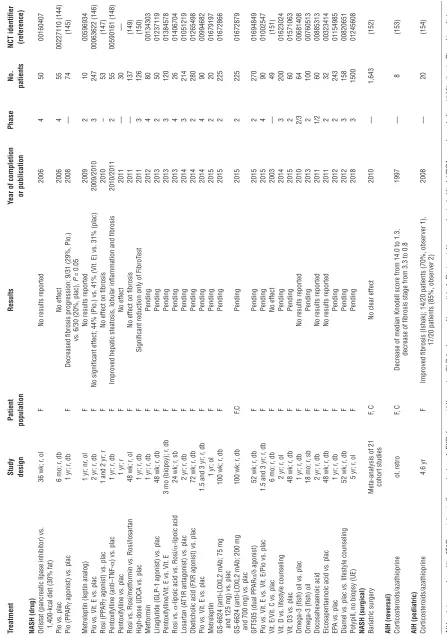
Several other molecular targets are of interest, and some have already entered clinical studies. ECM cross-linking, mainly of fibrillar collagen, is largely mediated by lysyl oxidase (LOXL2). LOXL2 likely impedes ECM degradation during fibrosis reversal, and antifibrotic activity has been seen in a small study of CCL4-induced liver fibrosis (98). A humanized antibody that blocks LOXL2 activity is currently assessed in the largest clinical study for liver fibrosis (Tables 1 And 2).
TLRs are sensors of bacteria, viruses, and foreign antigens. TLRs are expressed ubiquitously but are prominent on cells of the innate immune system, creating a proinflammatory environment and activating adaptive immunity to promote pathogen elimination. As the major interface between the gut and systemic circulation, liver cells are equipped with a variety of TLRs that are central to both maintaining immune tolerance and initiating inflammation and repair when confronted with (microbial) danger signals (99). A direct link exists between liver fibrosis and bacterial LPS, and activation of its receptor TLR4. LPS enters the portal hepatic cir-culation in conditions of enhanced intestinal permeability, such as in ASH, NASH, and other intestinal and liver diseases. LPS upregulates chemokine secretion of monocytes and macrophages/ Kupffer cells and downregulates the inhibitory TGF-β1 pseudo-receptor Bambi, which cumulatively sensitizes hepatic stellate cells/myofibroblasts to fibrogenic activation (99, 100). Prevention of excessive TLR4 activation or inhibition of TLR4 are therefore attractive strategies to inhibit fibrogenesis. Currently only the par-enteral TLR4 antagonist, eritoran tetrasodium, is being studied for the treatment of sepsis (101). Other interesting but little explored targets include TLR3, a double-stranded RNA sensor whose activa-tion by polyI:C attenuates liver fibrosis via activaactiva-tion of NK cells
(102), and TLR9, a receptor for double-stranded bacterial DNA that enhances fibrogenic immune activation via release of CCL2 (103). In addition, inhibitors of broadly expressed chemokine systems other than CCR2/CCL2, mainly CXCL4 and CCL5 (and their receptors CXCR4 and CCR5, respectively) on myofibroblasts, T cells, and macrophages, have been shown to attenuate liver fibro-sis (104–106). Furthermore, the recent explosion of data related to microRNAs (miRs) has uncovered miRs that inhibit (miR-29b) or promote fibrogenesis (miR-199, miR-200, and others) (107–109). While these miRs appear to have some specificity for myofibro-blasts, their efficient in vivo delivery poses a problem.
TGF-β and, to a lesser degree, its downstream mediator, con-nective tissue growth factor (CTGF), are potent profibrogenic cytokines for hepatic stellate cells/myofibroblasts (1, 2, 6, 9, 10, 18, 19). However, their general and untargeted inhibition poses risks, especially for TGF-β–neutralizing agents, given that this cytokine is central to cellular differentiation, immune regulation (dampen-ing excessive T cell activation), and regulated wound heal(dampen-ing, such as in vascular plaque stabilization in atherosclerosis (110).
Preclinical proof of concept
Before entering clinical studies, best preclinical proof of antifi-brotic activity needs to be obtained in complementary rodent models that reflect different aspects of human liver fibrosis (6). Moreover, drug testing in cultures of precision-cut human liver slices obtained from operations permit a first translation toward the human in vivo system (111).
Combination therapies
Combination therapies that address liver fibrosis in a multi-pronged approach hold much promise for future treatment, ideal-ly targeting interactions between cells, soluble mediators, the ECM and its receptors, and/or relevant intracellular signaling. Combi-nations of targeted antifibrotic agents have yet to be thoroughly tested in preclinical studies. Significant expense and effort will be required to rigorously validate combinations at different doses and in several rodent fibrosis models. However, combinations of specific drugs can be anticipated that interfere with fibrogenesis, induce fibrolysis, or address different cell types.
Clinical development of combination therapies that could guar-antee thorough efficacy and low toxicity is only feasible with the advent of improved noninvasive biomarkers and technologies to measure fibrosis, and especially fibrogenesis. Moreover, the neces-sary personalized approach to the patient with liver fibrosis or cir-rhosis will only be possible with such biomarkers, permitting the adjustment of different medications and their dose according to a readily measurable treatment effect.
Testing antifibrotics in clinical trials
Tab
le 3
Studies in pulmonar
y, renal, and other fibrosis with fibrosis as primar
y endpoint
Drug
Study design
Results
Year of completion
Phase No. NCT identifier or publication patients (reference) Pulmonar y fibrosis Etaner cept (anti–TNF-α
) vs. plac
48 wk; r
, db No effect 2005/2008 2 88 00063869 (155)
NAC (antioxidant) vs. plac
1 yr; r
, db
Reduction in FVC and DL
CO
in NAC group, no change in mortality
2005 1/2 182 (156) Tetrathiomolybdate (antiproliferative) nr , ol
No results reported
2005 1/2 20 00189176 IFN-α oral lozenge
1 yr; nr
, ol
Minimal/no progression (HRCT), 12/18 patients
2007
2
18
01442779
Thalidomide (anti-TNF)
1 yr; nr
, ol
No results reported
2007
2
19
00162760
Bosentan (dual ET1
A
R and ET1
B
R
1 yr; r
, db
W
orsening PFT
; two-thirds exhibited reduction in FVC (>10%),
2005/2008
2/3
158
00071461
antagonist) vs. plac
DL
CO
(>15%), O
2
saturation (>4%)
Bosentan vs. plac
12 mo, 21 mo, and
No significant effect
2010/2011
3
616
00391443
3 yr (biopsy); r
, db
Losartan (A
T1R)
1 yr; ol
Stable or improved FVC in 12/17 patients
2012
—
20
(157)
Imatinib (kinase inhibitor) vs. plac
92 wk; r
, db No effect 2010 2/3 120 00131274 (158) Ambrisentan (ET1 A
R antagonist) vs. plac
92 wk; r
, db
Terminated due to lack of efficacy
2012 3 600 00768300 (159) Pir fenidone (anti–TGF-β , anti–TNF-α ,
72 wk; r
, db
Study 004: reduced decline in FVC (
P
= 0.001); –8.0% vs. –12.4%
2008 3 435 00287716 (160) anti–IL-1 β
) vs. plac
in high-dose pir
fenidone group at week 72
Pir fenidone (anti–TGF-β , anti–TNF-α ,
72 wk; r
, db
Study 006: difference in FVC change at week 72 not significant (
P = 0.501) 2008 3 344 00287729 (160) anti–IL-1 β
) vs. plac
Pir
fenidone vs. plac
52 wk; r
, db
Significant worsening of FVC; +0.009 L (Pir
fenidone) vs. –0.16 (plac),
P = 0.0416 2010 3 275 (161)
BIBF1120 (nintendanib, multi-R
TK inhibitor
1 yr; r
, db
FVC decline 68.4% reduction with BIBF1120 vs. plac (
P = 0.01); 2010/2011 2 432 00514683 (162)
and angiokinase inhibitor; low dose) vs. plac
lower incidence of acute exacerbations vs. plac per 100 patient-years (
P
= 0.02)
BIBF1120 vs. plac
52 wk; r
, db Pending 2013 3 551 01335477
BIBF1120 vs. plac
52 wk; r
, db Pending 2013 3 515 01335464 BIBF1120
5 yr; ol
Pending 2015 2 20 01417156 BIBF1120
3 yr; nr
, ol Pending 2015 2 198 01170065
CNTO888 (anti-MCP1/CCL2 mAb) vs. plac
74 wk; r
, db
No results reported
2012
2
126
00786201
QAX576 (anti–IL-13 mAb)
4 wk; nr
, ol
No results reported
2009
2
52
00532233
QAX576 vs. plac
1 yr; r
, db Pending 2013 2 40 01266135
SAR156597 (anti–IL-4/13 mAb) vs. plac
6 mo; r
, db Pending 2014 1/2 24 01529853
Octreotide (somatostatin mimetic)
1 yr; nr
, ol Pending 2012 1/2 25 00463983
CC-930 (JNK inhibitor) vs. plac
4 wk; r
, db Pending 2013 2 28 01203943 STX-100 (anti-α V β
6 mAb) vs. plac
24 wk; r
, db Pending 2013 2 32 01371305
FG-3019 (anti-CTGF mAb)
109 wk; ol
Pending
2014
2
84
01262001
Cystic fibrosis (lung) SB656933 (CXCR2 antagonist) vs. plac
28 d; r
, db
No results reported
2010
2
100
00903201
Simvastatin (HMGR inhibitor) vs. plac
12 wk; r
, db Pending 2012 1/2 120 01092572
Renal fibrosis Pirfenidone
1 yr; ol
Decreased GFR in focal glomerulosclerosis and nephrotic syndrome; not significant
2008
2
21
00001959
Everolimus (mTOR inhibitor)/EC-MP
A/corticosteroids
12 mo; r
, ol Pending 2012 3 235 01079143
vs. cyclosporine A/EC-MP
A/corticosteroids
Pioglitazone vs. plac
48 wk; r
, db Pending 2012 4 160 00745225
Skin fibrosis Pravastatin (HMGCoAR inhibitor)
1 yr; nr
, ol Pending 2013 2 40 01268202
Myelofibrosis GS-6624 (anti-LOXL2 mAb)
24 wk; r
, ol Pending 2013 2 54 01369498
EE QAX576 (anti–IL-13 mAb) vs. plac
13 wk; r
, db Pending 2012 2 25 01022970 AT1R, angiot
ensin II r
ecept or t ype 1; DL CO , dif fusing capacit
y of the lungs f
or carbon mono
xide; EC-MP A, ent er ic-coat ed m ycophenolat e sodium;
EE, eosinophilic esophagitis;
ET1 A R, ET -1 r ecept or t ype A; FV C , f or ced vit al capacit y; GFR, glomer ular fi ltr ation r at e; HMGC oAR, 3-h ydr oxy -3-meth yl-glut ar yl-coenzyme A r educt ase; HRCT , high-r esolution comput ed t omog raph y; NA C , N-acet ylc yst eine; PF T, pulmonar
y function t
est; R TK, r ecept or tyr osine kinase . “Skin ” indicat es r adiation-induced cut
aneous and subcut
aneous fibr
osis
[image:10.585.62.519.96.734.2]Tab
le 4
Drugs in clinical trials for other indications that have antifibrotic potential for liver fibrosis Type of agent
Drug
Manufacturer
Disease target or
Year of completion
Phase
NCT identifier
clinical endpoint
[no. studies] (reference)
Anti–TGF-β mAb Fresolimumab (GC1008) Genzyme IPF 2008 1 00125385 Anti–TGF-β mAb Fresolimumab (GC1008) Genzyme FSGS A 2014 2 01665391 Anti-α V β 6 mAb STX-100 Biogen-Stromedix IPF A 2013 2 01371305
Anti-CTGF mAb, anti-CTGF antisense DNA
FG-3019 FibroGen T2DM (DN A) 2010 2 00913393
Anti-CTGF mAb, anti-CTGF antisense DNA
FG-3019 FibroGen IPF A 2014 2 01262001
Anti-CTGF mAb, anti-CTGF antisense DNA
EXC001 (anti-CTGF antisense DNA)
Pfizer -Excaliard Scar prevention 2010–2012 2 [4] PRM
PRM-151 (recombinant pentraxin-2)
Promedior IPF 2012 1 01254409 (163) PRM
PRM-151 (recombinant pentraxin-2)
Promedior Glaucoma 2012 2 01064817 Anti–IL-4/13 mAb SAR156597 Sanofi-A ventis IPF A 2014 2 01529853 Anti–IL-13 mAb QAX576 Novartis IPF
, EE, CD, AR
2009–2013 2 [4] Anti–IL-13 mAb QAX576 Novartis Asthma 2012–2013 1/2 [4] Neurochemical receptor S-777469 Shionogi Pharmaceuticals AD 2008–2009 1/2–2 — Neurochemical receptor
LH-21 (peripherally active CB1R antagonist)
— Obesity — Preclinical — Neurochemical receptor
CB2R agonist (HU-308, JWH 133, A-836339, BML-190, AM1241)
— — — Preclinical — Neurochemical receptor
Naltrexone (opioid receptor antagonist)
Dupont Liver fibrosis — Preclinical — Anti–IL-17A, anti–IL-17R
Secukinumab (AIN 457, anti–IL-17A mAb)
Novartis
PP
, AS, P
A, RA, asthma, uveitis, CD
—
2–3
[45] (164)
Anti–IL-17A, anti–IL-17R
Ixekizumab (L
Y2439821, anti–IL-17A mAb)
Eli Lilly PP , P A, RA — 2–3 [9] (164) Anti–IL-17A, anti–IL-17R
Brodalumab (AMG827, anti–IL-17RA)
Amgen-AstraZeneca
PA, PP
, CD, RA, asthma
—
2–3
[13] (164)
Anti–IL-17A, anti–IL-17R
RG4934 (humanized anti–IL-17A mAb)
Roche
PA
No information available
1
No information available
Anti–IL-17A, anti–IL-17R
NI-1401 (RG7624, anti–IL-17A/F mAb)
Roche
Autoimmune disease
No information available
1
No information available
Anti–IL-17A, anti–IL-17R
SCH 900117(anti–IL-17A mAb)
Schering-Plough
RA
Anti–IL-17A, anti–IL-17R
ABT
-122 (anti–IL-17A and TNF mAb)
Abbott
RA
Hh or Hh(R) (SMO) antagonist
Vismodegib (GDC-0449, Hh(R) blocker)
Genentech
Various cancers; MM
—
1–2
[38]
Hh or Hh(R) (SMO) antagonist
LY2940680 (SMO antagonist)
Eli Lilly and Company
NSCLC, MB, other cancers
—
1–1/2
[5]
CCR5 antagonist
TBR 652 (small-molecule inhibitor)
Tobira Therapeutics HIV Completed 1/2–2 [3] (165) CCR5 antagonist
Vicriviroc (SCH-D, SCH 417690, small-molecule inhibitor)
Schering-Plough HIV Completed 1–3 [11] (165) CCR5 antagonist
Aplaviroc (GW873140, small-molecule inhibitor)
GlaxoSmithKline HIV — 2–3 [8] (165) CCR5 antagonist
Maraviroc (small-molecule inhibitor)
Pfizer
HIV
, ED, cancer
Approved
1–4
[104] (165)
CCR5 antagonist
INCB009471 (small-molecule inhibitor)
Incyte
HIV
2007
1/2
00393120 (165)
CXCR4 antagonist, anti-CXCR4 mAb
BKT140 (small-molecule inhibitor)
Biokine Therapeutics
MM, leukemia
2010
1/2
01010880 (166)
CXCR4 antagonist, anti-CXCR4 mAb
TG-0054 (small-molecule inhibitor)
TaiGen Biotechnology
MM, leukemia
Completed
1–2
[3] (166)
CXCR4 antagonist, anti-CXCR4 mAb
Plerixafor (AMD3100, small-molecule inhibitor)
Genzyme
MM, NHL, ML
—
1–4
[94] (166)
CXCR4 antagonist, anti-CXCR4 mAb
POL6326 (small-molecule inhibitor)
Polyphor Ag
MM, leukemia
2015
1/2
01413568 (166)
CXCR4 antagonist, anti-CXCR4 mAb
MDX-1338 (BMS-936564, anti-CXCR4 mAb)
Bristol-Myers Squibb MM, ML 2014 1 [2] (166) CXCR3 antagonist
SCH 546738 (small-molecule antagonist)
Mer ck RA — 1 — CXCR3 antagonist
AMG487 (T487, small-molecule antagonist)
Amgen RA — 1–2 — NOX inhibitor
NOX inhibitor (GKT137831, dual NOX1/4 inhibitor)
Genkyotex Healthy individuals Completed 1 — Copaxone Copaxone Teva Pharmaceuticals MS — 2–4 [44] Adiponectin, AMPK
Recombinant adiponectin/AdipoR1/2 and AMPK agonist
— Liver fibrosis — Preclinical (167) YB-1 TGF-β pathway inhibitor — Liver fibrosis — Preclinical — LP A1R
AM152 (myofibroblast recruitment inhibitor)
Amira Pharmaceuticals
IPF
, liver fibrosis
—
2, —
—
Anti-Th17 MMP inducer
Halofuginone Collgard Kaposi sar coma Completed 1, 2 [2] (168) Adenosine receptors
ZM241385 (adenosine receptor A2A antagonist)
— Liver fibrosis — Preclinical (169) miR miR-29 Mirage Cardiac fibrosis 2008 Preclinical — miR Anti–miR-21 Regulus
Cardiac and renal fibrosis
—
Preclinical
—
miR
Anti–miR-122 (SPC3649, Miravirsen)
Santaris Pharm A/S
Hepatitis C — 1–2 [5] AF ibr
osis as secondar
y endpoint. AD , at opic der matitis; A
dipoR, adiponectin r
ecept
or
; AR, aller
gic rhinitis;
A
S
, ank
ylosing spondylitis;
CB1(2)R, cannabinoid r
ecept or t ype 1(2); CD , Cr ohn ’s disease;
DN, diabetic nephr
opath y; ED , endothelial dy sfunction;
EE, eosinophilic esophagitis;
FSGS , f ocal segment al glomer uloscler osis; IPF
, idiopathic pulmonar
y fibr
osis;
LP
A1R, l
ysoposphatidic acid r
ecept or 1; MB , medullob last oma; ML, m yeloid leuk emia; MM, m ultiple m yeloma; NHL, non-Hodgkin ’s l ymphoma; NSCLC
, non-small cell lung cancer
; P A, psor iatic ar thr itis; PP
, plaque psor
iasis;
PRM, r
ecombinant human ser
um am yloid P; RA, rheumat oid ar thr itis; SMO , smoothened; T2DM, t
ype 2 diabet
es mellitus; YB-1, Y-bo x binding pr ot ein-1 . C opax
one is a
T cell dif
fer
entiation–inhibiting glatir
amer acet
at
e mixtur
e of oligopeptides
, and a
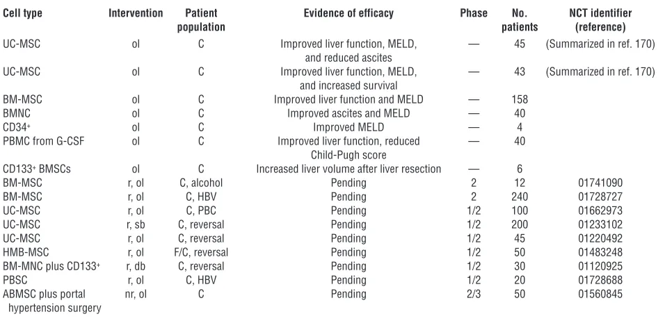
[image:11.585.62.509.98.731.2]While some additional human studies also suggested antifibrotic activities of tenofovir, others failed to show an effect. Tables 1 and 2 list past and current clinical studies with liver fibrosis as primary endpoint, and Supplemental Table 1 (supplemental mate-rial available online with this article; doi:10.1172/JCI66028DS1) shows studies with liver fibrosis as a secondary endpoint. Table 3 highlights clinical studies for other fibrotic diseases that are rel-evant for liver. Clinical studies targeting major multicellular fibro-genic pathways are shown in Table 4. We also summarize trials employing stem cells, an approach that is attractive in combina-tion with pharmacological therapies (Table 5). Notably, develop-ment efforts have largely focused on extracellular targets because intracellular targets are less accessible and tend to lack specificity for the fibrogenic cells.
Optimal selection and stratification of patients. Subjects should be matched according to etiology, age, gender, signs of the meta-bolic syndrome, medications, and risk factors such as alcohol or tobacco consumption (7–9). Preferably, patients should be in an intermediate fibrosis stage, where dynamic changes of fibrosis are best detectable. Patients with chronic HCV infection following liver transplant are considered a preferred study population because up to 30% experience an accelerated fibrosis progression to cirrhosis within three to five years (60). Patients should be further stratified according to their genetic risk to progress to cirrhosis (7, 8, 60, 113).
Assessing fibrosis
Assessment of fibrosis progression is far more difficult for liver than for lungs or kidneys because transaminases do not correlate with fibrosis or fibrogenesis and liver function parameters such as albumin or prothrombin time (protein synthesis) are usually only altered in cirrhosis. Liver biopsy remains the standard for
studies with antifibrotics. However, liver biopsy is invasive and risky (2, 6, 8, 60, 114) and prone to considerable sampling error, and its interpretation is subject to interobserver variability. Even in well-stratified cohorts, given the usually slow fibrosis progres-sion, conventional fibrosis staging (Metavir, Ishak) may require approximately 200 patients studied over a period of two to three years to detect a 20%–30% difference in fibrosis between treatment groups. However, by including current surrogates of fibrosis pro-gression, it appears feasible to conduct proof-of-concept trials in approximately 100 patients within 12 months or less.
Refined liver biopsy readouts. Predictive value may be improved using dynamic biopsy-derived parameters, such as semiquan-tification of activated α-SMA–positive myofibroblasts and the fibrogenic cytokine TGF-β after immunostaining (115), or quan-titative PCR quantification of transcripts that are related to fibro-genesis or fibrolysis (116).
Radiological imaging. Conventional and contrast ultrasonography, computerized tomography, and MRI, PET, single-photon emission computerized tomography, and diffusion-weighted MRI cannot differentiate fibrosis stages. However, magnetic resonance texture analysis, which requires sophisticated instrumentation and soft-ware, may permit semiquantitative fibrosis assessment (4, 8).
[image:12.585.52.530.113.344.2]Elastography. Ultrasound elastography (UE) and axial radiation force imaging (ARFI) measure hepatic stiffness and elasticity. These techniques sample a 100-fold-larger volume than biopsy and can differentiate mild (Metavir F0–F1) from significant fibro-sis (F2–F4) and cirrhofibro-sis (F4), with diagnostic accuracies (area under receiver operating characteristics [AUROC] curves) around and above 0.90, which is considered good (8, 114, 116). Magnetic resonance elastography assesses the whole liver and may be supe-rior to UE/ARFI, but is not generally available, and studies are
Table 5
Stem cell therapies for liver fibrosis and cirrhosis
Cell type Intervention Patient Evidence of efficacy Phase No. NCT identifier
population patients (reference)
UC-MSC ol C Improved liver function, MELD, — 45 (Summarized in ref. 170) and reduced ascites
UC-MSC ol C Improved liver function, MELD, — 43 (Summarized in ref. 170) and increased survival
BM-MSC ol C Improved liver function and MELD — 158
BMNC ol C Improved ascites and MELD — 40
CD34+ ol C Improved MELD — 4
PBMC from G-CSF ol C Improved liver function, reduced — 40 Child-Pugh score
CD133+ BMSCs ol C Increased liver volume after liver resection — 6
BM-MSC r, ol C, alcohol Pending 2 12 01741090
BM-MSC r, ol C, HBV Pending 2 240 01728727
UC-MSC r, ol C, PBC Pending 1/2 100 01662973
UC-MSC r, sb C, reversal Pending 1/2 200 01233102
UC-MSC r, ol C, reversal Pending 1/2 45 01220492
HMB-MSC r, ol F/C, reversal Pending 1/2 50 01483248
BM-MNC plus CD133+ r, db C, reversal Pending 1/2 30 01120925
PBSC r, ol C, HBV Pending 1/2 20 01728688
ABMSC plus portal nr, ol C Pending 2/3 50 01560845
hypertension surgery
small (8, 117). UE is useful for initial stratification of patients according to fibrosis stage.
Serum fibrosis markers. More than 2,000 studies in the last five years have employed serological markers to assess liver fibrosis, mostly in patients with HCV (2, 6, 8, 60, 118–121). These mark-ers reflect liver function (indirect markmark-ers), are related to matrix metabolism (direct markers), or both. The best marker panels show AUROCs around 0.8–0.85 to differentiate between no/mild fibrosis (Metavir F0–F1) and moderate/severe fibrosis (F2–F4). Fibrosis markers have almost exclusively been validated as predic-tors of fibrosis stage, while especially the direct parameters may rather reflect the dynamics of fibrogenesis and/or fibrolysis (2, 8). Recent studies suggest that certain marker combinations such as the (indirect) Fibrotest (122) and the (direct) enhanced liver fibro-sis (ELF) test can predict hard endpoints. Thus ELF was superi-or to fibrosis stage, the Child-Pugh superi-or the Model fsuperi-or End-Stage Liver Disease (MELD) score, to predict hepatic decompensation or death in long-term, retrospective follow-up studies of patients with advanced chronic HCV or PBC (123–125).
Combination of methods. The combination of serum fibrosis mark-ers with elastography increases diagnostic accuracy, permitting a clear allocation to either no/mild (F0–F1) or significant (F2–F4) fibrosis in 70% of patients (8, 114).
Measurement of portal hypertension and quantitative liver function. The hepatic vein pressure gradient (HVPG) is an excellent predic-tor of decompensation or death in patients with cirrhosis (126). A non-invasive alternative, the hepatic vein arrival time of an injected ultrasound contrast agent, needs further validation (127).
Tests that measure the metabolic capacity of the liver, such as demethylation of ingested methacetin and quantification of the exhaled metabolite 13CO
2, correlate inversely with the severity of
liver inflammation and fibrosis, and the results of such tests can complement antifibrotic drug trials (128).
Quantitative imaging of liver fibrosis and fibrogenesis. Methods that employ a small molecular ligand for fibrillar collagen, elastin, or a cell-associated molecule coupled to a radio-imaging or MRI agent are in development (4, 8). Examples include an elastin-specific MRI probe for imaging of fibrosis (129) and probes for quantify-ing fibrogenic cells via the cholangiocyte integrin αvβ6 or the myo-fibroblast-specific PDGFRβ (4, 8). When improved, such
method-ology could serve as a novel gold standard for the assessment of fibrosis/fibrogenesis and permit short-term testing of potential antifibrotics before and after a single dose of the drug.
Novel biomarkers. Apart from ongoing efforts to find and validate better serum markers of fibrosis, fibrogenesis, and fibrolysis (4, 8), three methodologies will likely become relevant for antifibrotic drug trials: (a) urinary assessment of proteolytic activities in the fibrotic liver could be monitored after injection of mass-encoded protease-sensitive peptides conjugated to nanoparticles and multiplexed detection of cleavage products by mass spectrometry (2, 130); (b) membrane microparticles, which are shed from activated or apop-totic cells, can be quantified in the bloodstream via their cell-specific surface molecules; microparticles represent a novel set of quantita-tive diagnostic markers to monitor cell-specific activation in liver inflammation and fibrosis (3, 131); and (c) circulating miRs that can reflect liver-specific pathology, including hepatocyte differentiation and activation, cancer growth, and liver fibrosis (109).
Conclusions
We have gained remarkable insight into the cellular and molec-ular mechanisms of liver fibrosis and reversal, and even reversal of cirrhosis appears feasible in preclinical models. Currently, the field has progressed toward clinical translation. As antifibrotics address mechanisms that are embedded in a complex multicellu-lar network, their efficacy is predicted to be context dependent. Combination therapies hold most promise, but their development and use require a personalized medicine approach that depends on the development and validation of novel noninvasive markers and techniques to quantify liver fibrosis and especially fibrogenesis.
Acknowledgments
D. Schuppan received funding from the NIH, the European Union, the state of Rhineland-Palatinate, the German Research Foundation, and the German Ministry of Education and Research.
Address correspondence to: Detlef Schuppan, Institute of Molec-ular and Translational Medicine and Department of Medicine I, Johannes Gutenberg University, Langenbeckstr.1, 55131 Mainz, Germany. Phone: 49.6131.177356; Fax: 49.6131.177357; E-mail: [email protected].
1. Friedman SL. Mechanisms of hepatic fibrogenesis.
Gastroenterology. 2008;134(6):1655–1669. 2. Schuppan D, Afdhal NH. Liver cirrhosis. Lancet.
2008;371(9615):838–851.
3. Gao B, Bataller R. Alcoholic liver disease: pathogen-esis and new therapeutic targets. Gastroenterology. 2011;141(5):1572–1585.
4. Tilg H, Moschen AR. Evolution of inflammation in nonalcoholic fatty liver disease: the multiple parallel hits hypothesis. Hepatology. 2010;52(5):1836–1846. 5. Bohinc BN, Diehl AM. Mechanisms of disease
pro-gression in NASH: new paradigms. Clin Liver Dis. 2012;16(3):549–565.
6. Popov Y, Schuppan D. Targeting liver fibrosis: strategies for development and validation of antifi-brotic therapies. Hepatology. 2009;50(4):1294–1306. 7. Friedman SL. Evolving challenges in hepatic fibro-sis. Nat Rev Gastroenterol Hepatol. 2010;7(8):425–436. 8. Schuppan D, Pinzani M. Anti-fibrotic therapy: lost in translation? J Hepatol. 2012;56(suppl 1):S66–S74. 9. Friedman SL, Sheppard D, Duffield JS, Violette S.
Therapy for fibrotic diseases: nearing the starting line. Sci Transl Med. 2013;5(167):1–17.
10. Hinz B, et al. Recent developments in myofibroblast biology paradigms for connective tissue
remodel-ing. Am J Pathol. 2012;180(4):1340–1355. 11. Dranoff JA, Wells RG. Portal fibroblasts:
underap-preciated mediators of biliary fibrosis. Hepatology. 2010;51(4):1438–1444.
12. Kisseleva T, Brenner DA. The phenotypic fate and functional role for bone marrow-derived stem cells in liver fibrosis. J Hepatol. 2012;56(4):965–972. 13. Gao B, et al. Innate immunity in alcoholic liver
disease. Am J Physiol Gastrointest Liver Physiol. 2011; 300(4):G516–G525.
14. Chiang DAJ, Pritchard MT, Nagy LE. Obesity, dia-betes mellitus, and liver fibrosis. Am J Physiol Gastro-intest Liver Physiol. 2011;300(5):G697–G702. 15. Mehal W, Imaeda A. Cell death and fibrogenesis.
Semin Liver Dis. 2010;30(3):226–231.
16. Wasmuth HE, Tacke F, Trautwein C. Chemokines in liver inflammation and fibrosis. Semin Liver Dis. 2010;30(3):215–225.
17. Forbes SJ, Newsome PN. New horizons for stem cell therapy in liver disease. J Hepatol. 2012;56(2):496–499. 18. Wynn TA, Ramalingam TR. Mechanisms of fibro-sis: therapeutic translation for fibrotic disease. Nat Med. 2012;18(7):1028–1040.
19. Fallowfield JA. Therapeutic targets in liver fibro-sis. Am J Physiol Gastrointest Liver Physiol. 2011;
300(5):G709–G715.
20. Schuppan D, Ruehl M, Somasundaram R, Hahn EG. Matrix as a modulator of hepatic fibrogenesis.
Semin Liver Dis. 2001;21(3):351–372.
21. Fernandes DJ, Bonacci JV, Stewart AG. Extracellular matrix, integrins, and mesenchymal cell function in the airways. Curr Drug Targets. 2006;7(5):567–577. 22. Pozzi A, Voziyan PA, Hudson BG, Zent R.
Regula-tion of matrix synthesis, remodeling and accumu-lation in glomerulosclerosis. Curr Pharm Des. 2009; 15(12):1318–1333.
23. Popov Y, et al. Macrophage-mediated phagocytosis of apoptotic cholangiocytes contributes to reversal of experimental biliary fibrosis. Am J Physiol Gastro-intest Liver Physiol. 2010;298(3):G323–G334. 24. Friedman SL, Bansal MB. Reversal of hepatic
fibrosis – fact or fantasy? Hepatology. 2006; 43(2 suppl 1):S82–S88.
25. Ellis EL, Mann DA. Clinical evidence for the regres-sion of liver fibrosis. J Hepatol. 2012;56(5):1171–1180. 26. Issa R, et al. Spontaneous recovery from micronod-ular cirrhosis: evidence for incomplete resolution associated with matrix cross-linking. Gastroenterol-ogy. 2004;126(7):1795–1808.