that modulate immune responses in a
TLR2-dependent manner in mice
Rafael Prados-Rosales, … , Steven A. Porcelli, Arturo
Casadevall
J Clin Invest.
2011;121(4):1471-1483. https://doi.org/10.1172/JCI44261.
Bacteria naturally release membrane vesicles (MVs) under a variety of growth
environments. Their production is associated with virulence due to their capacity to
concentrate toxins and immunomodulatory molecules. In this report, we show that the 2
medically important species of mycobacteria,
Mycobacterium tuberculosis
and
Mycobacterium bovis
bacille Calmette-Guérin, release MVs when growing in both liquid
culture and within murine phagocytic cells in vitro and in vivo. We documented MV
production in a variety of virulent and nonvirulent mycobacterial species, indicating that
release of MVs is a property conserved among mycobacterial species. Extensive proteomic
analysis revealed that only MVs from the virulent strains contained TLR2 lipoprotein
agonists. The interaction of MVs with macrophages isolated from mice stimulated the
release of cytokines and chemokines in a TLR2-dependent fashion, and infusion of MVs
into mouse lungs elicited a florid inflammatory response in WT but not TLR2-deficient mice.
When MVs were administered to mice before
M. tuberculosis
pulmonary infection, an
accelerated local inflammatory response with increased bacterial replication was seen in
the lungs and spleens. Our results provide strong evidence that actively released
mycobacterial vesicles are a delivery mechanism for immunologically active molecules that
contribute to mycobacterial virulence. These findings may open up new horizons for
understanding the pathogenesis of tuberculosis and developing vaccines.
Research Article
Infectious disease
Find the latest version:
Mycobacteria release active membrane
vesicles that modulate immune responses
in a TLR2-dependent manner in mice
Rafael Prados-Rosales,1 Andres Baena,1 Luis R. Martinez,2 Jose Luque-Garcia,3
Rainer Kalscheuer,4 Usha Veeraraghavan,5 Carmen Camara,3 Joshua D. Nosanchuk,1,2
Gurdyal S. Besra,5 Bing Chen,1,6 Juan Jimenez,7 Aharona Glatman-Freedman,8
William R. Jacobs Jr.,1,6,9 Steven A. Porcelli,1,2 and Arturo Casadevall1,2
1Department of Microbiology and Immunology, and 2Department of Medicine, Albert Einstein College of Medicine, New York, New York, USA. 3Department of Analytical Chemistry, Universidad Complutense de Madrid, Madrid, Spain. 4Institute for Medical Microbiology and Hospital Hygiene, Heinrich-Heine-University Düsseldorf, Düsseldorf, Germany. 5School of Chemistry, University of Birmingham, Edgbaston, Birmingham, United Kingdom.
6Department of Genetics, 7Analytical Imaging Facility, Department of Anatomy and Structural Biology, 8Department of Pediatrics, and 9Howard Hughes Medical Institute, Albert Einstein College of Medicine, New York, New York, USA.
Bacteria naturally release membrane vesicles (MVs) under a variety of growth environments. Their
produc-tion is associated with virulence due to their capacity to concentrate toxins and immunomodulatory
mol-ecules. In this report, we show that the 2 medically important species of mycobacteria,
Mycobacterium
tubercu-losis
and
Mycobacterium bovis
bacille Calmette-Guérin, release MVs when growing in both liquid culture and
within murine phagocytic cells in vitro and in vivo. We documented MV production in a variety of virulent
and nonvirulent mycobacterial species, indicating that release of MVs is a property conserved among
myco-bacterial species. Extensive proteomic analysis revealed that only MVs from the virulent strains contained
TLR2 lipoprotein agonists. The interaction of MVs with macrophages isolated from mice stimulated the
release of cytokines and chemokines in a TLR2-dependent fashion, and infusion of MVs into mouse lungs
elicited a florid inflammatory response in WT but not TLR2-deficient mice. When MVs were administered to
mice before
M. tuberculosis
pulmonary infection, an accelerated local inflammatory response with increased
bacterial replication was seen in the lungs and spleens. Our results provide strong evidence that actively
released mycobacterial vesicles are a delivery mechanism for immunologically active molecules that
contrib-ute to mycobacterial virulence. These findings may open up new horizons for understanding the pathogenesis
of tuberculosis and developing vaccines.
Introduction
Many intracellular bacterial pathogens use membrane vesicles (MVs) as an alternative way to deliver ligands that can be recog-nized by host cells (1, 2). These MVs have been extensively studied in Gram-negative bacteria, which release MVs into the surround-ing environment by a process that involves budding and “pinching off” from the outer membrane (3). The vesicles vary in size from 50 to 250 nm in diameter and contain phospholipids, proteins, and LPS as well as periplasmic components (3). Certain MVs can carry additional virulence factors, such as toxins, adhesins, or immuno-modulatory compounds that are important for pathogenesis (1). Recently, a vesicular transport system has been described in the Gram-positive bacterium Bacillus anthracis (4) and in fungal patho-gens whereby MVs function as so-called “virulence bags” to deliver molecules that modulate the host cell response (5). For mycobac-teria, there is a report of MV production by Mycobacterium ulcer-ans, which implies that vesicular transport and delivery systems are also found among mycobacteria (6). Mycobacterium tuberculosis (Mtb) infects a large proportion of the human population, yet only a small percentage of those infected develop clinically apparent
disease (7). This epidemiological fact implies that most human hosts mount effective responses to Mtb that control infection. The innate immune response against Mtb begins with the inhala-tion of bacilli into the lungs where it is recognized by the pattern recognition receptors (PRRs) of alveolar macrophages, dendritic cells, and monocytes. Moreover, the internalization of the bacte- ria leads to the activation and rapid production of proinflamma-tory cytokines and chemokines along with other molecules, such as MMPs, which are involved in the recruitment of neutrophils, natural killer and T cells, and granuloma formation (8, 9).
Recognition of Mtb by PRRs is mediated primarily by TLRs, especially TLR2, TLR4, and TLR9 (10). In fact, the host immune response to Mtb in macrophages has been reported to be medi-ated primarily by TLR2 (10). Bacilli in macrophage phagosomes secrete abundant cell-surface components that include a surpris-ingly large number of TLR2 ligands, such as bacterial lipoproteins (19-kDa lipoprotein, LprA, and LprG) and the immunologically active glycolipids LAM, PIM2, and PIM6 (11–15). Paradoxically,
the TLR2 interactions with the Mtb ligands can activate func-tions that promote the killing of the bacteria but also appear to be part of the bacterium’s strategy to escape the host immune response. TLR2 lipid ligands traffic actively inside host macro-phages and dendritic cells by incorporating into their endocytic networks and into exosomes (16). However, the mechanisms by which the bacilli shed these ligands are unknown.
Authorship note: Rafael Prados-Rosales and Andres Baena contributed equally to this work.
Conflict of interest: The authors have declared that no conflict of interest exists.
In this study, we have demonstrated MVs in many mycobacterium species, including the medically important Mycobacterium bovis bacillus Calmette-Guérin (BCG) attenuated vaccine strain and virulent Mtb. We show that mycobacterial MVs transport lipids and proteins previously known to be involved in the subversion of the immune response of the host. In addition, we demonstrate that these vesicles can trigger an inflammatory response in a TLR2-dependent man-ner that directly modulates the outcome of the interaction with the host, contributing to virulence and pathogenesis in Mtb infection.
Results
Release of MVs by multiple species of mycobacteria. We first investigated whether the M. bovis BCG and Mtb H37Rv strains release vesicles. We applied techniques developed for vesicle recovery from Gram-negative bacteria (17, 18) that used differential sedimentation and density gradient fractionation in an attempt to recover vesicles from mycobacterial culture supernatants. Transmission electron micrographic (TEM) analysis of pellets recovered from mycobacte-rial culture supernatants revealed vesicular structures associated to and pinching off the bacteria (Figure 1, A–D). The mass of this vesicular fraction was 250 μg ± 50 μg/l of minimal media (MM) (n = 4) and 135 μg ± 34 μg (n
= 4) for BCG and Mtb, respectively. Elec-tron micrographs revealed that more than 90% of the vesicles were closed forms (i.e., intact vesicles) (Figure 1, E and F), with diam-eters distributed between 60 and 300 nm (Figure 1, G and H).
Given that lipids released from dead or dying cells can self aggre-gate to form vesicles, we carefully assessed the viability of bacterial cultures from which vesicles were isolated and studied the effect of heat killing of the bacteria on vesicle generation. The viability of cells after 10 days of growth in 7H11 media was greater than 99%, as deter-mined by comparing direct counts of bacterial particles with CFUs (Figure 1I). Although high viability argued against the vesicular mate-rial originating from dead cells, we could not completely exclude the possibility that vesicles originated from a minor fraction of dead or dying cells. Hence, we investigated alternate methods to establish an origin for the vesicles from live cells using metabolic labeling. Live and heat-inactivated mycobacterial cells previously radiolabeled with
14C-acetate (Merck) (19) were suspended in fresh MM and incubated
for 5 additional days. The extent of the heat inactivation was deter-mined by counting CFUs on 7H11 media (Figure 1I). Vesicles were isolated and radioactivity was determined as the measure of lipid synthesis. The level of radioactivity found in material recovered from heat-inactivated cultures was similar to the background recovered
from sterile medium, whereas it was significantly greater in super-Figure 1
Dependence of MV production by mycobacteria on cell viability. TEM showing production of MVs by BCG (A and C) or Mtb strain H37Rv (B and D). In A and B, vesicles are seen budding from the surface of the bacteria (arrows). In C and D, arrows indicate vesicles that appear to have separated from the bacterial cell. The inset shows a vesicle associated to the bacterial surface. Scale bars: 50 nm. (E and F) TEM of MVs isolated from a day-14 culture of BCG (E) or Mtb H37Rv (F) grown in MM. Scale bars: 100 nm. The size distribution of isolated vesicles from BCG (G) or Mtb H37Rv (H) was determined by electron microscopy. (I) BCG cultures were labeled with 14C-acetate (2 μCi ml–1), harvested on day 5, and
[image:3.585.48.524.86.368.2]natants of cultures with live cells (Figure 1J). Furthermore, electron microscopy of mycobacteria revealed putative vesicles emerging from the bacterial cell wall with dimensions that were similar to isolated vesicles (Figure 1, A–D). We also investigated vesicle production in other mycobacterial species, including fast (Mycobacterium smegmatis,
Mycobacterium phlei) and slow growers (Mycobacterium avium, Mycobac-terium kansasii , and Mtb avirulent strain H37Ra) (Figure 2). All myco-bacterial strains studied released vesicles in liquid cultures, and their size distribution as measured by TEM was similar, ranging from 40 to 250 nm in diameter (Figure 2).
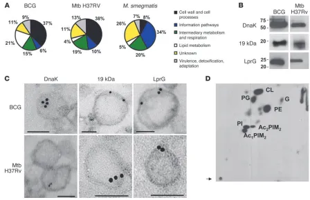
Analysis of proteins associated with vesicles . To gain insight in the pro-tein composition of MVs from both pathogenic (BCG and Mtb H37Rv) and nonpathogenic strains (M. smegmatis), vesicles were subjected to proteomic analysis using tryptic digestion prior to analysis by nano-LC-MS/MS. From 3 independent experiments, we identified 66 proteins in BCG, 48 in Mtb, and 64 in M. smeg-matis vesicular preparations (Supplemental Tables 1–3; supple-mental material available online with this article; doi:10.1172/ JCI44261DS1). All proteins detected were classified according to the functional category given by the databases TubercuList v2.3, BoviList v3.1, and SmegmaList v1.0 (Figure 3A). Overall, BCG and Mtb MVs had similar protein compositions, while M. smegmatis MVs contained a markedly different set of proteins. In Mtb, most vesicular proteins were classified as belonging to “cell wall and cell processes” (38%) or “intermediary metabolism and respiration” (19%). Proteins annotated as being involved in “lipid metabolism” were detected at a lower frequency (4%). Similarly, MVs BCG pro-teins belonging to cell wall and cell processes also represented 37%. Conversely, in M. smegmatis MVs, this category contained only 8% of the proteins identified. MVs from BCG were enriched in the “lipid metabolism” category (21% vs. 4% for Mtb and 5% for
M. smegmatis ; Figure 3A and Supplemental Tables 1–3). Remark-ably, both BCG and Mtb MVs showed a prominent component of lipoproteins representing 20% and 10% of the total proteins, respectively (Tables 1 and 2). Since lipoproteins typically represent between 1% and 2% of mycobacterial genomes (20), this repre-sented a major enrichment in the MVs. In order to experimentally confirm this enrichment in lipoproteins in MVs of BCG and Mtb, we analyzed the protein profiles of cellular lysates of these bacte-ria grown under the same culture conditions that were used for generation of MVs (Supplemental Tables 4 and 5). We found that lipoproteins represented 1.4% and 2.6% of the total cell-associated proteins in BCG and Mtb, respectively. This result established that MVs from BCG and Mtb strains were enriched in lipoproteins, a finding consistent with active release. In contrast, no lipoproteins were found in M. smegmatis MVs, despite the fact that M. smegmatis has 68 putative lipoproteins in the genome, representing 1.03% of the total protein in M. smegmatis (Supplemental Table 6).
The proteomic approach was validated by demonstrating the presence of some of the proteins identified by MS with serological techniques. In this regard, DnaK, 19 kDa (Lpqh), and LprG were detected by immunoblot analysis of vesicular preparations (Fig-ure 3B) and visualized by electron microscopy as vesicle associated by ImmunoGold electron microscopy (Figure 3C). Lipoproteins 19 kDa and LprG were located at the vesicle membrane, as expected from their lipidic nature. In contrast, DnaK localized to the lumen of the vesicles. We detected the presence of known secreted com-pounds such as LAM or α-glucan in the vesicle preparations by ELISA (Supplemental Figure 1A). Based on TEM images, LAM was detected both associated with the vesicles and unassociated, while
[image:4.585.48.286.84.617.2]α-glucan was clearly outside and apparently not associated with the vesicles (Supplemental Figure 1B). Since the molecular weight of α-glucan is around 100 kDa, it can be retained in the membrane
Figure 2
used to concentrate the supernatants. Consequently, we analyzed the fractions recovered after Optiprep density gradient purifica-tion by Western blot for the presence of LAM and by ELISA for
α-glucan (Supplemental Figure 1C). The 19-kDa lipoprotein was used as a vesicle marker, and the presence of this protein corre-sponded to fractions containing LAM. In contrast, the α-glucan was associated with the heaviest fractions of the gradient consist-ing of protein aggregates and polysaccharides. In summary, MVs from pathogenic mycobacteria were enriched in lipoproteins, some of them well-known TLR2 ligands, along with other major cell surface compounds such as LAM, but did not contain α-glucan.
Analysis of vesicle-associated lipids. To evaluate the lipid profile of the vesicles, we performed 2D-TLC analyses of total lipid extracts from BCG and H37Rv (data not shown) vesicles isolated from
14C-acetate–labeled cultures. The vesicular lipids predominantly
consisted of polar lipids (Figure 3D) with only low amounts of apo-lar lipids detectable (Supplemental Figure 2), consistent with the cell membrane being the likely origin of vesicles. The most abun-dant polar lipid species were phosphatidylinositol (PI) and acylated phosphatidylinositol dimannosides (PIM2) along with cardiolipin
(CL) and phosphatidylethanolamine (PE) (Figure 3D). To validate the 2D-TLC data, the BCG and Mtb vesicle samples were subjected
to MALDI-TOF–MS analyses (Supplemental Figure 3). In addition to PI and acylated PIM2, PIM6
was also detected in the vesicle prepa-ration. The results also showed that vesicles were enriched in polar lipids consistent with their origin from the cell membrane.
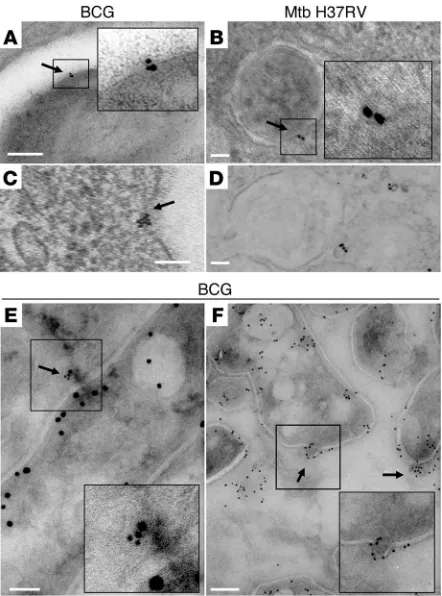
[image:5.585.61.509.81.371.2]Production of MVs by BCG and Mtb during intracellular infection of mac-rophages in vitro and in vivo. The abundance of 19-kDa lipoprotein and LAM in vesicles provided an opportunity to use monoclonal antibodies to these components in immunoelectron microscopy for identification of vesicles in mycobacteria-infected macrophages in vitro and in vivo. This revealed vesicles shedding from the bac-teria within phagosomes after 3 hours of infection (Figure 4A). In addition, MVs were seen trafficking at different locations in BCG-infected macrophages at 24 hours (Figure 4C). Similar findings were obtained with Mtb-infected macrophages at the same time intervals (Figure 4, B and D). To investigate whether the vesicle-release phe-nomenon could be documented in vivo, we infected mice with a high dose of CFSE-labeled bacteria. After 2 days, lungs were extracted and infected lung cells were sorted by FACS. Samples were processed for microscopy in the same manner as in vitro–infected macrophages and labeled with ImmunoGold. Images from this direct ex vivo analysis showed that BCG produced MVs within the phagosomes of macrophages that had been infected in vivo (Figure 4, E and F).
Figure 3
Mycobacterium vesicle–associated proteins and lipids. (A) Pie charts showing the BCG, Mtb, and M. smegmatis vesicle–associated proteins grouped into functional categories as in TubercuList, BoviList, and SmegmaList. Note that the “lipid metabolism” functional category is increased in BCG vesi-cles and that “cell wall and cell processes”–associated proteins are overrepresented in both BCG and Mtb compared with M. smegmatis. (B) Vesicles from the supernatant of the BCG culture were loaded onto a SDS-polyacrylamide gel, blotted on a nitrocellulose membrane, and incubated with mAbs with the indicated specificities. (C) ImmunoGold electron microscopy of thin sections of BCG and Mtb H37Rv vesicles treated with the mAbs specific for the indicated proteins and detected with a 10-nm IgG gold-labeled anti-mouse antibody. Scale bars: 100 nm. (D) Total lipids of vesicles isolated from 14C-acetate–labeled cells were extracted with chloroform/methanol (2:1, v/v). Lipid extracts were separated by 2D TLC by applying an amount
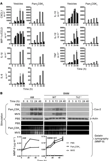
Vesicles stimulated macrophages in a TLR2-dependent manner . To evalu-ate the capacity of mycobacterial MVs to trigger a proinflammatory response, we added BCG vesicle preparations to freshly isolated BMM from WT and TLR2-knockout mice and measured cytokine and chemokine expression at different times after vesicle addition (Fig-ure 5A). The TLR1/2 ligand Pam3CSK4 was used as a positive control.
The MVs induced the production of IL-1β, IL-6, IL-10, IL-12, TNF, CXCL1, and MIP-1α/CCL3, and the majority of these cytokines were induced in a TLR2-dependent manner, since their production from MV-stimulated Tlr2–/–
cells was markedly reduced. However, induc-tion of MIP-1α/CCL3 and IL-10 by MVs was preserved in Tlr2–/–
cells, indicating that MVs also contained components that could activate macrophages through receptors different from TLR2. In contrast, Pam3CSK4 induced all cytokines and chemokines studied
in a TLR2-dependent manner. We were able to detect cytokines as early as 3 hours after MV stimulation, and the response peaked at 24 hours. The induction of MIP-1α/CCL3 and IL-10 by MVs had faster kinetics than the other inflammatory mediators, consistent with induction through a different, non–TLR2-related mechanism. Levels of IL-12 and TNF were not completely abolished in the TLR2-defi-cient macrophages. The residual levels of TNF could have been due to an alternative pathway of activation involving MIP-1α (21). The fact that the majority of cytokines remained persistently elevated at 48 hours suggested a prolonged effect of the MV-associated ligands. Subsequently, we analyzed the levels of cytokines produced by macro-phages stimulated with the same amount of M. smegmatis MVs at the same time points and found detectable levels of cytokines including
IL-10, IL-12, and IL-1β, but their concentrations were significantly lower than observed with BCG MVs. In addition, no chemokines or TNF were detected for M. smegmatis MV-stimulated macrophages at any time point analyzed in the study (Supplemental Figure 6A).
The cyclooxygenase Cox-2 is known to regulate production of MMP-9 in a TLR2-dependent manner (22). Since vesicles were enriched in TLR2 ligands, we analyzed the levels of Cox-2 in WT and TLR2-deficient BMM and alveolar macrophages by Western blot (Figure 5B). Again, Pam3CSK4 was used as a positive control for
TLR2 stimulation. BCG MVs triggered expression of Cox-2 in a time-dependent manner in WT BMM, whereas Cox-2 was not induced in macrophages from Tlr2–/– mice. The timing was very similar to that
of induction of cytokines by MVs, with detectable levels appearing as early as 3 hours. The effect of MVs on Cox-2 expression was still pres-ent at 48 hours, indicating persistent stimulation by MVs as seen for cytokine secretion. Accordingly, the enhanced expression of Cox-2 protein resulted in increased levels of MMP-9 in the culture superna-tants from BMMs stimulated with Pam3CSK4 or MVs, as measured
by gelatin zymography (Figure 5B). The identity of the band was con-firmed when the same experiment was carried out with BMMs from an mmp-9–knockout mouse, for which this gelatinolytic band was absent at 24 and 48 hours (Supplemental Figure 4). Neither Cox-2 nor MMP-9 was induced after stimulation of macrophages with
[image:6.585.58.534.109.264.2]M. smegmatis MVs (Supplemental Figure 6B). These results estab-lished that MVs from pathogenic mycobacteria were able to elicit production of a broad range of cytokines and other inflammatory mediators by macrophages in a TLR2-dependent manner.
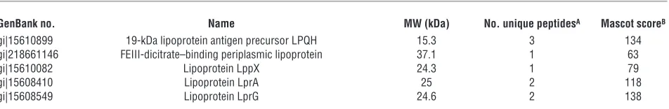
Table 1
BCG Pasteur vesicle–associated lipoproteins
GenBank no. Name MW (kDa) No. unique peptidesA Mascot scoreB
gi|15610899 19-kDa lipoprotein antigen precursor LPQH 15.3 3 108
gi|31794050 Cell-surface lipoprotein MPB83 11.1 1 69
gi|31794222 FEIII-dicitrate–binding periplasmic lipoprotein 37.1 1 72
gi|31791596 Lipoprotein aminopeptidase LpqL 52.2 1 70
gi|31794122 Lipoprotein LppX 24.3 2 131
gi|31794183 Lipoprotein LppZ 39.1 1 70
gi|31791765 Lipoprotein LpqN 23.7 2 69
gi|31792462 Lipoprotein LprA 25 1 66
gi|31792564 Lipoprotein LprF 27 1 66
gi|31792605 Lipoprotein LprG 24.6 1 66
gi|31792123 Periplasmic phosphate-binding lipoprotein PSTS1 (PBP-1) 38.4 2 58
gi|31792115 Periplasmic phosphate-binding lipoprotein PSTS3 38 3 225
gi|31617703 phoS1 34.1 1 97
AProteins identified with only 1 peptide were inspected manually. BAccurate identifications were considered when Mascot scores were above 45 (P < 0.05)
for peptides or 51 (P < 0.01) for proteins.
Table 2
Mtb vesicle–associated lipoproteins
GenBank no. Name MW (kDa) No. unique peptidesA Mascot scoreB
gi|15610899 19-kDa lipoprotein antigen precursor LPQH 15.3 3 134
gi|218661146 FEIII-dicitrate–binding periplasmic lipoprotein 37.1 1 63
gi|15610082 Lipoprotein LppX 24.3 1 79
gi|15608410 Lipoprotein LprA 25 2 118
gi|15608549 Lipoprotein LprG 24.6 2 138
AProteins identified with only 1 peptide were inspected manually. BAccurate identifications were considered when Mascot scores were above 45 (P < 0.05)
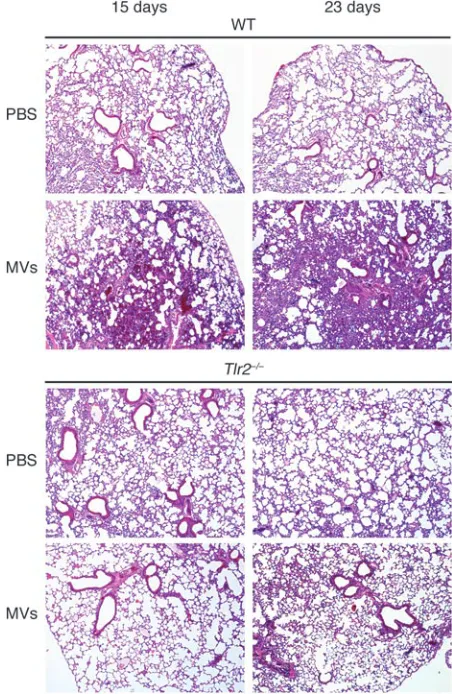
[image:6.585.56.532.643.721.2]Stimulation of TLR2-dependent inflammatory responses in the lung by MVs. To evaluate the effect of MVs on tissue inflammatory responses and the role for TLR2 in such responses, we adminis- tered a single intratracheal (i.t.) injection of MVs to WT or TLR2-deficient mice and evaluated the lungs at different times for inflammatory responses. On the fifteenth day after MV infusion, the lungs of the WT mice showed marked alterations compared with PBS-infused controls, including dense areas of mononuclear cell infiltration and interstitial pneumonia (Figure 6). In addi-tion, the MV-injected mice also showed a predominantly diffuse alveolar damage pattern of injury with more compaction of alveo-lar spaces. In contrast, no pathological changes were detected in TLR2-deficient mice. By day 23, the lesions in lungs of WT mice had worsened, but there was still little inflammatory change in the lungs of MV-injected TLR2-knockout mice, which appeared similar to the lungs of PBS-treated controls. Lung inflammation still occurred when lower doses of vesicles were administered (Supplemental Figure 7). Administration of the same amount of
M. smegmatis MVs also triggered an inflammatory response at day 15 and day 23 (Supplemental Figure 6C), but it was not as intense as observed with pathogenic MVs (Figure 6).
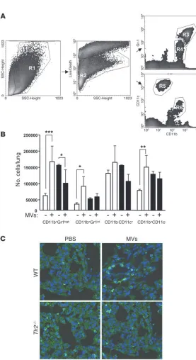
Since myeloid cells are known to be recruited as mediators of inflammatory processes in the lungs, we next asked whether i.t. injection of MVs was associated with changes in the numbers and phenotypic subsets of these myeloid cells. To analyze this, total lung cell suspensions were analyzed for the expression of CD11b, CD11c, and Gr1 (Figure 7A). A significant increase in the abso-lute numbers of pulmonary CD11b+Gr1hi, CD11b+Gr1int, and
CD11b+CD11c– cells (Figure 7B) was observed in the MV-treated
relative to the PBS-treated WT mice. These increases were not observed in the TLR2-deficient mice that received i.t. MVs. The
increase in the total number of cells expressing the CD11b marker was also apparent by confocal microscopy at day 15 (Figure 7C). When M. smegmatis MVs were administrated, we observed a signifi-cant increase in the absolute numbers of pulmonary CD11b+Gr1hi
population only on day 15, no change in CD11b+Gr1int or
CD11b+CD11c– cells, and decrease in CD11b–CD11c+
cells (Sup- plemental Figure 6D). These observations indicated that the ves- icle treatment resulted in a profound alteration in the composi-tion of the pulmonary myeloid populations in a TLR2-dependent manner. However, at day 23, only the CD11b+Gr1hi
cell popula-tion showed statistically significant differences (Supplemental Figure 5). Together, the above observations indicated that intro-duction of MVs into the lungs resulted in a profound alteration in the composition of the pulmonary myeloid populations in a TLR2-dependent manner.
[image:7.585.54.275.83.381.2]Vesicles impaired the control of Mtb infection. Next, we investigated whether exposure to MVs modified the course of experimental Mtb infection. Mice received an i.t. injection of either MVs or PBS and 15 days later were infected via aerosol with a low dose of Mtb H37Rv. Lung histology was examined at 15 and 23 days after infection (Figure 8A). As expected, inflammatory infiltrates were observed in lungs of both groups of animals at day 15, but the MV-treated mice showed more discrete granulomatous struc-tures, suggesting a more rapid progression of the inflammatory response. The difference between MV-treated and untreated mice was more pronounced at day 23, with MV-treated mice show-ing larger and more numerous granulomas compared with the PBS-treated control mice (Figure 8C). In contrast, the amount of inflammation observed for mice given M. smegmatis MVs at days 15 and 23 was significantly lower compared with that of the BCG-treated mice (Supplemental Figure 6E).
Figure 4
We also studied BCG-vaccinated mice to determine whether vesicles can alter the outcome of the infection in animals with a recall response induced by previous vaccination (Figure 8B). The BCG-vaccinated mice that received MV injections showed mark-edly increased granulomatous inflammation (Figure 8B), and the number of granulomas per lung was higher compared with BCG-vaccinated mice that received only PBS injections (Figure 8C). The number of CFUs in the lung of unvaccinated mice at day 23 following Mtb infection was significantly higher in mice treated with vesicles (Figure 8D). This was also true when CFUs
[image:8.585.43.405.77.608.2]were counted in spleen (Figure 8E), suggesting that vesicles can promote the dissemination of the bacilli form the lung to other lymphoid organs. Similar trends were observed for BCG-vacci-nated mice, although this did not reach statistical significance (Figure 8D). Conversely, the bacterial counts in the lung of mice treated with M. smegmatis MVs were significantly lower at day 23 compared with the PBS-treated mice, suggesting an alternative response that may be beneficial for the host (Supplemental Fig-ure 6F) and indicating specificity in the inflammatory response triggered by pathogenic mycobacterial MVs.
Figure 5
Responses of macrophages to BCG MVs. Freshly isolated BMM and alveolar macrophage cell line (AMJ2-C8) were stimulated with MVs and with Pam3CSK4 in vitro
(500,000 cells/500 μl of media). Supernatants and cell lysates were collected at time points ranging from 0 to 48 hours after stimulation (x axes of all graphs indicate time in hours). (A) The supernatants of cultures of WT (white bars) or TLR2-knockout (black bars) isolated BMMs were assayed for cytokines IL-1β, IL-10, IL-12p70, TNF-α, and chemo-kines CXCL1 by multiplex ELISA (MSD Systems). Cytokines IL-6 and MIP-1α were analyzed by standard ELISA. Data represent mean ± SEM. (B) The levels of Cox-2 and MMP-9 were analyzed by Western blot and zymography, respectively. For Cox-2 analysis (top), lysates were made from cells (AMJ2-C8 alveolar macrophage cell line (AM), WT BMM, or Tlr2–/– BMM collected at
the indicated times, ranging from 0 to 48 hours after addition of PBS (unstimulated), Pam3CSK4, or BCG
Discussion
The present work represents the first study, to our knowledge, of production of MVs in 2 of the most clinically significant species of mycobacteria, M. bovis BCG and Mtb, along with other representa-tives of the mycobacterial genus including nonpathogenic strains. The results extend a prior report on vesicle production by M. ulcerans (6) and further define a newly recognized aspect of the host-patho- gen interaction in mycobacterial infections with clear implica-tions for pathogenesis, since MVs from nonpathogenic strains do not drive the same responses in the host. Furthermore, our results complement the observation that another Gram-positive bacte-rium, B. anthracis, produces MV (4), extending this mechanism into the subset of the Gram-positive group defined by the mycobacteria. Our results indicated that BCG and Mtb vesicles were physiologic products of live bacilli growing in liquid culture or within macro-phages. Using different independent lines of experimentation, we showed that the production of vesicles by BCG depended on cell viability. Thus, vesicles were recovered only from live cultures, vesicle fractions were metabolically labeled only by live cells, and vesicular structures could be identified on the surface of mycobacteria in elec-tron micrographs. Hence, we conclude that mycobacterial species, such as Gram-negative bacteria (1) and fungi (5), produce MVs that may facilitate the export of a variety of potential virulence factors.
The earlier study of vesicles released by M. ulcerans showed these to be enriched in mycolactone, a known toxin produced by this microorganism (6). This suggests that like many Gram-negative
bacterial pathogens, mycobacteria produce MVs as carriers for vir-ulence factors that modify the responses of host cells to infection (1). Consistent with this idea, our proteomic analyses of BCG, Mtb, and M. smegmatis MVs showed that only BCG and Mtb MVs were enriched in proteins associated with virulence, including a remark- ably high content of putative TLR2 ligands such as the lipopro-teins 19 kDa, LprA, and LprG. These molecules have been shown to downregulate the levels of MHCII molecules on macrophages and dendritic cells through a mechanism involving recognition by TLR2 (11–13). The lipid profile of BCG and Mtb MVs suggested that they originated from the bacterial cell membrane, since polar lipids typically found in the plasma membrane were the most abundant species. Interestingly, some of these polar lipids, such as PIM2 or PIM6, are known to be ligands of TLR2. As suggested
by the nature of their protein and lipid cargo, mycobacterial MVs produced a profound TLR2-dependent inflammatory response in vitro and in vivo. In addition, MVs induced a range of inflammato-ry cytokines as well as the expression of key molecules implicated in early events of granuloma formation such as Cox-2 or MMP-9. Our in vivo studies indicated that MVs stimulated myeloid cell recruitment, leading to an increase in the CD11b+Gr1int
popula-tion subset in the lung. Remarkably, when vesicle-treated mice were challenged with Mtb H37Rv, they showed an increase in granulomatous inflammation in the lungs as well as an increase in the number of bacilli in both their lungs and spleens.
Mycobacterial lipids, including those originating from the cell wall, and proteins shed by phagocytosed mycobacteria are incorpo-rated into released vesicles known as exosomes, which are produced by exocytic machinery of the mammalian host cells (16, 23). The way in which these surface compounds are released from the bac-teria remains elusive. Given our observations using TEM showing the production of vesicles by the bacteria within the phagosomes of BMMs in vitro and in vivo and BMDCs (Rafael Prados-Rosales, unpublished observations), we propose that direct release by the bacteria of MVs provides a mechanism for transport of immuno-modulatory compounds such as lipoproteins. This may lead to the distribution of these compounds to various compartments within infected host cells or to their incorporation into host cell–derived exosomes. Thus, generation and release of MVs may allow myco-bacteria confined within phagosomes to deliver virulence factors to other cellular compartments or into the extracellular milieu to affect neighboring cells.
In addition to lipoproteins, our lipidomic studies showed that PI and PIM2 along with CL and PE were the most prevalent polar
[image:9.585.43.269.85.432.2]lipids in MVs. Moreover, LAM was also detected as a part of the vesicles. LAM, lipomannan (LM), and PIMs are major lipoglycans that are noncovalently attached to the plasma membrane through their phosphatidyl-myo-inositol anchor and extend to the exterior of the cell wall (24). LAM is an important ligand in the interaction between Mtb and macrophages (25) and contributes to modulating the inflammatory response by downregulating IL-12 production (26). In addition, LAM inhibits phosphadityl inositol-3-phosphate
Figure 6
TLR2-dependent inflammation in mouse lungs after vesicle injection. Representative images of H&E-stained lung sections of i.t. vesicle or PBS-treated WT and Tlr2–/– C57BL/6 mice. Lungs were analyzed at 15
(PI3P) association with Mtb phagosomes (27) and blocks calcium flux in infected cells (28). Conversely, LM and PIMs can induce a proinflammatory response by inducing TNF-α, IL-8, and IL-12 secretion (29). Other important mycobacterial cell wall compo-nents, such as the glycolipid trehalose dimycolate (TDM), have been implicated in the development of an inflammatory response and in granuloma formation (30, 31). However, we did not find TDM associated with MVs in our 2D-TLC analysis. Recently, mycobacte-rial PIM2
was reported to induce the expression of Cox-2 and subse-quently MMP-9 in a TLR2-dependent manner (22), which parallels our findings in cells exposed to isolated MVs. These enzymes modify the host response, as Cox-2 generates prostaglandins that modulate the cell death response to infection (32) and MMP-9 is a
metalloprotease known to be involved in the remodel-ing of the extracellular matrix during the early events of granuloma formation (33, 34). We did not explore the contribution of other TLRs, such as TLR4, TLR6, or TLR9, which have been shown to have a role in the recognition of Mtb in mice and human (35–38). How- ever, we detected DNA in purified and intact mycobacte-rial MVs (our unpublished data), suggesting a possible interaction of MVs with TLR9.
One of the key features of mycobacterial infections is the production of an inflammatory Th1 response that promotes the secretion of selected cytokines such as TNF-α, INF-γ, and IL-12 (39, 40). However, our analysis of cytokines produced by BMMs stimu-lated with MVs did not indicate the induction of a typical Th1 priming milieu, since cytokines that oppose Th1 priming such as IL-10 were also induced (41). IL-10 has been shown to block activation of mac-rophages, which may increase the dissemination of the bacteria. In addition, the chemokines CXCL1 and CCL3 were induced in BMMs by MVs, which along with other cytokines are critically involved in recruit-ment of multiple effector cells at the site of infection. The analysis of the type of myeloid cells recruited after a BCG MV treatment showed an increase in the CD11b+Gr1hi, CD11b+ Gr1int, and CD11b+CD11c– cell
populations. This is relevant because a strong inflam- matory response and an increase in the cell popula-tion CD11b+Gr1int have been recently associated with
progression of tuberculosis (42), providing support for the hypothesis that MVs contribute to the viru-lence of Mycobacterium. Moreover, this population has been shown to be permissive for mycobacterial replication in mice (43). Although more phenotypic markers are required to define this CD11b+Gr1int population, we believe that they
may be a subset of myeloid-derived suppressor cells (MDSC), which may have inhibitory properties in the context of infec-tion by intracellular bacterial pathogens, including Mtb (44). Remarkably, this CD11b+Gr1int myeloid cell population did not
[image:10.585.42.319.67.575.2]increase upon treatment with M. smegmatis MVs, suggesting that its recruitment may be specifically associated with pathogenic mycobacteria. We note that there is increasing evidence that macrophage and dendritic cell responses to microbial antigens can differ (45) and the interaction of vesicles with dendritic cells should be a priority for future studies.
Figure 7
TLR2-dependent increase in myeloid cells in the lungs following i.t. MV injection. (A) Representative flow cytometry dot plots showing the gating for analysis of different myeloid populations (R3, CD11b+Gr1hi; R4, CD11b+Gr1int; R5,
CD11b–CD11c+; and R6, CD11b+CD11c–). (B) Bar graphs
indicating the mean absolute numbers of the different popu-lations of lung cells of WT (white bars) and Tlr2–/– (black bars)
mice. Data represent mean ± SEM. *P < 0.05; **P < 0.01; ***P < 0.001. (C) Confocal inmmunofluorescence micros-copy of cryosections stained with DAPI and CD11b–Alexa Fluor 647 showing an increase in CD11b+ cells after vesicle
treatment at 15 days in WT but not in the Tlr2–/– mice. Data
Our findings point to MVs as a factor that is detrimental for the host during Mtb infection. In support of this, we note the observations that mice pretreated with pathogenic MVs developed a more acute inflammation and higher bacterial loads in the lungs compared with control and nonpathogenic MV-treated mice and that preexposure to MVs also increased dissemination of bacilli from the lung to other lymphoid organs such as the spleen. Furthermore, we observed MVs in mycobacteria recovered from mice, providing evidence for the formation of these structures in vivo and lending support to the view that MVs represent a bacterial release mechanism relevant to pathogenesis.
In summary, we have confirmed the existence of a vesicular system in mycobacteria and demonstrate that the vesicles pro- duced contain numerous components associated with viru-lence. The finding that MVs are released inside macrophages in vivo suggests that they have the capacity to influence the outcome of the macrophage-mycobacteria interaction. Myco- bacterial vesicles are proinflammatory through a TLR2-depen-dent mechanism, providing additional evidence for the key role of the innate immune receptor in host defense against Mtb. The work reported in the current study raises many important questions about the relevance of MVs to the adaptive immune response against Mtb, which ultimately may be resolved through the isolation of bacterial mutants that are deficient in vesicle production. The discovery of a vesicular transport sys- tem in mycobacteria opens up new horizons for the pathogen-esis of tuberculosis and vaccine development.
Methods
Mycobacteria and bacterial culture. M. bovis BCG (Pasteur strain) and other mycobacteria (Mtb strains H37Rv [virulent] and H37Ra [avirulent], M. kan-sasii, M. phlei, M. smegmatis, and M. avium) were grown in an MM consisting of 1 g/l KH2PO4, 2.5 g/l Na2HPO4
, 0.5 g/l asparagine, 50 mg/l ferric ammo-nium citrate, 0.5 g/l MgSO4 × 7 H2O, 0.5 mg/l CaCl2, 0.1 mg/l ZnSO4, with
or without 0.05% tyloxapol (v/v), containing 0.1% (v/v) glycerol, pH 7.0. In some experiments as stated, cultures were grown in Middlebrook 7H9 medium (M7H9) supplemented with 10% (v/v) OADC enrichment (BD Microbiology Systems), 0.5% (v/v) glycerol, and with or without tyloxapol 0.05% (v/v; Sigma-Aldrich). Cultures were grown for up to 10 days as indi-cated in roller bottles at 37°C. To study the requirement for cell viability, mycobacterial precultures were used to inoculate fresh MM supplemented with 1 μCi ml−1 [1,2-14C]-acetate (50–62 mCi mmol−1; Merck). Cultures were
harvested at day 5 and bacterial cells were killed by heating to 80°C for 2 hours. After this, live cells and heat-killed cells were again inoculated in fresh MM and incubated for 5 additional days at 37°C. Vesicles were then isolated and radioactivity measured in a scintillation counter. To assess cell viability at day 10, number of cells was determined directly by man-ual counting using a Petroff-Hausser Counting Chamber and standard microscope with ×40 magnification and then compared with the number of CFUs obtained by plating serial dilutions on 7H11 agar media.
[image:11.585.43.374.83.412.2]Vesicle isolation and purification. Vesicles were isolated as described with minor modifications (17). The basic protocol involved a combination of serial differential sedimentation and density gradient purification. Briefly, cells were pelleted (3,450 g for 15 minutes, 4°C) from 1000 ml cultures and the supernatants were filtered through a 0.45-mm–pore size polyvinylidene difluoride filter (Millipore). The supernatant volumes were
Figure 8
then concentrated approximately 20-fold using an Amicon Ultrafiltration System (Millipore) with a 100-kDa exclusion filter. The concentrate was then sequentially centrifuged at 4,000 and 15,000 g (15 minutes, 4°C) to remove cell debris and aggregates, and the remaining supernatant was then centrifuged at 100,000 g for 1 hour at 4°C to sediment the vesicu-lar fraction into a pellet. The supernatant was then discarded, the pellet was suspended in 1 ml of 10 mM HEPES, 0.15 M NaCl, and mixed with 2 ml of Optiprep solution (Sigma-Aldrich) in 10 mM HEPES and 0.15 M NaCl (yielding 35% [w/v] Optiprep final concentration). The crude vesicle sample was then overlayed with a series of Optiprep gradient layers with concentrations ranging from 30%–35% (w/v). The gradients were centri-fuged (100,000 g, 16 hours), and 1 ml fractions were removed from the top. The fractions were then dialyzed separately in PBS overnight and again recovered by sedimentation at 100,000 g for 1 hour. Finally, the vesicles were suspended in LPS-free PBS. For quantitation of vesicles, aliquots of preparations isolated from MM were stained with CFSE (Invitrogen) at 10 μM final concentration in PBS for 30 minutes at 37°C. After 2 washes in PBS, labeled vesicles were diluted and counted by fluorescence micros-copy (Axiovert 200M; Carl Zeiss Inc.). Fluorescent images consisted of 1344 × 1024 pixels and were taken at ×40 (0.101 μm × pixel). The concen-tration of vesicles was calculated as a function of the field area and then corrected for the loaded volume. Five different fields were counted.
Electron microscopy . TEM was used to analyze the morphology and integ- rity of extracellular vesicles isolated from culture supernatants and to visu-alize the interaction of MVs with mycobacteria and macrophages. MVs were fixed with 2% glutaraldehyde in 0.1 M cacodylate at room temperature for 2 hours and then incubated overnight in 4% formaldehyde, 1% glutaralde-hyde, and 0.1% PBS. After fixation, the samples were stained for 90 minutes in 2% osmium, then serially dehydrated in ethanol and embedded in Spurrs epoxy resin. Thin sections were obtained on an Ultracut UCT (Reichert) and stained with 0.5% uranyl acetate and 0.5% lead citrate (Reichert). Samples were observed in a JEOL 1200EX transmission electron microscope operat- ing at 80 kV. For ImmunoGold labeling of BCG- or H37Rv-infected mac-rophages or vesicles, samples were first fixed in 0.1 M sodium cacodylate buffer (pH 7.2) containing 4% paraformaldehyde, 0.2% glutaraldehyde, infil- trated in 25% polyvinylpyrrolidone and 2.1 M sucrose and then rapidly fro- zen by immersion in liquid nitrogen. Cryosections were obtained in a tem-perature range of −70 to −90°C using an Ultracut UCT. After blocking with PBS-BSA with 50 mM NH4Cl, the cryosections were incubated overnight in
the presence of the mouse monoclonal antibodies specific for DnaK (IT-42; IgG1), lipoprotein 19 kDa (IT-12; IgG1), lipoprotein LprG (Rv1411, IgG1), LAM (CS-35, IgG1), or α-glucan (24c5; IgG1; ref. 46). After incubation with ImmunoGold-labeled anti-mouse IgG, specimens were observed in a JEOL 1200EX transmission electron microscope operating at 80 kV.
Protein identification by liquid chromatography-tandem mass spectrometry . Pro-teins were precipitated from vesicle suspensions and bacterial lysates by adding 6 volumes of ice-cold acetone and the pellets dissolved in 25 mM ammonium bicarbonate. Proteins were reduced with 10 mM DTT at room temperature for 1 hour, and the cysteine residues were subsequently alkyl-ated with 10 mM iodoacetamide at room temperature for 1 hour in the dark. Protein digestion was carried out with trypsin (Promega, 1:20; w/w) at 37°C for 16 hours. The peptide mixtures from in-solution tryptic digestions were analyzed using Nanoflow LC-MS/MS. The peptides were loaded onto a 0.3 × 5 mm C18 precolumn (New Objective) and then eluted with a linear gradient of 2%–90% acetonitrile in 0.1% aqueous solution of formic acid. The gradient was delivered over 120 minutes by a NanoLC 1D Plus (Eksigent) at a flow-rate of 200 nl/min, through a 75-μm × 15-cm fused silica capillary C18 HPLC PepMap column (LC Packings) to a stainless steel Nanobore emitter (Proxeon). The peptides were scanned and fragmented with an LTQ
XL linear ion trap mass spectrometer (ThermoFinnigan) operated in data- dependent and MS/MS switching mode using the 3 most intense precur-sors detected in a survey scan from 400 to 1600 amu (3 μscans). Normalized collision energy was set to 35%, and dynamic exclusion was applied during 3-minute periods to avoid repetitive fragmentation of ions. Generated .raw files were converted to .dta files for Mascot database. A database containing the NCBInr Bacteria sequences (9,940,348 sequences, as of April 15, 2010) was searched using Mascot Software (version 2.3; Matrix Science) for pro-tein identification. Search criteria included trypsin specificity with 1 missed cleavage allowed and methionine oxidation as variable modification. Search criteria included (a) trypsin specificity with 1 missed cleavage allowed, (b) methione oxidation as variable modification, and (c) a minimum precur-sor and fragment-ion mass accuracy of 1.2 and 0.3 Da, respectively, and a requirement of at least 1 bold red peptide (i.e., highest scoring peptide matching to protein with highest total score). Cut-off values for Mascot scores of peptides and proteins were set to 45 (P < 0.05) and 51 (P < 0.01), respectively, for consideration as accurate identifications. Proteins identi-fied with only 1 peptide were inspected manually. The false-positive rate (1.68%) was calculated searching the same spectra against the NCBInr Bac-teria decoy database, using the version updated April 15, 2010. A combined list of proteins identified in all replicates was condensed in order to remove redundant IDs such as orthologous sequences, redundant database entries, and indistinguishable isoforms based on observed peptide coverage.
Vesicle-associated protein analysis. Proteins (5 μg) of MVs and mycobacterial fractions were separated with 12% SDS-PAGE, and gels were stained with Coomassie blue. To validate the proteomic approach, the presence of some proteins of interest in the vesicle preparation was confirmed by Western blot. Proteins were transferred to nitrocellulose membranes and blocked with 5% (w/v) nonfat dry milk in PBS for 1 hour at room temperature before incubating with the mAbs specific for DnaK (IT-42; IgG1, BEI Resources), 19-kDa lipoprotein (IT-12; IgG1, BEI Resources) and LprG (Rv1411; IgG1, BEI Resources) overnight at 4°C. After washing with PBS plus 0.05% Tween-20 (PBST), the membranes were incubated with an anti-mouse IgG (H+L) HRP (Invitrogen) as a secondary antibody for 1 hour. The immunoreactive bands were visualized with a chemiluminescent substrate.
Vesicle-associated lipid analysis. Total lipids of vesicles isolated from 14
C-ace-tate–labeled cells (2 μCi ml–1) for 12 days were extracted with chloroform:
methanol (2:1; v/v). Lipid extracts were separated by a series of 2D TLC systems as previously published (47) by applying equal amounts of lip-ids corresponding to 10,000 DPM per TLC plate and by using the solvent systems A–D for separation of apolar lipids and E for separation of polar lipids, respectively. MALDI-TOF mass spectrometry analysis of lipids was performed as previously described (48).
Cox-2 and MMP-9 analysis . Macrophage lysates were separated by SDS-PAGE and then transferred electrophoretically to PVDF membranes. The blots were blocked with 5% (w/v) nonfat dry milk in PBS for 1 hour at room temperature and subsequently incubated overnight at 4°C with a Cox-2–spe-cific primary antibody (Cayman) in PBST with 5% (w/v) nonfat dry milk. Following 3 washes of 5 minutes each with PBST, the blots were incubated with horseradish peroxidase–conjugated goat anti-rabbit IgG (Southern Bio-tech) in blocking buffer for 1 hour at room temperature. After 3 washes with PBST, the blots were developed by chemiluminescence using the ECL-pico Western Detection Kit (Pierce) and exposed to x-ray film (Kodak XAR5).
10 minutes. Finally, gels were incubated in 5 mM CaCl2, 150 mM NaCl,
and 0.01% Triton X-100 for 18 hours at 37°C. Gelatinolytic activity was visualized as clear areas in the Coomassie blue–stained gel. Recombi-nant mouse MMP-9 (R&D Systems) was used as a standard for the assay. Quantification of MMP-9 activity in gels was done by comparing the pixel intensity of the white areas in the gel.
Mouse immunizations and infections. C57BL/6 female mice between 6 and 8 weeks old were purchased from Jackson Laboratories. All procedures involving mice were reviewed and approved by the Animal Use and Care Committee of the Albert Einstein College of Medicine. Animals were main-tained in a specific pathogen–free animal facility under animal biosafety level 2 conditions for all experiments, except for those involving infection with virulent Mtb, for which animal biosafety level 3 conditions were used. TLR2-deficient mice on a C57BL/6 background were a gift of S. Akira (49). For i.t. immunization, anesthetized mice received an i.t. injection of 25 μg of vesicles in 50 μl of PBS, corresponding to 2 × 10⁸ vesicles according to
previously published methods (9). Control mice received i.t. injections of PBS. Aerogenic challenge was done using a whole-body exposure aerosol chamber (Mechanical Engineering Workshop) custom fitted to a class 3 biosafety cabinet (Baker) to deliver 10–50 CFU per animal of Mtb (H37Rv). For BCG immunizations, each mouse received s.c. 1 × 10⁶ CFUs. Mice were
euthanized at 15 or 30 days after challenge. Lungs and spleens of indi-vidual mice were aseptically removed and homogenized separately in 5 ml PBS plus 0.05% tyloxapol using a Seward Stomacher 80 blender (Tekmar). The homogenates were diluted serially and plated on Middlebrook 7H11 agar to determine CFU of Mtb. The number of granuloma per lung was assessed by counting 4 sections from each lung cut at intervals of 100 μm in depth at a magnification of × 40. Pictures were taken using a digital cam-era COOLSCOPE (Nikon). Five lungs per treatment were analyzed.
BM macrophages. BM macrophages (BMMs) were derived from the femur and tibia of mice, as described (50). Briefly, leg bones were removed and the attached muscle tissue was removed. After disinfection in 70% ethanol, both ends were cut and the marrow flushed with PBS using a syringe with a 0.45-mm diameter needle. At day 0, BM precursors were seeded at 2 × 10⁶
per 100-mm dish in 10 ml complete medium (DMEM; Gibco, Invitrogen, supplemented with decomplemented 10% FCS, l-glutamine, HEPES, β2-mercaptoethanol, and penicillin/streptomycin) with 20% conditioned
medium (containing M-CSF) isolated from a L929 fibroblast cell line cul-ture (50). The BMM were harvested at day 7 for use in experiments.
Cytokine ELISAs. Macrophage culture supernatants were assayed for a panel of mouse Th1/Th2 cytokines and chemokines using a 9-plex mul- tiplex ELISA system that uses signal development based on an electroche-miluminescence reaction (Mesoscale Discovery Systems Inc.), according to the manufacturer’s specifications. The cytokines IL-6 and MIP-1α were analyzed using standard capture ELISAs (from BD Biosciences and R&D Systems, respectively) according to the manufacturers’ instructions.
Macrophage infections. Monolayers of BMMs (5 × 10⁵ cells) were infected with
BCG or Mtb H37Rv at an MOI of 10:1 or treated with 4.5 × 10⁷ vesicles. After
3 hours, cells were treated with gentamicin (100 μg/ml; Life Technologies) for 1 hour to inhibit the growth of extracellular bacteria, and the cells were then washed 2 times with PBS and cultured in fresh medium containing 20 μg/ml of gentamicin. Cells were collected at different time intervals (3, 6, 24, and 48 hours) after infection and processed for analysis. At the same time intervals, cell-free supernatants were collected and stored at –80°C until assayed for cytokines by ELISA or for MMP-9 activity by gelatin zymography.
Histological analysis. The lungs were removed aseptically from mice, fixed in 10% buffered formalin phosphate (Fisher), and embedded in paraffin. Serial 4-μm sections were cut and stained with H&E. For immunohistochemistry, lungs were fixed with 2% paraformaldehyde in PBS for 2 hours at 4°C. After fixation, the tissues were transferred and
incubated in 30% sucrose overnight at 4°C. Tissues were then embedded in OCT (Sakura Finetek USA Inc.) and frozen in –70°C isopentane. Sections of 4-micron thickness were cut and stained with Alexa Fluor 647–labeled anti-CD11b and DAPI. Staining was analyzed using a LEICA SP5 confocal microscope (Leica Inc.) and Leica LAS-AF software.
Analysis of lung cell populations. The composition of lung inflammatory infiltrates was analyzed by flow cytometry. Briefly, lungs were perfused with heparin in DMEM after excision and incubated with 0.4 mg/ml Liberase CI solution (Roche Biochemicals) and DNase I for 1 hour at 37°C. Single-cell suspensions were prepared by gently pressing the tissue through a 70-μm nylon mesh cell strainer with a syringe plunger. Red cells were lysed for 5 minutes in 3 ml of erythrocyte lysis buffer (Sigma-Aldrich) diluted 1:1 with PBS. Cells were centrifuged and resuspended in FACS buffer (2% FCS, 0.05% sodium azide in PBS). Clumps were removed by passing the cell suspensions through a 50-μm cell strainer. To analyze the phenotype of pulmonary subpopulations, cell suspensions were first blocked with 2.4G2 mAbs to block Fc receptors and then stained with LIVE/DEAD dye (Invitrogen) followed by staining with fluorochrome-conjugated mAbs specific for CD11b (Alexa Fluor 647; eBioscience), CD11c (PE; BD Biosciences), and Gr1 (FITC; BD Biosciences). The FACS data was analyzed using WinMDI 2.9 (free software; Scripps Institute).
Analysis of production of MVs in vivo. M. bovis BCG Pasteur was stained with CFSE (Invitrogen) at a 10-μM final concentration in PBS for 30 minutes at 37°C. Cells were washed extensively in PBS plus 0.05% tylox-apol (Sigma-Aldrich), and OD was measured at 600 nm. An amount corresponding to 5 × 10⁷
CFSE-labeled bacteria was injected intrave- nously into mice by the lateral tail vein. Two days later, mice were sac-rificed and lungs were extracted and processed as above to obtain a single-cell suspension. Lung cells were sorted using a high-speed cell sorter (MoFlo; DAKO-Cytomation) with gating on CFSE-positive cells. Samples were further submitted to ImmunoGold labeling and cryosec-tioning for TEM analysis as above.
Statistics . ANOVA was used to assess statistical significance of differ-ences in the means. Student’s t test (2-tailed) was applied for experiments involving pairwise comparisons. P ≤ 0.05 was considered significant.
Acknowledgments
A. Casadevall is supported by NIH awards HL059842, AI033774, AI052733, AI033142, and the AERAS foundation for vaccine development. A. Casadevall is also supported by the Northeastern Biodefense Center under grant U54-AI057158-Lipkin. S.A. Porcelli and W.R. Jacobs Jr. were supported by the NIH Program Project grant AI-063537. R. Kalscheuer acknowledges support from the Juergen Manchot Foundation. Flow cytometry studies were carried out using core facilities supported by the Einstein Cancer Center (NIH/NCI CA013330) and the Einstein Center for AIDS Research (NIH AI-51519). Special thanks are given to C.A. Duque and V. Cabezon for their helpful discussion, support, and inspiration.
Received for publication July 6, 2010, and accepted in revised form January 5, 2011.
Address correspondence to: Arturo Casadevall, Albert Einstein College of Medicine, Jack and Pearl Resnick Campus, 1300 Morris Park Avenue, Forchheimer Building, Room 411, New York, New York 10461, USA. Phone: 718.430.2811; Fax: 718.430.8771; E-mail: [email protected].