CASS SCHOOL OF EDUCATION
COMPETENCY BASED ASSESSMENT USING VIRTUAL
REALITY (VERT): IS IT A REALISTIC POSIBILITY?
D
AVID
M. F
LINTON
A Thesis submitted in partial fulfillment of the requirements for the degree of a professional Doctorate in Education
i
Abstract
The education of the radiography profession is based within higher education establishments, yet a critical part of all radiography programmes is the clinical component where students learn the practical skills of the profession. Assessments therefore not only have to assess a student’s knowledge, but also their clinical competence and core skills in line with both Health and Care Professions Council and the Society and College of Radiographers requirements. This timely thesis examines the possibility of using the Virtual Environment for RadioTherapy (VERT) as an assessment tool to evaluate a student’s competence so giving the advantage of a standard assessment and relieving time pressures in the clinical department.
A mixed methods approach was taken which can be described as a Quantitative Qualitative design with the emphasis being on the Quantitative element; a so called QUAN qual design. The quantitative evaluation compared two simulations, one in the virtual reality environment and another in the department using a real treatment machine. Students were asked to perform two electron setups in each simulation; the order being randomly decided and so the study would be described as a randomised cross-over design. Following this, qualitative data was collected in student focus groups to explore student perspectives in more depth.
ii
Declaration
I hereby declare that, except where explicit attribution is made, the work presented in this thesis is entirely my own.
Word count (exclusive of appendices, the list of references and bibliographies, but including footnotes, endnotes, glossary, maps, diagrams and tables): 54,902 words.
iii
Table of Contents
Abstract ... i
Declaration ... ii
Table of Contents ... iii
List of Figures ... vii
List of Tables ... viii
List of Abbreviations/Glossary ... ix
Acknowledgements ... xiii
Dedication ... xiv
Chapter 1 - Introduction ... 1
1.1 Background ... 1
1.2 VERT ... 4
1.3 Simulation & Assessment ... 6
1.4 Problem Statement ... 8
1.5 Aims and Objectives ... 9
1.6 Significance of the Study ... 10
1.7 Positionality and Reflexivity ... 10
1.8 Outline of the Thesis ... 13
Chapter 2 - Radiotherapy and Education ... 15
2.1 Introduction ... 15
2.2 Professionalization and Institutionalisation ... 16
2.3 The Practice of Radiotherapy ... 20
2.3.1 Electron Beams ... 23
2.3.2 Patient care ... 29
2.4 The simulations ... 30
2.4.1 The Phantom ... 30
2.4.2 The equipment ... 31
2.4.3 The room ... 32
Chapter 3 - Literature Review ... 33
3.1 Introduction ... 33
3.2 Structure of the Literature Review ... 34
3.3 Constructivism ... 34
iv
3.4.1 Classification of Educational Simulators ... 39
3.5 Simulation and VR ... 42
3.5.1 Fidelity ... 42
3.5.2 Immersion ... 44
3.5.3 Presence... 45
3.6 Immersion and Presence in Education ... 47
3.6.1 Affordance theory and VR ... 47
3.7 Simulation and Transfer of Learning ... 50
3.8 Competence and competency based assessment ... 52
3.9 Assessment with Simulation and Virtual Reality... 54
3.9.1 Reliability of VR simulations ... 56
3.9.2 Validity of simulations including VR simulation. ... 58
Chapter 4 - Research Design and Methodology ... 64
4.0 Chapter Summary... 64
4.1 Introduction and epistemological stance ... 64
4.2 Study Phases ... 66
4.3 Phase 1: The Pilot Study ... 66
4.3.1 The electron set-up ... 67
4.3.2 Electron measurements ... 68
4.3.4 Focus groups ... 69
4.4 Phase 2: The pre-experimental stage... 69
4.4.1 The electron set-up ... 70
4.5 Phase 3: The experimental stage (QUAN) ... 71
4.5.1 The Setup ... 71
4.5.2 Stratified Randomisation ... 71
4.5.3 Observation study ... 74
4.6 The experimental stage (qual) ... 77
4.6.1 Qualitative Procedures ... 77
4.6.2 Quantitative Analyses ... 78
4.6.3 Qualitative Analyses ... 79
4.7 Ethical considerations and Approval ... 80
Chapter 5 - Results ... 83
5.1 Background ... 83
v
5.2.1 Unpaired data ... 85
5.2.2 Paired data ... 86
5.3 Inferential Statistics ... 87
5.4 Aim 1. To discover if Immersive tendency predicts the feeling of presence in both simulators. ... 88
5.5 Aim 2. To assess if Presence will be higher on the LINAC simulation compared to the VERT simulation. ... 89
5.6 Aim 3. What student characteristics moderate the presence scores? ... 91
5.7 Aim 4. Do the outcomes of the two simulated setups utilising different equipment agree with each other? ... 92
5.7.1 Time taken to perform the setups ... 92
5.7.2 Difference between setups ... 93
5.8 Paired analysis ... 95
5.8.1 Time taken to perform the setups ... 95
5.8.2 Difference between setups ... 96
5.9 Aim 5. What factors moderate the simulation scores? ... 99
5.10 Aim 6. Do participants utilise the same cognitive process on both systems? ... 102
5.10.1 Machine movement at the start and end of the setup. ... 102
5.10.2 Student inactivity during the setup. ... 105
5.10.3 Multiple operations. ... 107
Chapter 6 - Qualitative Analysis ... 110
6.1 Identified Themes... 110
6.2 Theme #1 Equipment use ... 110
6.3 Theme #2 Reality ... 114
6.3.1 Reality: Environment and senses ... 115
6.4 Theme #3 Learning opportunities ... 118
6.5 Theme #4 Assessment of competence ... 120
Chapter 7 - Discussion of Results ... 123
7.1 Chapter Overview ... 123
7.2 Discussion of the results for Aim 1 ... 123
7.3 Discussion of the results for Aim 2. ... 124
7.4 Discussion of the results for Aim 3 ... 127
vi
7.6 Discussion of the results for Aim 5 ... 132
7.7 Discussion of the results for Aim 6 ... 134
7.8 Additional Discussion of Qualitative Findings ... 138
7.8.1 Equipment use ... 138
7.8.2 Reality ... 140
7.8.3 Learning Opportunities ... 143
7.8.4 Assessment of Competence ... 144
7.8.5 Presence, Immersion and Competency ... 145
7.8.6 Content Validity ... 150
7.8.7 Final thoughts and Chapter Summary ... 150
Chapter 8 - Key findings and implications for research ... 152
8.1 Limitations and Boundaries ... 154
8.2 Future Directions ... 155
References ... 158 Appendix I – Ethics Committee Approval Letters ...I Appendix II – Dissemination and Publications ... IV Appendix III – Letter of Invitation, Subject Information Sheet and Consent Letter ... V Appendix IV – Survey Instruments ... VI Appendix V – Data Collection Sheet ... XVIII
vii
List of Figures
Figure 1.1 Simplified representation of a RVC. ... 3
Figure 2.1 Central axis depth doses of x-ray and electron beams. ... 24
Figure 2.2 Electron setup and skin apposition ... 25
Figure 2.3 Penumbra variation for 2 different electron beams at three distances. ... 26
Figure 2.4 Depth dose curves for two electron energies and obliquity angles. ... 27
Figure 2.5 Electron treatment on a phantom. ... 29
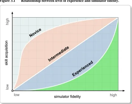
Figure 3.1 Relationship between level of experience and simulator fidelity. ... 43
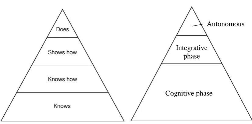
Figure 3.2 Conceptual Clinical competence pyramids. ... 53
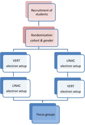
Figure 4.1 Flow chart of the Pilot Study ... 68
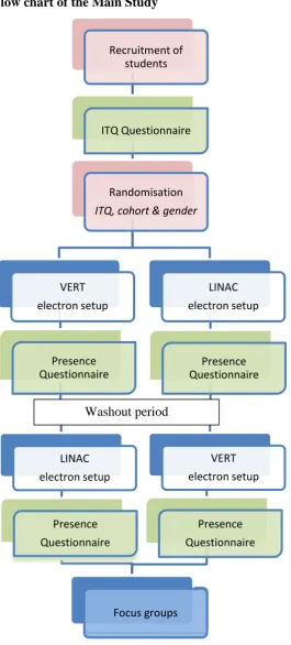
Figure 4.2 Flow chart of the Main Study ... 73
Figure 5.1 Presence scores for the VERT and LINAC simulations. ... 90
Figure 5.2 Comparison of setup components. ... 94
Figure 5.3 Competency score for setups. ... 95
Figure 5.4 Bland Altman plot of overall competency scores ... 99
Figure 5.5 Machine parameter movements during the first 30 seconds. 1st setup. . 103
Figure 5.6 Machine parameter movements during the final 30 seconds. 1st setup. 104 Figure 5.7 Machine parameter movements during the first 30 seconds. 2nd setup. 104 Figure 5.8 Machine parameter movements during the last 30 seconds. 2nd setup. . 105
Figure 5.9 User inactivity. ... 106
Figure 5.10 Periods of user inactivity. ... 107
Figure 5.11 Students utilising multiple movements. ... 108
Figure 5.12 Paired movements used. ... 109
viii
List of Tables
Table 3.1 Drivers for uptake of simulated patient-based education. ... 38
Table 3.2 Typology of simulation methodologies. ... 40
Table 4.1 Presence questionnaires. ... 76
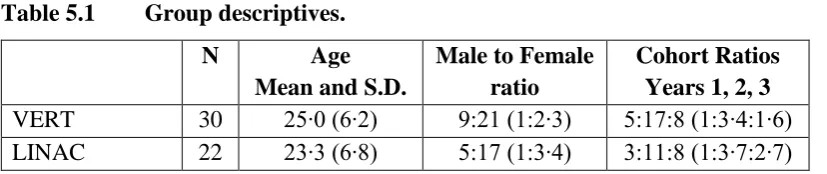
Table 5.1 Group descriptives. ... 85
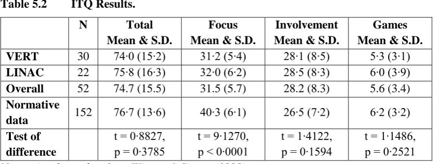
Table 5.2 ITQ Results. ... 86
Table 5.3 Group descriptives (Paired data) ... 86
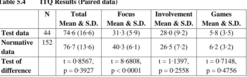
Table 5.4 ITQ Results (Paired data) ... 87
Table 5.5 Correlations between ITQ and PQ scores: All students. ... 88
Table 5.6 Correlations between ITQ and PQ scores: Unit dependent scores. ... 89
Table 5.7 Simulation Presence scores. ... 90
Table 5.8 The effect of gender on Presence score (LINAC). ... 91
Table 5.9 The effect of gender on Presence score (VERT). ... 91
Table 5.10 Correlations between Age and PQ scores: Gender dependent scores. (LINAC) ……….92
Table 5.11 Correlations between Age and PQ scores: Gender dependent scores. (VERT)………92
Table 5.12 Time taken to undertake the electron setups. ... 93
Table 5.13 Comparison of setup competency components. ... 94
Table 5.14 Time taken to undertake the electron setups. ... 96
Table 5.15 Prescott’s contingency table. ... 97
Table 5.16 Prescott’s test for setup competence components. (Paired data) ... 98
Table 5.17 Prescott’s test results for period effects. (Paired data) ... 98
Table 5.18 The effect of gender on setup time. ... 100
Table 5.19 The effect of gender on setup score. ... 101
Table 5.20 Competency score by Cohort. ... 101
Table 5.21 Correlations between inactivity and time to undertake the setup. ... 107
ix
List of Abbreviations/Glossary
app This abbreviation is used in two forms. The first use is an abbreviation for a software application used on an IPad. The second use of app is found in the student quotes where app is a shortened from of apposition and is used in radiotherapy to describe bringing the applicator close to and parallel to the skin, “skin app”.
AR Augmented Reality.
A real-world environment that is supplemented or overlaid with computer-generated sensory input(s).
ARRT American Registry of Radiological Technologists.
Credential organisation for radiographers within the USA whose function is similar to that of the COR, promoting high quality patient care in the field of radiography.
AV Augmented Virtuality.
A virtual world with a degree of reality in it.
COR College of Radiographers
A charitable subsidiary of the Society of Radiographers. Its main objectives are directed towards education and research in the support of the practice of radiography.
CPD Continuing professional development.
Education of health care professionals following completion of formal training. It is an expectation of all radiographers and an HCPC requirement.
CPF Career Progression Framework
Framework initially launched to progress radiography careers that introduced four levels of clinical practice ranging from assistant practitioner to consultant practitioner.
DCR Diploma of the College of Radiographers
Registerable qualification for radiographers that was replaced by an undergraduate degree around 1992/3.
DICOM Digital Imaging and Communications in Medicine
A standard file format developed by The American College of Radiology together with the National Electrical Manufacturers to support the communication of digital image information, regardless of device manufacturer.
DoH Department of Health
x dmax Dose Maximum
The maximum dose point of a beam or treatment. It can be talked about in relative terms when it will be quoted in Gray, but is more often normalized to 100%.
EBRT External Beam Radiotherapy
The most common form of radiotherapy consisting of a beam of radiation directed towards the patient from a distance.
ERCP Endoscopic Retrograde Cholangiopancreatography
A surgical technique to diagnose and treat conditions of the bile ducts and main pancreatic duct.
FSD Focus to Skin Distance
Distance between the focal spot (where x-rays are produced) of the x-ray unit to the skin surface of the patient.
GLM Generalised Linear Model
A flexible generalization of ordinary linear regression that allows for the transformation of the expected response or as a nonlinear regression model for the response.
HCPC Health and Care Professions Council
Regulatory body for health professions including radiographers. Anyone using the title radiographer must be registered with the council in order to practise in the UK.
HEI(s) Higher Education Institute(s)
Usually Universities that can either award a bachelor’s degree or, provides not less than 1 year of training towards gainful employment or, offers a vocational program that provides training for gainful employment and has been in existence for at least two years.
H0 Null hypothesis
A general statement or default position that there is no statistical difference/relationship between two measured phenomena.
ICRU International Commission on Radiation Units & Measurements
A committee that exists to develop and promote internationally accepted recommendations in the field of radiography, including radiation related quantities, units, terminology and procedures.
ITQ Immersive Tendencies Questionnaire
Questionnaire developed by Witmer and Singer to measure presence in virtual environments.
KSF Knowledge Skills Framework
xi LCS Liquid Crystal Shutter
Liquid Crystal Shutter glasses consist of a liquid crystal layer that can turn opaque so as to alternately block one eye. This occurs in
synchronisation with the refresh rate of the screen can give the impression to the brain that the person is looking at a 3D image.
LINAC Linear accelerator
Treatment machine that can produce high energy x-rays and electrons by using radiofrequency waves to accelerate electrons down a linear evacuated tube. Used almost exclusively in the treatment of cancer.
LMPA Low Melting Point Alloy
A range of alloys that can be used to create customisable blocks and cut-outs which are used to shield parts of the radiation beam to protect parts of the body from radiation.
MAR Missing at Random
Data values that are missing that are related to a particular variable, but are not related to the value of the variable that has missing data.
MCAR Missing Completely at Random
Data values that are missing that are independent both of observable variables and of unobservable parameters of interest, and occur entirely at random.
MNAR Missing Not at Random
A values in a data set that is missing for a specific reason (non-random).
NHS National Health Service
A publicly funded health care system that was originally launched in 1948. It is often used to encompass the four publicly funded health care systems in each country of the United Kingdom, but officially it refers just to England.
NRAG National Radiotherapy Advisory Group
A group set up in 2004 to advise the Government on the current position of radiotherapy services in England in order to ensure resources are deployed to best effect and to advise on future directions.
PTSD Post-traumatic Stress Disorder
This is a mental health condition that is usually triggered by either experiencing or witnessing a terrifying event that usually involves physical harm or the threat of physical harm. Symptoms are typically classed into three types, re-experience, avoidance and hyper-arousal.
PIXY® A type of anthropomorphic phantom.
xii PQ Presence Questionnaire
A questionnaire designed to measure presence in virtual environments.
RVC Reality-Virtuality Continuum
Framework suggested by Milgram to look at the concepts of reality and virtual.
SCOR Society and College of Radiographers
Trade union and professional body representing radiographers in the UK.
SOR Society of Radiographers
The professional body and trade union that represents radiographers in the UK.
TLRP The Teaching and Learning Research Programme
The UK’s largest educational research programme that coordinated research and investment in research between 2000 and 2012.
t(d) Welch’s t-test
An adaptation of the standard t-test, which is more reliable when the two samples have unequal variance.
TURP Transurethral Resection of the Prostate
A surgical procedure under anaesthetic that involves inserting a resectoscope into and up the urethra to cut away section(s) of the prostate gland.
VE Virtual Environment
A computer-generated, three-dimensional representation of a real life settingwhich can be explored and interacted with.
VERT Virtual Environment for Radiotherapy
A virtual environment consisting of a radiotherapy treatment room, treatment machine and patient.
VR Virtual Reality
xiii
Acknowledgements
Many people have contributed to my learning experience during the process of my studies at UEL. I am thankful to my two Directors of Studies, Dr. J. Truschel and Professor J. Preston, for their insight, thought provoking questions and guidance for my thesis.
In addition, I would like to acknowledge my PhD thesis supervisor Dr Chadderton for her help and reassurance at various stages of the work.
I would also like to thank my work colleagues in the Department of Radiography, Mrs P. Cherry, Miss C. Raymond-Barker, Mr R. Thorne and Mr R. Khine. They have kindly taken on extra work themselves in order to allow me time to undertake the necessary data collection and subsequent write up. Perhaps most importantly they were always there to bounce ideas off and offer encouragement and support, especially when times were bad.
Dr James Ward, Managing Director, Head of Research and Development and Founder of VERT who found the time to help with the electron field graphic overlays for the VERT phantom.
xiv
Dedication
I am especially indebted to my wife, Helen and my two sons Alan and Peter for allowing me to put part of my life on hold during the last few busy years whilst I struggled with the development of this thesis, which was largely for my own personal development.
Finally in memory of Dave Wood, Therapeutic Radiographer. A student in the 2012 cohort who was tragically killed in a road traffic accident in 2015 shortly after qualifying.
“Well some say life will beat you down. Break your heart, steal your crown. So I've started out for God knows where.
I guess I'll know when I get there. I'm learning to fly.”
David Flinton Page 1 Student no. 0845690
Chapter 1 - Introduction
1.1
Background
Recently there has been a paradigm shift in higher education with students being increasingly placed at the centre of the learning process. This view of learning is diametrically opposed to the traditional approach that tended to consider students as passive receptors of information. Students would typically sit in lecture theatres and not be an active participant in the learning process, (TLRP 2010). The didactic lectures would be the students’ main source of knowledge, there being little additional material to support learning, and students were rarely expected to contribute, ask questions or challenge the expertise of the academic, (McCarthy & Anderson, 2000).
With the move towards a more student centred approach to learning educators are facing the challenge of finding ways of putting the student at the centre of the learning experience. Students have to be engaged in order that the student should learn by doing, and it is in this respect that technology may help. One recent technology that has been identified as potential enhancement pedagogy is that of Virtual Reality (VR), a computer technology that was developed in the early sixties and allows the creation of a Virtual Environment (VE) which users can interact with in real time in an autonomous way.
David Flinton Page 2 Student no. 0845690 Supporting this stance, Bell & Fogler (1995) stated that VR addressed each of the dimensions of those learning styles proposed by Felder & Silverman (1988), i.e. sensory/intuitive, visual/auditory, inductive/deductive, active/reflexive and sequentially/ globally (ibid.: 675). On the sensory/intuitive scale, VR can provide a tangible representation of abstract concepts. VR is highly visual and therefore meets the needs on the visual/auditory scale, and can provide non-verbal auditory stimulus which is important to the realism of the overall experience and can also be used to provide educational sound cues, such as the sound of bonds breaking as atoms interact. On the inductive/deductive scale, VR can provide a medium for exploration and learning by observational experience. Virtual reality is highly active and immersive, the main value of VR is that the subjects are inside the simulation and so are active participants. The final component of Felder and Silverman’s learning styles is the sequential/global scale. VR can address the needs of the global learners by showing the inter-relationships of the real and abstract concepts, as VR can suspend or work within the physical reality so allowing the learner to work within the big picture.
However, as with all technologies, there are potential disadvantages. Whilst VR systems are getting cheaper, they are by no means cheap and some of the devices can be clumsy for the user which can detract from the immersive experience. Other issues that can affect the use of VR and the impression of reality are immersion, presence, cybersickness, lag, and restriction of view. Dalgarno & Lee (2012) broadly support the concept that benefits are offered by VR, but also counselled caution as, although their study provided a strong support for the idea that benefits of VR occurred, the results suggested that the links between the learning tasks and learning reported benefits from the perspective of the user were minimal and not statistically significant. Although presence, the sense of being in the virtual world is considered a cornerstone of virtual reality, Hodges et al., (1994), and Whitelock et al. (2000) express reservations for fully immersive environments as their findings suggest that a subject’s performance can be negatively affected by cognitive overload.
David Flinton Page 3 Student no. 0845690 potential for VR systems to seamlessly integrate imagination and interaction with computer generated reality shown in science fiction television programmes such as Star Trek: The Next Generation; Red Dwarf and films like The Matrix has not yet been achieved. VR can still therefore be considered to be in its infancy and is still principally seen as a training tool, it being primarily associated with skills development, rather than gaining knowledge (Walsh et al., 2010). Proponents might consider this view of virtual reality use as being rather limited and other uses are currently being investigated by various researchers. At present the use of VR in general, and especially these newer areas of study, must be considered as being at an early stage of development rather than an established educational tool.
VR is one extreme on the reality-virtuality continuum (RVC) all of which may have uses in education. The RVC was first proposed by Milgram et al. (1994) and is shown below in Figure 1.1. It has on the left one extreme of an environment consisting only of real objects, and on the right the opposing extreme of virtual environments consisting exclusively of virtual objects, such as those described above. In between these two extremes we have mixed reality, sometimes referred to hybrid reality, realities that consist of a mixture of real and virtual objects. Mixed reality itself can be further divided into Augmented Reality (AR) and Augmented Virtuality (AV). Both AR and AV allow real and virtual objects to coexist in the same space and be interacted with in real time, the difference being that AR is built around a real environment, adding virtual media to the environment, (Ternier et al. 2012) whereas AV is a virtual world with a degree of reality present in it.
Figure 1.1 Simplified representation of a RVC.
Milgram et al. (1994: 283)
David Flinton Page 4 Student no. 0845690 of uses including education, Bower et al. (2014: 1) stating that “Augmented Reality is
poised to profoundly transform Education as we know it.” The main advantage offered by all these methods is that they are able to root the learning in experience. Because of this virtual and mixed reality environments have specific opportunities to be beneficial for children with both mental and physical disabilities, VR minimising the effects of a disability, allowing learning through social participation also potentially improving the child’s quality of life, (McComas, Pivik, & Laflamme, 1998). This method therefore suits itself to both primary education and medical and paramedical fields where experiential learning is important, the reported main advantages of such systems being learning and motivational gains, (Bacca et al. 2014). As with VR it is the immersive nature of the mixed reality systems that is cited as a major advantage, enhancing the student’s educational experience, (Lindgren & Johnson-Glenberg, 2013; Kamphuis et al. 2014).
Outside of the education forum virtual reality is also still being used in the military as a training aid. Its use however, has also expanded into therapies, particularly psychological conditions such as anxiety disorders for example arachnophobia, pteromerhanophobia and post-traumatic stress disorder (PTSD), where patients are exposed to the triggers for their condition, which they gradually become acclimatized to. This has the effect of decreasing their symptoms and enabling them to cope to better when challenged by their anxiety trigger, and in the case of PTSD a change in their fear response, (Opriş et al., 2012).
1.2
VERT
David Flinton Page 5 Student no. 0845690 automatically as they change their position within the “real” room. These variants offer increasing immersion within the virtual world although at the expense of increasing cost.
Initially only two institutions, University Aarhus Hospital (Denmark) and Birmingham City University had invested in the VERT system when the UK government launched the Cancer Reform Strategy (2007). Central to the strategy was the consideration of the predicted demands on the existing service. Currently it is estimated that approximately 1 in 3 people within the UK will develop a cancer sometime during their life and, although modellers believe there will be no change in the age standardised incidence rate of cancers, there will be a change in the actual number of cancers being reported. This increase is due to the aging population and it is predicted that, over a twenty year period covering 2010 to 2030, there will be a 55% and 35% increase in male and female cancers respectively (Mistry et al., 2011). Subsequent to this prediction, the Government allocated £500 million for new and replacement equipment including 167 new linear accelerators (LINACs). In order to prepare the necessary radiographers to staff the new units - and to address the shortfall in radiographers at the time a further five million pounds of capital funding was allocated to provide both Higher Education Institutes (HEIs) and hospitals with radiotherapy departments with the VERT system. The purchase of the VERT systems detailed in the Government report was based on the recommendations of the earlier UK National Radiotherapy Advisory Group (NRAG) report (2007) that highlighted the need to introduce VERT as a means to prepare more radiographers. VERT’s role would be dual fold: firstly, it was proposed that it would combat the high student attrition on radiotherapy programmes by improving the learning experience; and, secondly, it would increase training capacity.
David Flinton Page 6 Student no. 0845690 which the Government invested was untested at the time for the goals for which it was purchased.
1.3
Simulation & Assessment
Simulation and simulators such as VERT allow educators to both train and undertake assessment of students. Simulators can be designed to address a number of clinical scenarios on demand and can be readily available at any time irrespective of patient throughput. According to Haycock (2011) the main potential offered by simulators is that unlike real life they can provide a number of reproducible cases for assessment. Also in the case of computer driven simulation they also have the potential to provide an objective rather than subjective evaluation of procedural skills so providing a standardized platform for assessment. Because of these reasons health educational leaders are suggesting that simulation based assessments are essential, however before a tool is widely implemented, the evidence of its usefulness and validity needs to be established, (Brydges et al., 2015).
Assessments within higher education can essentially be divided into two broad types, formative and summative. The former is essentially assessment for learning, intended to provide the teacher and more importantly the student with feedback which is essential for improving knowledge and skills acquisition, (Schute, 2007) and therefore can be considered as a process to actively engaging students in their own learning, (Looney, 2011). Despite this potential for assessment influencing learning the evidence of any effect is sparse, (Schuwirth & van der Vleuten, 2011); for example, Grosas et al., (2014) found that many students fail to acknowledge that formative testing is a learning process.
Summative assessment is often referred to as assessment of learning. They can resemble formative assessments, but are typically placed at the end of a learning cycle rather than during. However, the essential difference is in the use of the evidence derived from the assessment procedure. In this instance the assessment typically does not contribute to the students’ learning, and there is no feedback into the teaching of the material, (Gardner, 2012).
David Flinton Page 7 Student no. 0845690 constructed construct between both the learners and assessors/teachers, (Boud & Soler, 2015). Assessments are now considered to have a multiple emphasis, being expected to fulfil several purposes regardless of whether they are predominantly summative or formative in function. This multiple emphasis on assessments consists of the following components, identified by (Boud 2010: 253-254).
the assessment actively engages students;
it is comprised of authentic activities;
involving students in the design of assessments;
integrating tasks to give an assessment that takes a holistic view of what they have been learning and know what they need to know;
allows students to engage with model answers and practice so that they can see improvement in their work;
working with peers to foster team work;
giving and receiving feedback.
Trying to effectively meet all these components when designing an assessment creates tensions and compromises, and because of the importance of assessment, the task design becomes very important, (Carless, 2009). This view is supported by Ramsden (2003) and Gibbs and Simpson (2004) who both emphasise the importance of finding a suitable assessment method to ensure that the students take the correct approach to the learning tasks and have quality student engagement.
David Flinton Page 8 Student no. 0845690
1.4
Problem Statement
Since the installation of VERT facilities at the researcher’s host institution in 2008, the challenge of the programme team has been to look at what is offered by the new system and to integrate the VERT system into the curriculum in order to aid and enhance both education and training. Although the technology was originally given to the HEIs, the course teams need to explore its potential to improve teaching and learning. In other words, a social constructivism stance is being taken by the lecturers and students with both being given opportunities to adopt or reject the various uses that they can see for VERT regardless of the reasons for which it was supplied. One potential area that has been identified where VERT may be of use is in electron setups. Within radiotherapy, most treatment setups use predefined positions for the collimator and gantry alignment but others, such as those generally used for skin treatments, require the positioning of an applicator parallel to the surface of the patient “skin apposition”. The accuracy of this type of “free” setup is determined by the expertise and skill of the radiographer, requiring very good hand eye coordination and 3D spatial ability and so requires a different set of skills to most treatment setups. These techniques are not common and are sometimes pre-calculated by planning radiographers, with the implication that learning opportunities and occasions to obtain competencies on this type of setup are limited. VERT potentially could be used in the preparation of students for a competency based assessment by allowing the students to learn and practice simulations of electron setups or alternatively as a method of competency based assessment for the “free setup” of electron fields. The stimulus for consideration of the latter not only being the relative infrequency of this type of setup, but also being able to standardise the assessment, not only in terms of standardising the setup, but also to reduce the perceived variation in marks between assessors and hospital sites which had been highlighted as an issue of concern by the students their course feedback in previous years.
David Flinton Page 9 Student no. 0845690 As well as looking at the use of VERT to measure competency, the study also investigated if the system was acceptable to students as a method of assessment and asked how they felt the system should be used.
1.5 Aims and Objectives
Simulation is a key tool in learning and assessment and is used frequently in many health based programmes. The overall focus of this study was to compare the use of two simulation methods to investigate firstly if similar results were obtained on both simulations and secondly to discover the students’ perceptions of both methods.
In order to investigate these overarching aims the following research questions were answered. The first six aims were investigated using quantitative data and as such the enquiry was based on looking at the null hypothesis (H0) where possible. The final two aims were investigated by looking at the qualitative data.
1. To discover if Immersive tendency predicts the feeling of presence in both simulators.
H01 There is no relationship between immersive tendency and presence.
2. To assess if Presence will be higher on the LINAC simulation compared to the VERT simulation.
H02 There will be no difference in Presence scores between the two simulation methods.
3. What student characteristics moderate the presence scores? H03 Age and gender will not affect the presence score.
4. Do the outcomes of the two simulated setups utilising different equipment agree with each other?
H04 There will be no difference in simulation setup parameters for the two simulation methods.
David Flinton Page 10 Student no. 0845690 H05 Cohort, age, gender and immersive tendency will not affect the simulation
score.
6. Do participants utilise the same cognitive process on both systems?
7. What do students think about the appropriateness of both methods as an assessment tool?
8. What do students think about the use of VERT as a method of learning?
1.6
Significance of the Study
Most of the published work considers the use of VR systems as an educational tool for the delivery of information to the user, the VE being used as a learning environment allowing users to develop and practice skills rather than as a means of measuring competence. This study undertakes the next logical step, if research indicates that we can train students using virtual reality and VERT, can we assess the students’ competency via this technology as well?
1.7
Positionality and Reflexivity
Bartell & Johnson (2013) state that we cannot separate ourselves from whom we are, and that what we are has been influenced and shaped by factors such as race, gender, class as well as what society has placed upon us and our own life experiences. All researchers are positioned whether they acknowledge it or not, and their positionality will affect how they perceive their role and practice, which is what makes it so important for qualitative studies where the researcher is part of the process. Part of the ongoing debate on positionality is that of the concept of insider/outsider positionality, and the apparent advantages and disadvantages offered by each status such as insiders possessing deeper insight into field at the expense of being more biased, whereas an outsider would be more objective, possibly at the expense of lack of understanding, (Chavez, 2008).
David Flinton Page 11 Student no. 0845690 preferences reflections and preconceptions.” Polit & Tatano Beck (2012: 740), so as to offer a transparency to the methods used.
The majority of the researcher’s work to date has utilised quantitative methodologies and have been firmly positioned in positivism. This position predominantly stems from earlier experiences; the researcher having first studied biological sciences and worked for a period as a medical laboratory scientific officer and microbiologist, all of which was based firmly in rigour of method and standardised interpretation of results. Because of this background there is a natural tendency of the author to work with quantitative methods, presumably due to experience, familiarisation and security over any actual methodological requirement.
However, positionality goes beyond an understanding of just one’s self and it must be acknowledged that the students themselves are positioned and that the researcher’s position with them in not just based on the experience of researcher’s past, but on other factors. As the study was undertaken in the researcher’s host institution there would be “insider” positionality, which would be varied for different students. The researcher has different roles with different students, personal tutor and link lecturer to some, lecturer and programme manager to others. Depending on which role the student felt was dominant could affect how the student would perceive the researcher, affecting the researcher/subject interaction on a number of levels. Firstly it might have led to a selective bias based on expectation, which could have posed a challenge to the research process. Secondly the different relationships could negatively affect the data collection process, the students who knew the researcher better being more relaxed in the researcher’s presence during data collection, so finding it easier to perform the tasks and talk more freely in the focus groups than other students so impacting on the nature of the narrative form. This familiarity and closeness to certain student groups did appear to affect the sample. Not all students volunteered for the study, but all the students who were on the researcher’s clinical sites did, perhaps because they were more sympathetic to the researcher’s personal position or alternatively because of the belief they may be more disadvantaged in some way if they didn’t volunteer.
David Flinton Page 12 Student no. 0845690 implied that they felt that that there was an expectation in undertaking the study, rationalising during the study that the expectation of the research was to have VERT succeed as an assessment tool during the work-down after the simulation set-up. These comments came from students close to the researcher and were made outside of the data collection process. From a quantitative perspective this could be considered as an overt manifestation of bias within the study. The most likely bias that would be introduced in this situation is a type of bias known as expectancy bias or the “Hawthorne effect”. The Hawthorne effect is a known issue with any participatory observational research, (Coombs, 2003), the effect occurring when subjects adjust their behaviour simply because they are being observed, and is time dependent, the effect fading with familiarisation of the observation over time, (Walker, 2005).
The Hawthorne effect is rarely quantified as part of the research process, (Fernald et al. 2012) however, a number of established methods exist in order to mitigate or at least reduce the effect. Oswald, Sherratt & Smith (2014) suggest two major mitigating techniques based on existing work, firstly building a relationship and establishing a rapport with the subjects being observed. In this work this relationship and rapport was already established and it is because of this rapport that this specific element of the Hawthorne effect came to light. The second suggestion is the process of triangulation, which is possible in this instance because of the study design which allows a cross check of the findings between the two different types of data collected. In this case the results show students performing a lot poorer on VERT despite the students’ expectation and from the qualitative feedback the feeling that the VERT simulation was not as good as the LINAC based one. The likely inference from this is that the direction of difference found within the study is unaffected and correct, but that the actual degree of difference being observed might not be as large as in actuality.
David Flinton Page 13 Student no. 0845690 data collection methods particularly in the quantitative data collection where checklists were used for the observation of the participant’s setups. In the focus groups the researcher was cautious to avoid his influence in the research process and used interview guides to help ensure that both the relevant data were collected and the researcher’s involvement in the construction of the data and its meanings was reduced.
1.8
Outline of the Thesis
This thesis is presented in eight chapters. In Chapter 1 the potential of VR for educational assessment is posed, specifically the use of VERT as a competency based assessment tool for electron setups in radiotherapy. The specific research questions are presented and an indication of whether the questions would be answered through analysis of quantitative or qualitative data.
Chapter 2 gives a short background introducing the reader to the profession that is radiotherapy. First the chapter looks at the development of both the profession and the education of its members. In the latter sections of this chapter the reader is given a brief overview of radiotherapy, especially that of electron treatments in order to help understand the clinical context of the study.
Chapter 3 reviews the relevant literature on virtual reality and explains what VR is. Firstly it considers the types of virtual reality and how virtual reality and other methods of simulation are being utilised in education, with particularly emphasis on their use in health care education. The chapter then returns to VR and considers the key features of the systems, fidelity, immersion and presence considering how these factors impact on learning. The final sections in this chapter consider assessment of competence and simulation, especially VR considering the validity and reliability of VR as an assessment tool.
David Flinton Page 14 Student no. 0845690 procedures and data analysis techniques to ensure the reliability and validity of the study. The chapter concludes with a section on the positionality of the researcher within the study.
Chapter 5 presents analyses of the quantitative data collection, based on all the empirical data collected comparing the use of both simulation methods. In order not to waste data this was done firstly by comparing the first use VERT and LINAC data allowing an unpaired analysis. A second paired analysis was then undertaken on subjects who completed both simulations.
Chapter 6 focuses on presenting the data from the focus groups using thematic analysis. This section focuses on the students’ perceptions of the two simulations, the use of the two simulations and their appropriateness as an assessment method considering the four identified themes, equipment; reality; the learning opportunities afforded by the VERT system; and assessment of competence.
Chapter 7 discusses the results presented in Chapters 5 and 6 relating them back to the study’s aims whilst attempting to triangulate the data looking for reasons in the qualitative data to explain the quantitative data more fully.
David Flinton Page 15 Student no. 0845690
Chapter 2 - Radiotherapy and Education
2.1
Introduction
Radiography is a relatively new profession, having come into existence soon after two major discoveries in the late nineteenth century. Firstly, on November 30th 1895, Wilhelm Roentgen announced the discovery of x-rays, (Lederman, 1981) and a few months later, on the 2nd of March 1896, Henri Becquerel reported the discovery of radioactivity, (Blaufox, 1996).
The medical profession quickly realised the potential of the two new types of radiation both for diagnosis and treatment, with the first treatment reportedly being undertaken by Grubbé in January 1896, (Orton, 2013). In order to try and organise radiological work the Roentgen Society was established in England in 1897, permitting - “after vehement discussion” (Pasveer, 1989: 364) - both medical and non-medical members to join, in order to achieve a non-clinical bias. This however was opposed by some medical members who split from the Society in 1902 and founded the British Electrotherapeutical Society, which became, in 1907, a section under the Royal Society of Medicine.
The use of ionising radiation for the treatment of disease (Radiotherapy) was originally undertaken on many different types of conditions including tuberculosis, excessive sweating and ringworm, but modern use is restricted almost exclusively to malignant tumours. This restrictive use of radiation is due to both an increase in the understanding of the radiobiology of x-rays and radioactivity, and the recognition of the potential side effects of radiotherapy treatments such as tumour induction, (Kunkler, 2003).
David Flinton Page 16 Student no. 0845690
2.2 Professionalization and Institutionalisation
With the almost instantaneous recognition of the medical potential of x-rays, implementation of this new technology occurred simultaneously within hospitals throughout the world. The next few years saw a rapidly expanding clinical demand for x-ray treatments, Suit & Loeffler (2011) reporting 923 radiation treatments being performed at the Massachusetts General hospital in 1924 which increased to 1,220 a year later.
In the early years of radiography both the purchase and operation of x-ray equipment was unregulated, and as equipment was purchased hospitals called on nurses, doctors, porters and in some instances handymen to use the equipment, (Witz, 2004). Brecher & Brecher (1969) observed in a US context that, in 1910, radiography “was staffed primarily with younger men, most of whom were still in school or medical school when Rontgen's discovery was announced” (ibid.: 104). Many of these young men “had begun working with the X-rays in 1896 or 1897 as physicists, engineers, electricians, photographers” or technicians and had qualified as doctors “specifically for the purpose of qualifying as radiologists” (ibid.).
The process of professionalization was disrupted by the outbreak of the First World War when, “anybody, whether lay, physician or engineer, who was in possession of the apparatus performed radiological work for war purposes” (Pasveer, 1999: 366), but resumed post-war when: “established radiologists started pleading for good practical and theoretical education for X-ray workers in the medical as well as the physical sciences, complaining about the low status of radiology and the inadequate location of Rontgen departments in hospitals” (ibid.). Eventually new professions evolved; on the diagnostic side the radiologist and the diagnostic radiographer; and on the therapeutic side the radiotherapist and therapeutic radiographer. The radiologist and radiotherapist professions were subsumed into the medical fraternity whereas for the two new radiography professions the Society of Radiographers was formed in 1920 to give professional status to the non-medical workers in the fields of radiography.
David Flinton Page 17 Student no. 0845690 qualification purposes who awarded the Diploma of the College of Radiographers (DCR) allowing state registration. However, the professional role of radiographers, particularly radiotherapy radiographers is not well understood by the public and radiotherapy itself has a low public profile, (YouGov, 2011). Sim & Radloff (2009) also suggest that radiographers have a lack of professional recognition and professional respect from other healthcare practitioners.
In 1977 the SOR became registered as a trade union and a new charitable company set up called the College of Radiographers to take over the educational and professional responsibilities (ibid). At about the same time the debate started about degree education for radiographers, one of the original proposals being that the degree course should be 4 years 5 months long and not only involve a degree qualification from the University, but also the DCR in order to confer state registration, (Jordan, 1995). The debate continued within radiography which together with fellow paramedical professions began to call for graduate courses. This proposal was opposed by the Department of Health and Social Security (DHSS) at the time stating that,
“there seems only the most limited scope in radiography for the academic orientation of degree training….. it would be difficult to keep a balance between academic and vocational training in radiography…..without further lengthening the training, with no real benefit to the NHS.” DHSS, (1979: 1).
David Flinton Page 18 Student no. 0845690 different institutions had the freedom to set their own curriculum content rather than use that set by the COR.
The world’s first radiography journal was the “Archives of Clinical Skiagraphy,” first
published in May 1896, (Mould, 2011). In the following years the number of journals devoted to radiography and radiotherapy increased, but the journals were aimed primarily at oncologists, radiologists and physicists rather than radiographers. With the move to a graduate programme the profession recognised the need for its own knowledge base as the vast majority of the existing knowledge base for radiography was built on evidence produced by medical practitioners and physicists (Nixon, 2001). As part of the effort for radiography to be seen as having a professional standing a peer reviewed journal aimed at radiographers was introduced in 1995 in order for radiographers to have a vehicle to publish their own research.
David Flinton Page 19 Student no. 0845690
2.3
Radiography Education
“Radiography education has followed the lead provided by nurse education whereby initial and post-registration provision are now located in institutions of higher education”, (Castle, Holloway & Rage, 1998: 333). Prior to radiography becoming a graduate entry profession, the distinction between radiographer and radiologist as drawn by Mackay, (2003) was that, “Radiographers were educated for two or three years up to diploma level whereas radiologists were medically qualified and had several years’ experience in medicine or surgery before becoming radiologists” (ibid.: 93).
Middlemiss (1973) noted the potential need for the development of the radiographer’s role due to a potential shortage of consultant radiologists, suggesting an advanced radiologic technologist who could, “perform some of the procedures and undertake some of the responsibilities at present performed or undertaken by radiologists” (ibid.: 804).
This shortfall in consultants did indeed come to pass, and the COR was keen to further develop the role of the radiographer. This brought about role extension possibilities such as radiographic reporting. It also brought about a number of changes to the staffing model in the UK and the introduction of a four tier structure, which is now referred to as the Career Progression Framework (CPF). Qualifying students would enter the profession as practitioners and could progress to advanced practitioners (MSc level) who would be autonomous (DoH, 2003) and eventually consultant practitioners (PhD level) each of which are state registered titles and regulated by the HCPC. A new level of assistant practitioner was introduced below the practitioner level which would be a sub-professional level that could only work under the supervision of a practitioner.
In order to support the structure Universities had to produce programmes for assistant practitioners and as the CPF stipulated a continuum between levels they also had to produce a route from assistant practitioner to practitioner status as a means of widening participation for the profession, (DoH, 2003). Also, in order for practitioners to progress through to advanced and consultant levels post graduate modules and programmes needed to be developed up to Doctoral level to suit this purpose.
David Flinton Page 20 Student no. 0845690 maintain and develop throughout their career the capacity to practice safely. Until 2005 there was no requirement for radiographers to undertake any personal development post qualification and there was a general ambivalence towards Continuing Professional Development (CPD), (Henwood, Yielder & Flinton, 2004). However, in 2005 for UK radiographers it was legislated as mandatory for continuing registration with the HCPC, and a sample of registrants are checked every two years to ensure compliance. Despite this legislative change the attitude of radiographers towards CPD activity remains largely unchanged, (Henwood & Flinton, 2012). Both CPD and the CPF could also be said to be key in the professional nature of radiography as both promote the application of advanced learning and expertise to provide a service to clients, which according to Madden & Mitchell (1993) are key concepts of a profession.
Presently within England and Wales most students who become eligible for HCPC registration upon qualification pursue a three year therapeutic radiography programme leading to a BSc degree, although a small number of institutions do offer a two year post-graduate route to registration. Although the curriculum varies between the numerous institutions most programmes essentially consist of a roughly 50:50 split between academic and clinical time. The academic learning is facilitated by radiographers with higher degrees and a teaching qualification, whereas the clinical learning is facilitated by the clinical staff, most of whom have no teaching qualification. The clinical part of the curriculum at City University London as with most other institutions offering radiography programmes takes the form of a portfolio that prescribes the objectives and competencies that students need to have achieved during each year of the students’ clinical education. The objectives and competencies are derived from HCPC and SCOR documentation.
2.3
The Practice of Radiotherapy
David Flinton Page 21 Student no. 0845690 EBRT is usually delivered by one of two different types of machine. Kilo-voltage units which, due to their limited penetration, tend to only be used for small superficial tumours such as skin lesions (Flinton, 2009), and the linear accelerator (LINAC) which is currently used for most treatments and is the unit utilised in this study.
The workflow for most radiotherapy treatments involves the patients undergoing four distinct stages, localisation, planning, treatment and verification. During each of these stages, although the patient will be under the care of a multidisciplinary team, the professionals with most patient contact would be the therapeutic radiographers.
Localisation is the first step in the treatment pathway and the first consideration is the patient position and whether immobilisation is needed (Griffiths & Short, 1994). The optimum patient position is important as it should allow entry and egress of the radiation beams whilst being reproducible and allowing the patient some degree of comfort. Some patients, particularly patients with head and neck tumours may also need immobilisation, and devices such as personalised thermoplastic immobilisation shells are necessary to help the patient maintain the correct position throughout their treatment (Barrett, Dobbs & Roques, 2009). The next part of the process is to find the tumour location, which is usually done via a Computed Tomography (CT) scanner. The CT scan will provide cross sectional images of the body which can provide information on the tumour size and its location in relation to the body’s normal organs, reference points and the surface of the patient (ibid).
David Flinton Page 22 Student no. 0845690 Treatment is usually undertaken on a LINAC using the patient position and beam parameters determined during the localisation and planning stages. The treatment is usually delivered over an extended time period, which is both tumour and site dependent, the dose being split into smaller daily doses called fractions. The basis of this approach lies in understanding the basic radiobiological principles called the 4 “R’s”, each of which will affect the efficacy of treatment, (Mitchell, 2013). It must be acknowledged at this point that some authors such as Steel, McMillan, & Peacock (1989) suggest that a fifth “R”, Radiosensitivity be included. Radiosensitivity acknowledges the inherent susceptibility of cells, tissues, organs to the harmful effect of ionizing radiation, however unlike the other “R’s” this R does not take place in the inter-fraction interval and is not considered further here.
Firstly, fractionating the dose allows cells to Repair between treatments, because tumour cells have less capacity to repair themselves compared to normal tissue. The radiation damage to the tumour cells builds up quicker leading to cell death whilst normal tissues recover (Nieder & Baumann, 2011). Cells also show a difference in sensitivity to radiation in different parts of their life cycle, and giving the dose in small fractions means that cells will be irradiated whilst in different parts of their life cycle i.e. they will Redistribute themselves between treatments, (McMillan, 2002), so increasing the effectiveness of the radiation.
Cells are more sensitive to radiation if well oxygenated (ibid). Small tumours are generally well oxygenated, but as tumours grow they tend to outgrow the blood supply leading to areas of poorly oxygenated cells, typically towards the middle of the tumour mass. With fractionated treatment, the outer oxygenated cells will be more sensitive to the radiation and they will be killed relatively quickly. This shrinks the tumour mass allowing the blood vessels to get closer to the poorly oxygenated area and so start to Re-oxygenate cells making the previously radio-resistant cells more sensitive to the radiation and so improving patient prognosis, so increasing tumour cell kill, (Brown, Carlson & Brenner 2014).
David Flinton Page 23 Student no. 0845690 death increases. This is desirable for the normal tissue being irradiated, but it also means that during the treatment the tumour cells will also repopulate and this can affect the treatment outcome (Fowler, 2010). The amount of tumour repopulation therefore has to be factored into the treatment dose and overall treatment time and the effect of any delays to the treatment must also be considered, (Wyatt, Jones & Dale 2008).
As the dose is being given over a protracted time, in some cases over 7 weeks, it is imperative that each day the fields are precisely placed in order that the dose is delivered accurately to the tumour. This needs to be checked on a frequent basis, a process called Verification which is the final stage of the process. Verification exists in two distinct forms, geometric - “is the dose going to the right place?” - and dosimetric - “is it the right dose?” It is currently recommended that geometric verification be undertaken for all
megavoltage external beam x-ray treatments, (Royal College of Radiologists et al., 2008) and is usually achieved by imaging the patient with x-rays whilst in the treatment position before treatment is given.
LINACs have the potential to produce two different types of radiotherapy beam, x-rays and electrons. X-ray beams are usually used to treat deep seated tumours, whereas electron beams are used for superficial treatments as their penetration is tissue is limited as explained in the following section.
2.3.1 Electron Beams
David Flinton Page 24 Student no. 0845690 Figure 2.1 Central axis depth doses of x-ray and electron beams.
(Herer et al., 2009: 532).
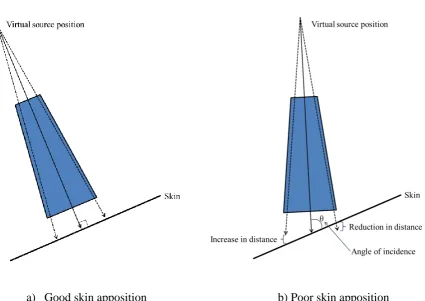
David Flinton Page 25 Student no. 0845690 Figure 2.2 Electron setup and skin apposition
a) Good skin apposition b) Poor skin apposition
David Flinton Page 26 Student no. 0845690 One problem with calculating the effect of changing distance on the beam is that the electron’s source position is a point in space that has to be found by experimentation rather than a physical point such as the electron scattering foils and because of this is known as the “virtual source position” (Strydom, Parker & Olivares, 2005). Changes to the dose as a result of the changes in distance are relatively small, but other effects, primarily to the penumbra of the beam can also be produced. The penumbra is an area of unwanted partial dose at the edge of the beam. At increased distances the penumbra for electron beams is larger nearer the skin’s surface due to the sideways scatter of electrons before reaching the surface of the patient so degrading the beam edge, (Arunkumar et al., 2010). At reduced treatment distances the effect is reversed and the penumbra is smaller, (Washington & Leaver, 2009). Figure 2.3 shows two beam profiles, the penumbra is the dose at the side of the beam represented by the vertical lines which can be seen is wider with increasing distances meaning that more normal tissue will be irradiated.
Figure 2.3 Penumbra variation for 2 different electron beams at three distances.
6MeV Energy beam 20MeV Energy beam
Arunkumar et al., (2010: 209)
David Flinton Page 27 Student no. 0845690 dmax changing the dose at depth also changes which is represented by the variation in the shape and slope of the line beyond dmax.
Figure 2.4 Depth dose curves for two electron energies and obliquity angles.
Adapted from Khan et al., (1985)
As irregular surfaces are a frequently encountered situations in radiotherapy corrections can be made to the dose at given points using the formula below, which accounts for both the change in distance and the obliquity.
𝐷(𝑆𝑆𝐷𝑒𝑓𝑓+ 𝑔, 𝑧) = 𝐷0(𝑆𝑆𝐷𝑒𝑓𝑓, 𝑧) (
𝑆𝑆𝐷𝑒𝑓𝑓+ 𝑧 𝑆𝑆𝐷𝑒𝑓𝑓 + 𝑔 + 𝑧)
2
× 𝑂𝐹(𝜃, 𝑧)
Taken from Strydom, Parker & Olivares (2005: 291)
Where: SSD eff is the effective source to skin distance. Note this is different from the FSD as indicated by the distance light as it is dependent on the virtual source position, which in turn is dependent on the energy and cut out used. g is the stand-off, z is the depth in the patient, 𝜃 is the obliquity angle between the tangent to the skin surface and the beam central axis. D0 (SSD eff, z) is the dose at depth z for a beam incident normally on a flat
David Flinton Page 28 Student no. 0845690 The International Commission on Radiological Units and Measurements (ICRU) recommends that for electron beams the dose delivered to a tumour should be within 10% of the prescribed dose. The distance, skin apposition and angle of incidence have only a small effect on the dose compared to other factors such as the presence of air cavities and density changes, but these other factors are beyond the control of the radiographer whereas distance, skin apposition and obliquity are.
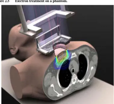
David Flinton Page 29 Student no. 0845690 Figure 2.5 Electron treatment on a phantom.
Adapted from: Northwest Radiation Oncology (2013)
The ability to fit the light beam to the skin marks at the correct treatment distance, and the amount of skin apposition can be influenced by the expertise of the treatment radiographer. All of these aspects need a good 3D perception. It is because of this and the nature of the electron set-up that this form of treatment is the focus of this study.
2.3.2 Patient care
David Flinton Page 30 Student no. 0845690 of the patient’s treatment. The radiographer must therefore not only be able to operate the equipment effectively, but also develop communication skills and a compassionate manner in order to handle these situations and give most benefit to the patient, (Martin & Hodgson, 2006).
2.4
The simulations
This section describes the two simulations, Virtual and LINAC based, explaining the similarities and differences between the two.
2.4.1 The Phantom
Both simulations used in this study represented a single patient with two electron fields, both on the chest wall. Field 1 was placed on the left anterior aspect of the chest wall and Field 2 on the right anterior aspect, see Appendix IX. Both fields were 10x7cm as currently this is the only size electron field that can be created in VERT. Field 1 was the simpler setup as the surface of the phantom is relatively uniform at this point, there being just a slight angling of the chest wall. Field two was more complicated as the surface of the phantom changes more in this area and the direction of slope changes, particularly in the supraclavicular area. There is also a further potential problem with Field 2 in that the neck and head can interfere with the applicator placement and this adds a further complexity to the setup.
David Flinton Page 31 Student no. 0845690 simulations students on the LINAC prior to commencement of the simulation were instructed to complete Field 1 first.
2.4.2 The equipment
The treatment machines used were identical for each setup, both being Varian 2100IX linear accelerators. Again the position of the start point was identical, the couch being lowered to its lowest point (-65cm), the lateral movement being set to 0 and the longitudinal position being 1.5cm.The collimators and gantry angle were both set to 0°. These positions are common start points for most patient setups. In both instances the electron applicator and lead cut out was in place before the subject started the setup. The handsets the subjects used to control each unit were identical and had identical functions allowing the subjects to control the couch and gantry movements as well as other functions such as toggling the room lights, FSD light, field light and lasers on and off. The only other physical difference in the equipment was that on VERT subjects had to wear LCS glasses in order to view the projected image in 3D. The glasses had a wire to a battery pack and sensor mount that the students clipped to a pocket. This equipment was not worn in the LINAC room.
Both simulations contained the setup parameters displayed on the LINAC centre stand. This information was repeated on monitors in the LINAC room and on the right hand side of the screen on VERT. The main difference in the equipment was that on VERT the equipment was virtual whereas in the LINAC room it was real. The couch also has controls on it that allowed movement of the couch top in the verti