Copyright © 2001, American Society for Microbiology. All Rights Reserved.
Relatedness of
Streptococcus suis
Isolates of Various Serotypes
and Clinical Backgrounds as Evaluated by Macrorestriction
Analysis and Expression of Potential Virulence Traits
ACHIM ALLGAIER,1RALPH GOETHE,1HENK J. WISSELINK,2HILDE E. SMITH,2 ANDPETER VALENTIN-WEIGAND1*
Institut fuer Mikrobiologie und Tierseuchen, Tieraerztliche Hochschule Hannover, Hannover, Germany,1and
Department of Bacteriology, Institute for Animal Science and Health, Lelystad, The Netherlands2
Received 14 July 2000/Returned for modification 29 September 2000/Accepted 7 November 2000
We evaluated the genetic diversity ofStreptococcus suis isolates of different serotypes by macrorestriction
analysis and elucidated possible relationships between the genetic background, expression of potential viru-lence traits, and source of isolation. Viruviru-lence traits included expression of serotype-specific polysaccharides, muramidase-released protein (MRP), extracellular protein factor (EF), hemolysin activity, and adherence to
epithelial cells. Macrorestriction analysis of streptococcal DNA digested with restriction enzymesSmaI and
ApaI allowed differentiation of single isolates that could be assigned to four major clusters, named A1, A2, B1, and B2. Comparison of the genotypic and phenotypic features of the isolates with their source of isolation
showed that (i) theS. suispopulation examined, which originated mainly from German pigs, exhibited a genetic
diversity and phenotypic patterns comparable to those found for isolates from other European countries; (ii) certain phenotypic features, such as the presence of capsular antigens of serotypes 2, 1, and 9, expression of MRP and EF, and hemolysin activity (and in particular, combinations of these features), were strongly associated with the clinical background of meningitis and septicemia; and (iii) isolates from pigs with meningitis and septicemia showed a significantly higher degree of genetic homogeneity compared to that for isolates from pigs with pneumonia and healthy pigs. Since the former isolates are considered highly virulent, this supports the theory of a clonal relationship among highly virulent strains.
Streptococcus suisis a major cause of meningitis, septicemia, arthritis, and bronchopneumonia in young pigs and can cause meningitis in humans (1, 2). Effective control of the disease is hampered by the poor knowledge about its epidemiology and pathogenesis. Since many healthy pigs harbor S. suis as an “early colonizer” (6, 18), identification of virulentS. suis iso-lates is of particular importance. This is, however, complicated by the pathogen’s extreme diversity, in particular with respect to its virulence.
Phenotypic markers used to distinguish highly virulent and avirulent isolates include the presence of serotype-specific cap-sular polysaccharides (25), expression of muramidase-relased protein (MRP) (27, 35) and extracellular protein factor (EF) (35), and hemolysin activity (10, 11, 16). The role of these factors as virulence markers, however, is still unclear. The capsular polysaccharides are the basis for classification into serotypes (of which 35 are currently known) and have recently been shown to prevent phagocytosis and, thus, have been pro-posed as an important virulence trait (25). Serotype 2 is con-sidered the most dominant one among highly virulent strains, but disease is also frequently caused by strains of other sero-types (24, 33, 36), suggesting that serotype-independent viru-lence markers must exist. MRP and EF have previously been found to be strongly associated with highly virulent isolates in
Europe (35, 36) but do not appear to confer virulence directly, as recently demonstrated by the use of gene knockout technol-ogy (28). Hemolysin activity has been characterized and asso-ciated with virulent strains, but its in vivo expression does not seem to correlate with virulence (11, 12, 15). Adherence to epithelial cells has also been suggested as a virulence trait (9), and adhesins that recognize erythrocytes have been found to induce opsonizing antibodies in mice (32). However, studies with adhesins were limited to a very few strains, and informa-tion concerning the adherence mechanisms and the possible role of adhesins in the virulence of the bacterium is yet very poor.
Different molecular typing methods such as ribotyping have been evaluated in comparison with conventional phenotyping, indicating that the latter is of limited value for epidemiological analyses (12, 21, 23, 25, 29). Results of these studies also revealed an association between virulence and ribotype, thus demonstrating that a close relationship between highly virulent strains is plausible (23, 26, 29). Nevertheless, most of these studies were restricted to serotype 2 isolates, and the hypoth-esis about a clonal lineage or relationship of highly virulent strains is still under debate (4, 12, 13, 19).
The purpose of the present study was to elucidate the ge-netic diversity within anS. suispopulation belonging to various serotypes, particularly by comparing isolates from pigs with invasive disease, i.e., meningitis and septicemia, versus isolates from pigs with pneumonia and healthy pigs. For this, we stud-ied a collection of 99 isolates, mostly recovered from German pigs, by assessing possible relationships between the genetic background, different phenotypic markers (serotype, expres-* Corresponding author. Mailing address: Institut fuer
Mikrobiolo-gie und Tierseuchen, Tieraerztliche Hochschule Hannover, Bischof-sholer Damm 15, 30171 Hannover, Germany. Phone: 49-511 9537362. Fax: 49-511 9537697. E-mail: [email protected].
445
on May 15, 2020 by guest
http://jcm.asm.org/
sion of MRP and EF, hemolysin activity, adherence to epithe-lial cells), and clinical backgrounds.
MATERIALS AND METHODS
If not stated otherwise, all chemicals were purchased from Sigma (Munich, Germany).
Bacteria.S. suisisolates had been randomly collected in the course of routine diagnostic procedures from tissues of healthy and diseased pigs over a period of 3 years (1996 to 1998). Most pigs were 4 to 12 weeks old and from different farms located in different geographic regions in (especially northern) Germany. Ac-cording to the source of isolation and clinical background, the isolates were assigned to three groups, i.e., (i) those from pigs with meningitis, septicemia, or arthritis (isolates from brains and joints of animals with respective clinical symp-toms [also referred to as meningitis-septicemia isolates]), (ii) those from pigs with pneumonia (S. suiswas isolated from lungs, either as a pure culture [40% of all pneumonia isolates] or in association with other respiratory pathogens such as Pasteurellaspp. andBordetella bronchiseptica[60% of all pneumonia isolates]), and (iii) those from healthy pigs (isolated from swabs of the upper respiratory or the genital tract). For comparison purposes, a number of reference and control strains were included, such as the suilysin control strain P1/7 described by Jacobs et al. (15), serotype 2 reference strain Henrichsen S735 (DSM 9682; German Culture Collection, Braunschweig, Germany), serotype 1 reference strain Hen-richsen S428 (DSM 9683), and MRP-EF control strains D282, T15, and 4005 (34). Bacteria were isolated on blood agar plates, identified asS. suisby standard biochemical testing, and cultured in Todd-Hewitt broth (Oxoid, Wesel, Germa-ny), on blood agar plates, or on tryptic soy agar plates for 18 h at 37°C.
Phenotyping. (i) Serotyping and expression of MRP and EF.Serotyping and determination of expression of MRP and EF were done as described previously (33). Serotype-specific antisera had been prepared in rabbits (8) against refer-ence strains of serotypes 1 to 28.
(ii) Hemolysin activity.Determination of hemolysin activity was done as de-scribed by Staats et al. (29), with some minor modifications. Briefly, streptococci from an overnight plate culture on tryptic soy agar were washed and suspended in phosphate-buffered saline (PBS) to an optical density (OD) at 560 nm of 1.6. The suspensions were incubated for 1 h at 40°C to optimize hemolysin produc-tion and centrifuged, and the supernatants were immediately tested for hemo-lysin activity by using 1% sheep erythrocytes in PBS. Tests were run in round-bottom 96-well microtiter plates as twofold serial dilutions beginning with 100l of supernatant. A total of 100l of erythrocytes was added to each dilution. After incubation for 2 h at 37°C in 6% CO2, the plates were centrifuged and the
supernatants were transferred to a new plate for spectrophotometric measure-ments at 410 nm. As controls, streptococcal supernatants were replaced by PBS (negative control) or H2O (positive control). Each experiment was run in
trip-licate and included a freshly prepared supernatant of hemolysin control strain P1/7 as an internal reference. Results were expressed as mean relative hemolysin activity as a percentage of the activity expressed by strain P1/7. According to the resulting activities, the isolates were grouped as hemolysin negative (⬍10% of the hemolysin activity of P1/7), intermediate (10 to 80% of the hemolysin activity of P1/7), or positive (⬎80% of the hemolysin activity of P1/7).
(iii) Adherence to epithelial cells.Adherence to epithelial cells was tested as described previously (31) by using human HEp-2 cells (ATCC CCL23) and a porcine testis epithelial cell line (kindly supplied by G. Herrler, Institut fuer Virologie, Tieraerztliche Hochschule Hannover) grown to confluency on glass coverslips. Approximately 100 streptococci per epithelial cell were incubated for 1 h at 37°C. After three washes with PBS, the coverslips were stained with Giemsa and examined microscopically by counting the number of adherent streptococci per 50 epithelial cells. The results were expressed as adherence negative (⬍25 adherent bacteria), intermediate (25 to 100 adherent bacteria), or positive (⬎100 adherent bacteria).
PFGE.Macrorestriction analysis by pulsed-field gel electrophoresis (PFGE) was done by using a modification of the method of Olsen and Skov (22). Strep-tococcal DNA was prepared from 18-h cultures in Todd-Hewitt broth. After centrifugation of the cultures, the pelleted bacteria were washed and adjusted to an OD at 576 nm of 0.3 with PFGE buffer (1 M NaCl, 10 mM EDTA, 10 mM Tris-HCl [pH 8.0]). Five milliliters of this suspension was pelleted, washed once in PFGE buffer, resuspended in 0.5 ml of PFGE buffer, mixed with 0.5 ml of low-melting-temperature high-purity agarose (1.2%; Bio-Rad, Munich, Germa-ny), and subsequently aliquoted into 100-l portions and placed into a plug mold. After solidification, five plugs each were placed in 3 ml of fresh lysis buffer (1 M NaCl, 200 mM EDTA, 10 mM Tris-HCl [pH 8.0], 0.5% sarcosyl, 0.2% sodium deoyxycholate, 1 mg of lysozyme per ml, 2 g of RNase per ml, 13 U of mutanolysin per ml). After incubation for 2 h at 37°C, the lysis solution was
replaced by 3 ml of ESP buffer (0.5 M EDTA, 1% sarcosyl, 1 mg of proteinase K per ml) and further incubated overnight at 56°C. Subsequently, the plugs were washed with TE buffer (10 mM Tris-HCl [pH 8.0], 1 mM EDTA) and incubated twice in TE buffer containing 1 mM phenylmethylsulfonic acid fluoride for 30 min at room temperature. Then the plugs were washed three times for 30 min each time with 3 ml of TE buffer. After the final washing, 5 ml of TE buffer was added and the plugs were stored overnight at 4°C. For digestion with the restric-tion enzymesSmaI andApaI (Boehringer Mannheim, Mannheim, Germany), approximately 0.5-mm slices of the plugs were cut and equilibrated in 250l of restriction enzyme reaction buffer (as supplied by the manufacturer). After 1 h of equilibration at room temperature, the buffer was replaced by 50l of freshly prepared restriction enzyme reaction buffer containing 10 U ofSmaI orApaI per ml. After incubation for 4 h at 37°C, the resulting DNA fragments were sepa-rated by PFGE for 14 h (SmaI digestion) or 18 h (ApaI digestion) at 6 V/cm with a CHEF DR II apparatus (Bio-Rad). Subsequently, the bands were visualized by staining of the gels with ethidium bromide and documented under UV illumi-nation with (the MulitAnalyst system (Bio-Rad).
Analysis of PFGE patterns.Fragment sizes were calculated for each lane by comparison with a size standard (ApaI-digested genomic DNA from Staphylo-coccus aureusstrain DSM 1104 [German Culture Collection]) which was run in parallel in each experiment. Bands of less than 50 kb were not included to avoid possible interference of plasmid DNA. On the basis of the calculated sizes of the resulting DNA fragments, the gels were analyzed with GelCompar software (Applied Maths, Kortrijr, Belgium, supplied by HeroLab, Wiesloch, Germany). Dendrograms were generated by using the Dice coefficient, and clustering was done by the unweighted pair group method with arithmetic averages (UPGMA) with a 3% tolerance in band size.
Statistical analysis.The statistical significance of probabilities of dependences between different phenotypes, assignment to PFGE clusters, and clinical back-grounds was analyzed by linear regression (odds ratio) by the chi-square test.P values of less than 0.01 were considered significant.
RESULTS
Macrorestriction analysis. Macrorestriction analysis was
done by PFGE of streptococcal DNA digested with restriction enzymesSmaI andApaI. The number of bands obtained with SmaI-digested DNA ranged from 6 to 13, and the number of bands obtained withApaI-digested DNA ranged from 15 to 23. Genome sizes calculated by addition of DNA fragments of various sizes varied; for most isolates a genome size of 1.4⫻
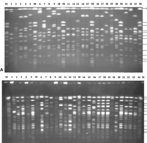
106to 2.0⫻106bp was calculated. This corresponded to the genome size of streptococci as indicated in the literature (30) (additional details can be found at the genome website Uni-versity of Oklahoma’s Advanced Center for Genome Technology (http://www.genome.ou.de/strep.html). Representative PFGE gels ofSmaI-digested DNAs of isolates mainly belonging to serotype 9 are shown in Fig. 1A, and PFGE gels of Apa I-digested DNAs of isolates mainly belonging to serotype 2 are shown in Fig. 1B. The data show that PFGE allowed differen-tiation of isolates between and within serotypes. Moreover, PFGE enabled us to monitor isolates from various locations of a single animal (Fig. 1A, lanes 11 and 12; Fig. 1B, lanes 17 to 19) as well as from different animals within the same herd (Fig. 1A, lanes 10, 16, and 20). Strongly related but not identical isolates could also easily be discriminated (Fig. 1A, lanes 21 and 22; Fig. 1B, lanes 20 to 24). Cluster analysis of PFGE patterns ofSmaI-digested DNA by UPGMA revealed a den-drogram with four major groups, designated groups A1, A2, B1, and B2, each comprising isolates with a similarity of at least 20% (Fig. 2). Isolates which appeared to be identical by visual inspection of the gels showed similarities of approximately 80 to 100% by cluster analysis. Examples are isolates I9841/1-3 (Fig. 2, cluster A1, corresponding to Fig. 1B, lanes 17 to 19) as well as isolates A3313/1/98 and A3313/3/98 (Fig. 2, cluster B1, corresponding to Fig. 1A, lanes 11 and 12). Closely related
on May 15, 2020 by guest
http://jcm.asm.org/
isolates showed similarities of approximately 70% by cluster analysis, e.g., strains P1/7 and D282 (Fig. 2, cluster A1, corre-sponding to Fig. 1A, lanes 21 and 22), as well as isolates B422/97 and 631/97 (Fig. 2, cluster A2, corresponding to Fig. 1B, lanes 14 and 15). All isolates determined to be closely related by PFGE with SmaI-digested DNA could be more clearly differentiated visually in PFGE gels ofApaI-digested DNA (data not shown). A dendrogram constructed from the PFGE patterns ofApaI-digested DNA was almost similar to
the dendrogram constructed from the PFGE patterns ofSma I-digested DNA, except that the degree of differentiation was higher (data not shown).
[image:3.612.65.556.75.550.2]Comparison of the clinical backgrounds of isolates with their assignment to the different clusters revealed that 20 of 48 isolates from pigs with meningitis-septicemia belonged to clus-ter A1 (Table 1). In contrast, isolates from pigs with pneumo-nia or from healthy pigs were almost equally distributed among the different clusters except for cluster A1 (Table 1). The FIG. 1. Macrorestriction analysis ofS. suisisolates by PFGE. (A) PFGE ofSmaI-digested DNA ofS. suisserotype 9 isolates (lanes 1 to 20 and 23) and, in addition, serotype 2 hemolysin activity-positive control strain P1/7 (lane 21) and serotype 2 MRP- and EF-positive control strain D282 (lane 22). Lane M, size standard (ApaI-digested genomic DNA fromS. aureusstrain DSM 1104) indicating molecular sizes (in kilobases). (B) PFGE ofApaI-digested DNA ofS. suisserotype 2 isolates (lanes 1 to 24) including the type 2 reference strain (lane 5), MRP- and EF-positive control strains 4005 (lane 7), T15 (lane 8), and D282 (lane 23), as well as hemolysin activity-positive control strain P1/7 (lane 20). Lane M, size standard (ApaI-digested genomic DNA fromS. aureus, strain DSM 1104) indicating molecular sizes (in kilobases).
on May 15, 2020 by guest
http://jcm.asm.org/
FIG. 2. Dendrogram of similarity among the observed PFGE macrorestriction patterns ofSmaI-digested DNAs from 99 S. suisisolates. Serotypes (ST) and clinical backgrounds (CB) are indicated on the right. Isolates originated either from pigs with typical invasive disease (meningitis [M], septicemia [S], arthritis [A]), from pigs with pneumonia (P), or from healthy pigs (H). Major clusters are indicated by A1, A2, B1, and B2. nt, nontypeable.
on May 15, 2020 by guest
http://jcm.asm.org/
phenotypic features of the cluster A1 isolates are described below.
Serotyping and analysis of potential virulence traits.Results
from serotyping and phenotyping of isolates are summarized in Tables 2 to 4. The distribution of serotypes within the different isolates shows that the majority belonged to serotypes 2 (25%) and 9 (20%), followed by serotypes 3, 7,1⁄2, 5, 4, and 1 (Table 2). The remaining isolates (indicated as “others” in Table 2) either belonged to serotype 8, 15, 16, or 25 or were nontype-able with the antisera used (i.e., antisera to serotypes 1 to 28). The nontypeable isolates represented 7% of the total number tested. The most dominant serotypes among all isolates tested, serotypes 2 and 9, also represented the majority of isolates from pigs with meningitis-septicemia (44% were serotype 2, 25% were serotype 9), followed by serotype 1 (8% of all men-ingitis-septicemia isolates) (Table 2). On the other hand, the serotypes of isolates from pigs with pneumonia showed a broader distribution, with most of these isolates belonging to serotype 3, 5, 7, or 9 (Table 2). Interestingly, most isolates from healthy pigs were nontypeable or belonged to one of the rarely represented serotypes (both indicated as “others” in Table 2). Analysis of expression of virulence-associated proteins MRP and EF (summarized in Table 3) demonstrated that the ma-jority (67%) of all isolates expressed MRP, either alone or in combination with expression of EF. None of the isolates ex-pressed solely EF. Isolates from pigs with meningitis-septice-mia mostly either expressed MRP alone (52%) or expressed
MRP in combination with EF (29%). Among isolates from pigs with pneumonia, only one expressed MRP and EF; the remain-ing isolates expressed either MRP only (52.5%) or neither of these proteins (45%). All isolates were also tested for hemo-lysin activity, another potential virulence marker. The results are shown in Table 4. Isolates were classified as hemolysin negative, intermediate, or positive with respect to their relative activity in comparison with the activity of control strain P1/7 (see Materials and Methods). A high proportion of all isolates were hemolysin negative (50%); only 27% were positive, and 22% were intermediate. This distribution was comparable among isolates from pigs with meningitis-septicemia and healthy pigs, whereas a significantly higher proportion (95%) of isolates from pigs with pneumonia were hemolysin negative or intermediate. Adherence of streptococci to epithelial cells was tested since adherence capacity and/or presence of ad-hesins has been related to the virulence ofS. suis(and other bacteria) in earlier studies (10, 32). Surprisingly, the adherence of all isolates to two commonly used cell lines, porcine testis cells and human HEp-2 epithelial cells, was generally very low. Substantial adherence was observed for only 25% of all isolates by using porcine cells and only 18% of all isolates by using HEp-2 cells. The isolates that adhered well to HEp-2 cells were the same ones that adhered well to porcine cells. There was no association of adherence capacities with source of isolation (data not shown).
[image:5.612.51.293.92.190.2]Taken together, these results showed that certain pheno-typic features were striking among isolates from pigs with men-ingitis-septicemia, such as the presence of capsular antigens of serotypes 2 and 9, expression of MRP and EF, and hemolysin TABLE 1. Assignment to PFGE clusters amongS. suisisolates
from pigs with various clinical backgrounds
PFGE
clustera isolatesNo. of
No. (%) of isolates from: Pigs with
meningitis-septicemia pneumoniaPigs with Healthy pigs
A1 26 20 (42) 5 (12.5) 1 (9)
A2 22 9 (19) 9 (22.5) 4 (36)
B1 32 13 (27) 15 (37.5) 4 (36)
B2 19 6 (12) 11 (27.5) 2 (18)
Total 99 48 (100) 40 (100) 11 (100)
[image:5.612.310.552.92.190.2]aIsolates were assigned to four different clusters as revealed from cluster analysis of macrorestriction patterns ofSmaI-digested streptococcal DNA.
TABLE 2. Distribution of serotypes amongS. suisisolates from pigs with various clinical backgrounds
Serotype isolatesNo. of
No. (%) of isolates from: Pigs with
meningitis-septicemia pneumoniaPigs with Healthypigs
1 5 4 (8) 0 1 (9)
1⁄2 8 2 (4) 5 (10) 1 (9)
2 25 21 (44) 3 (6) 1 (9)
3 9 1 (2) 8 (20) 0
4 6 1 (2) 4 (10) 1 (9)
5 7 2 (4) 5 (12.5) 0
7 8 3 (6) 5 (12.5) 0
9 20 12 (25) 7 (17.5) 1 (9)
Othersa 11 2 (4) 3 (6) 6 (55)
Total 99 48 (100) 40 (100) 11 (100)
aIsolates belonged to serotype 8, 15, 16, or 25 or were nontypeable.
TABLE 3. Expression of MRP and EF amongS. suisisolates from pigs with various clinical backgrounds
MRP/EF expression statusa isolatesNo. of
No. (%) of isolates from: Pigs with
meningitis-septicemia pneumoniaPigs with Healthypigs
⫹/⫹ 15 14 (29) 1 (2.5) 0
⫹/⫺ 52 25 (52) 21 (52.5) 6 (54.5)
⫺/⫹ 0 0 0 0
⫺/⫺ 32 9 (19) 18 (45) 5 (45.5)
Total 99 48 (100) 40 (100) 11 (100)
[image:5.612.53.294.574.718.2]aIsolates expressed MRP and EF (⫹/⫹), only MRP (⫹/⫺), or none of the two proteins (⫺/⫺); none of the isolates expressed only EF (⫺/⫹).
TABLE 4. Hemolysin activities amongS. suisisolates from pigs with various clinical backgrounds
Hemolysin
activitya isolatesNo. of
No. (%) of isolates from: Pigs with
meningitis-septicemia pneumoniaPigs with Healthypigs
⫹ 27 21 (43.7) 2 (5) 4 (36)
⫾ 22 8 (16.6) 13 (32.5) 1 (9)
⫺ 50 19 (39.5) 25 (62.5) 6 (55)
Total 99 48 (100) 40 (100) 11 (100)
aIsolates were grouped as hemolysin positive (⫹;⬎80% activity), intermedi-ate (⫾; 10 to 80% activity), or negative (⫺;⬍10% activity) in comparison to the activity of control strain P1/7.
on May 15, 2020 by guest
http://jcm.asm.org/
[image:5.612.313.552.622.702.2]activity. A more heterogeneous background seemed to exist within isolates from pigs with pneumonia or healthy pigs.
Relation of individual features of the isolates with clinical
background.We next calculated the probabilities that isolates
with certain features, alone or in combination with each other, originated from pigs with meningitis-septicemia, which would indicate a high degree of virulence. A meningitis-septicemia background was found among 4 of the 5 serotype 1 isolates, 21 of the 25 serotype 2 isolates, and 12 of the 19 serotype 9 isolates (Table 2), indicating a high probability that isolates of serotypes 2 and 1 and, to a lesser extent, of serotype 9 origi-nated from pigs with meningitis. On the other hand, most isolates belonging to serotypes1⁄2(five of eight), 3 (eight of nine), 4 (four of six), 5 (five of seven), or 7 (five of eight) were from pigs with pneumonia; most isolates designated “others” originated from healthy animals (Table 2). We also found a very high probability of the clinical background meningitis-septicemia among isolates expressing MRP and EF and hemo-lysin-positive isolates: 14 of the 15 MRP- and EF-positive iso-lates and 21 of the 27 hemolysin-positive isoiso-lates were from pigs with meningitis-septicemia (Tables 3 and 4). Interestingly, a high proportion of isolates that lacked hemolysin activity (25 of 50) or that expressed intermediate hemolysin activity (13 of 22) had been isolated from pigs with pneumonia (Table 4). Moreover, the majority of isolates of PFGE cluster A1 (20 of 26) had a meningitis-septicemia background, and most isolates of cluster B2 (11 of 19) were from pigs with pneumonia (Table 1). Taken together, these data show that there was a high probability that isolates exhibiting either the feature serotype 2 or serotype 1 capsular antigen expression, expression of MRP and EF, strong hemolysin activity, or assignment to PFGE cluster A1 had a clinical background of meningitis-septicemia. On the other hand, the features serotype 3, 4, or 5 capsular antigen expression and a lack of or intermediate hemolysin activity indicated a high probability of pneumonia as the clin-ical background.
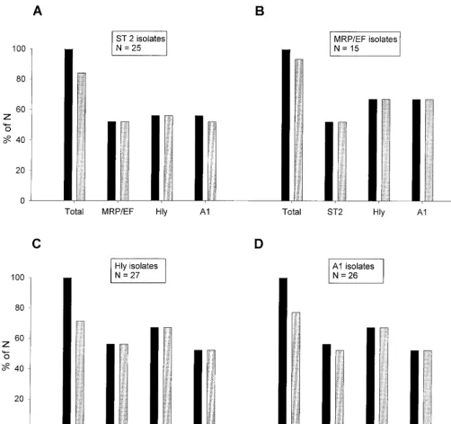
The probability of the clinical background meningitis-septi-cemia was even higher when some of the features mentioned above were found in combination (Fig. 3). We found that of all serotype 2 isolates, 52% also expressed MRP and EF, 56% were hemolysin positive, and 56% belonged to cluster A1. All serotype 2 isolates that were either MRP and EF positive or hemolysin positive as well as 93% of the serotype 2 isolates assigned to cluster A1 originated from pigs with meningitis (Fig. 3A). Of the MRP- and EF-positive isolates, 52% be-longed to serotype 2, 67% were hemolysin positive, and 67% belonged to cluster A1, and all isolates with any of these combinations had been isolated from pigs with meningitis-septicemia (Fig. 3B). Features of the hemolysin-positive and cluster A1 isolates in relation to the clinical background men-ingitis-septicemia are partially included in Fig. 3A and B but are also shown separately in Fig. 3C and D, respectively.
Taken together, the probability of having a clinical back-ground of meningitis-septicemia was highest (100%) among isolates with a combination of serotype 2 with MRP and EF positivity or hemolysin activity, of MRP and EF positivity with hemolysin activity or assignment to cluster A1, and of hemo-lysin activity with assignment to cluster A1. In addition, there was a striking correlation of serotypes 7 and 9 with a certain MRP-EF pattern, in that none of these isolates expressed EF.
Furthermore, none of the serotype 7 isolates expressed MRP, whereas all but one of the serotype 9 isolates expressed MRP, but most of these expressed a size variant of MRP (37). Only two (10%) of all serotype 9 isolates were hemolysin positive. Most serotype 9 isolates were clustered in clusters A2 and B1 (35% each). Only one of the five serotype 1 isolates expressed MRP and EF (the other four isolates were negative for both), and all were positive for hemolysin activity. There was no specific combination of features that was strongly associated with a background of pneumonia. Furthermore, there was no association of adherence capacity with a certain other feature.
DISCUSSION
S. suis is a major pathogen in swine and can also infect humans. The presence ofS. suisin pigs, however, is not a good indicator of disease, since carrier rates of this “early colonizer” can be up to 100%, with prevalences of disease generally being not more than 5% (5, 20). This indicates that the level of virulence varies extremely between strains. Therefore, many recent studies have been aimed at the identification of pheno-typic and/or genopheno-typic virulence markers which might be useful to distinguish virulent from less virulent or avirulent isolates (23, 26, 29, 35). As with many other pathogens, a single marker that is sufficient for the identification of all highly virulent strains has not yet been identified (and probably does not exist), but there is strong evidence that highly virulent strains share certain features and might be more related than others (4, 21, 23, 26). Some of these virulence-associated features seem to be production of serotype-specific capsular polysac-charides (25), expression of proteins MRP and EF (35), and expression of hemolysin activity and suilysin (10, 17, 18). How-ever, on the basis of the high prevalence of serotype 2 isolates, most studies have been restricted to serotype 2. Furthermore, some results might have been overinterpreted with respect to the terms virulent and avirulent, as recently discussed by Gottschalk et al. (M. Gottschalk, R. Higgins, and S. Quessy. Letter, J. Clin. Microbiol.37:4202–4203, 1999).
The ongoing debate about the relatedness of highly virulent strains and the poor knowledge about the features of theS. suis population in Germany prompted us to perform the present study, in which we characterized a collection of 99 S. suis isolates including some of the major reference strains. Isolates were first serotyped to ensure that serotypes other than sero-type 2 were also represented. The results of serotyping corre-late well with those of others (7), in that serotypes 2 and 9 are the dominant serotypes in most European countries except the United Kingdom, where serotype 1 is most prevalent. Further-more, we showed that other serotypes such as 1,1⁄2, 3, 4, 5, and 7 seem to be quite common in the GermanS. suispopulation, which is in agreement with recently published data (36).
Nearly half (48 of 99) of the isolates analyzed for the present study originated from pigs with typical signs of disease (men-ingitis, septicemia, arthritis [referred to below as “typical iso-lates”]); 40 isolates were from pigs with pneumonia. In contrast to the importance ofS. suisas a primary pathogen in pigs with meningitis, septicemia, or arthritis, it’s causative role in pneu-monia is not as clear since in many cases S. suis is isolated together with other lung pathogens such asPasteurellaspp.,B. bronchiseptica, orMycoplasmaspp. (18, 24). Similarly, in our study
on May 15, 2020 by guest
http://jcm.asm.org/
only 16 of 40S. suisisolates from pigs with pneumonia were isolated as pure cultures. In agreement with the current knowl-edge of the pathogenicity ofS. suis(14, 18, 24), we therefore assumed that the isolates from pigs with typical signs (i.e., meningitis, etc.) represented the most virulent strains, whereas isolates from pigs with pneumonia were less invasive and, thus, less virulent. The remaining 11 of the 99 isolates studied were from healthy pigs and therefore represented strains from car-riers. Certainly, it must be considered that the origin of
isola-tion can only give some hints as to an isolate’s capacity to cause disease but is never proof of an isolate’s virulence.
[image:7.612.57.560.69.542.2]Taking this into account, we analyzed threeS. suis subpopu-lations (i.e., typical isolates, isolates from pigs with pneumonia, and isolates from potential carriers) by examining all currently known putative virulence traits including adherence to epithe-lial cells. In addition, all isolates were genotyped by macror-estriction analysis by PFGE, a technique with a high resolution power which hitherto has not been applied toS. suis. In our
FIG. 3. Comparison of combinations of different features ofS. suisisolates with their clinical backgrounds. Additional features of serotype 2 isolates (A), isolates expressing MRP and EF (MRP/EF isolates) (B), hemolysin-positive isolates (Hly isolates) (C), and cluster A1 isolates (A1 isolates) (D) are shown. The leftmost pair of bars in each graph (labeled Total) refers to all isolates expressing the indicated feature; labeling of the remaining pairs of bars indicates the additional feature found in isolates of the respective group. Each pair of bars shows the proportion of isolates from pigs with meningitis-septicemia (gray bars) compared to the total proportion of isolates with this combination of features (black bars). Results are expressed as a percentage of the total number for each group (N), which is indicated in boxes above the respective groups.
on May 15, 2020 by guest
http://jcm.asm.org/
hands, this technique proved to be most suitable for differen-tiation of single isolates, even those of the same serotype within an infected herd, and thus should be most valuable in epidemiological studies withS. suis. Due to the high degree of sensitivity and the discriminatory power of this technique, how-ever, isolates that appeared to be identical by visual inspection could also be found in very closely related instead of identical branches after cluster analysis. Therefore, it seemed reason-able to define a cutoff value for identity (i.e.,⬎80%) to exclude possible overinterpretation of differentiation of two isolates.
The most prominent features among the typical isolates were the presence of capsular antigens of serotype 2, 9, or 1, expression of MRP and/or EF, and strong hemolysin activity. However, these features were found in only about half of this population; the remaining isolates had more heterogeneous backgrounds, suggesting that their virulence is attributed to other characteristics. PFGE and subsequent cluster analysis confirmed these assumptions, since most of the isolates exhib-iting one more of these most prominent features were found in one cluster (cluster A1), whereas the others were more homo-geneously distributed in the resulting dendrogram. A different picture was seen in isolates from pigs with pneumonia: type 2 was rarely found, and in addition to serotype 9, sero-types 3, 5, and 7 were most prominent. Moreover, only one of the isolates from pigs with pneumonia expressed both MRP and EF, and only two isolates from pigs with pneumonia ex-pressed strong hemolysin activity. This might suggest a lower capacity of isolates from pigs with pneumonia to express viru-lence properties, which would correlate well with the assump-tion that isolates from pigs with pneumonia are less invasive and virulent than the typical isolates. No significant clustering of the isolates from pigs with pneumonia was found by cluster analysis. This indicates that the isolates from pigs with pneu-monia had more heterogeneous phenotypic and genotypic backgrounds than typical isolates. Among the isolates from healthy animals that were potential carriers, the high propor-tion (55%) of rarely seen serotypes and nontypeable isolates was most striking, as was the high proportion (36%) of hemo-lysin-positive isolates. This suggests that S. suis strains from carriers are more diverse than virulent strains, but this must be confirmed by analysis of a larger population of isolates.
A surprising finding of the present study was that adherence to epithelial cells was not correlated at all to the clinical back-grounds of the isolates, independent of the epithelial cell type used (human or porcine). This raises the question of the sig-nificance of adherence as a virulence trait, as suggested by others (9, 32). An explanation for the poor adherence observed in our study might be either that adherence is in fact no virulence trait or that a more specific detection of adhesins is needed to evaluate the virulence potential of a given strain or isolate. Concerning the latter, other target cell types (e.g., porcine tonsillar epithelial or endothelial cells) and/or different conditions for bacterial cultivation might have to be tested. In addition, possible inhibitory effects of encapsulation on adher-ence might have to be ruled out in future studies, even though recently published data by Charland et al. (3) did not support an inhibitory role of the capsule in adherence.
As described above, the phenotypic and genotypic back-grounds of the isolates from diseased pigs revealed some va-riety but also confirmed features that were commonly found.
Therefore, we analyzed our data with respect to the probabil-ities that the commonly found features, alone or in combina-tion with others, were associated with the clinical background. The results showed that certain combinations of features such as the presence of the capsular antigen of serotype 2 and expression of MRP and EF or the presence of the capsular antigen of serotype 2 and strong hemolysin activity were highly correlated with the invasive clinical background meningitis-septicemia. Furthermore, the probability of such a clinical background was even higher when isolates expressing one of those features could be assigned to PFGE cluster A1. In agree-ment with the findings of others on the specific ribotype pro-files of virulent strains and isolates (21, 29), our clustering results strongly support the hypothesis of a relatively conserved genetic background of highly virulent strains. Together with our observations on the serotypes and potential virulence traits of the isolates tested, these data could indicate that certain highly virulent clones have established themselves successfully within the EuropeanS. suispopulation.
In conclusion, we have presented evidence that (i) PFGE analysis is a most valuable tool for epidemiological studies ofS. suis, (ii) several known and yet unknown virulence traits exist inS. suiswhich, when combined, can give important hints as to whether a given isolate is highly virulent, and (iii) highly viru-lent isolates or strains from pigs with clinical backgrounds of invasive disease seem to be more related than isolates from pigs with the less invasive pneumonia or typical isolates from carriers. This raises the perspective that in the near future it should be possible to develop strategies for rapid identification of highly virulent isolates and for monitoring of the epidemi-ology ofS. suisinfections.
ACKNOWLEDGMENTS
We are in debt to L. Kreienbrock and R. Meyer (Tieraerztliche Hochschule Hannover) for excellent help with statistical analyses and S. Schwarz (FAL, Celle, Germany) for great support with PFGE. We also thank G. Amtsberg and M. Ganter (both from the Tieraerztliche Hochschule Hannover) and C. Laemmler (FB Veterinaermedizin, JLU Giessen) for kindly providing bacterial isolates.
One of us (A.A.) was supported by a grant from the Impfstoffwerk Dessau-Tornau, Rosslau Germany.
REFERENCES
1.Arends, J. P., and H. C. Zanen.1988. Meningitis caused byStreptococcus suis in humans. Rev. Infect. Dis.10:131–137.
2.Chanter, N., P. W. Jones, and T. J. L. Alexander.1993. Meningitis in pigs caused byStreptococcus suis—a speculative review. Vet. Microbiol.36:39–55. 3.Charland, N., V. Nizet, C. E. Rubens, K. S. Kim, S. Lacouture, and M. Gottschalk.2000. Streptococcus suisserotype 2 interactions with human brain microvascular endothelial cells. Infect. Immun.68:637–643. 4.Chatellier, S., M. Gottschalk, R. Higgins, R. Brousseau, and J. Harel.1999.
Relatedness ofStreptococcus suisserotype 2 isolates from different geo-graphic origins as evaluated by molecular fingerprinting and phenotyping. J. Clin. Microbiol.37:362–366.
5.Clifton-Hadley, F. A. 1986. The epidemiology, diagnosis, treatment and control ofStreptococcus suistype 2 infection, p. 471–491.InJ. D. McKean (ed.), Proceedings of the American Association of Swine Practioners. Amer-ican Association of Swine Practioners, Minneapolis, Minn.
6.Clifton-Hadley, F. A, and T. J. L. Alexander.1980. The carrier site and carrier rate ofStreptococcus suistype II in pigs. Vet. Rec.107:40–41. 7.Esteopangestie, S., and C. Laemmler.1993. Distribution of capsular types 1
to 28 and further characteristics ofStreptococcus suisisolates from various European countries. Zentbl. Bakteriol. Parasitenkd. Infektkrank. Hyg. Abt. 1 Orig.279:394–403.
8.Gottschalk, M., R. Higgins, and M. Jacques.1993. Production of capsular material byStreptococcus suisserotype 2 under different growth conditions. Can. J. Vet. Res.57:49–52.
9.Gottschalk, M., S. Petitbois, R. Higgins, and M. Jaques.1991. Adherence of
on May 15, 2020 by guest
http://jcm.asm.org/
Streptococcus suiscapsular type 2 to porcine lung sections. Can. J. Vet. Res.
55:302–304.
10.Gottschalk, M., S. Lacouture, and J. D. Dubreull.1995. Characterization of Streptococcus suiscapsular type 2 haemolysin. Microbiology141:189–195. 11. Gottschalk, M., A. Lebrun, H. J. Wisselink, J. D. Dubreuil, H. E. Smith, and
U. Vecht.1998. Production of virulence-related proteins by Canadian strains ofStreptococcus suiscapsular type 2. Can. J. Vet. Res.62:75–79. 12. Hampson, D. J., D. J. Trott, I. L. Clarke, C. G. Mwaniki, and I. D.
Robert-son.1993. Population structure of Australian isolates ofStreptococcus suis. J. Clin. Microbiol.31:2895–2900.
13. Harel, J., R. Higgins, M. Gottschalk, and M. Bigras-Poulin.1994. Genomic relatedness among reference strains of differentStreptococcus suisserotypes. Can. J. Vet. Res.58:259–262.
14. Hoefling, D. C.1998. Tracking the incidence of porcine respiratory diseases. Vet. Med.93:391–398.
15. Jacobs, A. A. C., P. L. W. Loeffen, A. J. G. Van Den Berg, and P. K. Storm.
1994. Identification, purification, and characterization of a thiol-activated hemolysin (suilysin) ofStreptococcus suis. Infect. Immun.62:1742–1748. 16. Jacobs, A. A. C., A. J. G. Van Den Berg, J. C. Baars, B. Nielsen, and L. W.
Johannsen.1995. Production of suilysin, the thiol-activated haemolysin of Streptococcus suis, by field isolates from diseased pigs. Vet. Rec.139:295– 296.
17. Jacobs, A. A. C., A. J. G. Van Den Berg, and P. L. W. Loeffen.1996. Protection of experimentally infected pigs by suilysin, the thiol-activated haemolysin ofStreptococcus suis. Vet. Rec.139:225–228.
18. Macinnes, J. I., and R. Desrosiers.1999. Agents of the “suis-ide diseases” of swine:Actinobacillus suis, Haemophilus suis, andStreptococcus suis. Can. J. Vet. Res.63:83–89.
19. Mogollon, J. D., C. Pijoan, M. P. Murtaugh, J. E. Collins, and P. P. Cleary.
1991. Identification of epidemic strains ofStreptococcus suisby genomic fingerprinting. J. Clin. Microbiol.29:782–787.
20. Mwaniki, C. G., I. D. Robertson, D. J. Trott, R. F. Atyeo, B. J. Lee, and D. J. Hampson.1994. Clonal analysis and virulence of Australian isolates of Strep-tococcus suistype 2. Epidemiol. Infect.113:321–334.
21. Okwumabua, O., J. Staats, and M. M. Chengappa.1995. Detection of genomic heterogeneity inStreptococcus suis isolates by DNA restriction fragment length polymorphism of rRNA genes (ribotyping). J. Clin. Micro-biol.4:968–972.
22. Olsen, J. E., and M. Skov.1984. Genomic lineage ofSalmonella enterica serovar Dublin. Vet. Microbiol.40:271–282.
23. Rasmussen, S. R., F. M. Aarestrup, N. E. Jensen, and S. E. Jorsasl.1999. Associations ofStreptococcus suisserotype 2 ribotype profiles with clinical disease and antimicrobial resistance. J. Clin. Microbiol.37:404–408. 24. Reams, R. Y., D. D. Harrington, L. T. Glickman, H. L. Thacker, and T. L.
Bowersock.1996. Multiple serotypes and strains ofStreptococcus suisin naturally infected swine herds. J. Vet. Diagn. Investig.8:119–121.
25. Smith, H. E., M. Damman, J. Van Der Velde, F. Wagenaar, H. J. Wisselink, N. Stockhofe-Zurwieden, and M. A. Smits.1999. Identification and charac-terization of the cpslocus ofStreptococcus suis serotype 2: the capsule protects against phagocytosis and is an important virulence factor. Infect. Immun.67:1750–1756.
26. Smith, H. E., M. Rijnsburger, N. Stockhofe-Zurwieden, H. J. Wisselink, U. Vecht, and M. A. Smits.1997. Virulent strains ofStreptococcus suisserotype 2 and highly virulent strains ofStreptococcus suisserotype 1 can be recog-nized by a unique ribotype profile. J. Clin. Microbiol.35:1049–1053. 27. Smith, H. E., U. Vecht, A. L. J. Gielkens, and M. A. Smits.1996. Cloning and
nucleotide sequence of the gene encoding the 136-kilodalton surface protein (muraminidase-released protein) ofStreptococcus suistype 2. Infect. Immun.
60:2361–2367.
28. Smith, H. E., U. Vecht, H. J. Wisselink, N. Stockhofe-Zurwieden, Y. Bier-mann, and M. A. Smits.1996. Mutants ofStreptococcus suistypes 1 and 2 impaired in expression of muramidase-released protein and extracellular protein induce disease in newborn germfree pigs. Infect. Immun.64:4409– 4412.
29. Staats, J. J., B. L. Plattner, J. Nietfeld, S. Dritz, and M. M. Chengappa.1998. Use of ribotyping and hemolysin activity to identify highly virulent Strepto-coccus suistype 2 isolates. J. Clin. Microbiol.36:15–19.
30. Suvrov A. N., and J. J. Ferretti.1996. Physical and genetic map of an M type 1 strain ofStreptococcus pyogenes.J. Bacteriol.178:5546–5549.
31. Talay, S. R., P. Valentin-Weigand, P. G. Jerlstroem, K. N. Timmis, and G. S. Chhatwal.1992. Fibronectin binding protein ofStreptococcus pyogenes: se-quence of the binding domain involved in adherence of streptococci to epithelial cells. Infect. Immun.60:3837–3844.
32. Tikkanen, K., S. Haataja, and J. Finne.1996. The galactosyl-(1-4)-galactose-binding adhesin ofStreptococcus suis: occurrence in strains of different hem-agglutination activities and induction of opsonic antibodies. Infect. Immun.
64:3659–3665.
33. Vecht, U., J. P. Arends, E. J. Van Der Molen, and L. A. M. G. Van Leengoed.
1989. Differences in virulence between two strains ofStreptococcus suistype II after experimentally induced infection of newborn germ-free pigs. Am. J. Vet. Res.50:1037–1043.
34. Vecht, U., N. Stockhofe-Zurwieden, B. J. Tetenburg, H. J. Wisselink, and H. E. Smith.1997. Virulence ofStreptococcus suistype 2 for mice and pigs appeared host-specific. Vet. Microbiol.58:53–60.
35. Vecht, U., H. J. Wisselink, M. L. Jellema, and H. E. Smith.1991. Identifi-cation of two proteins associated with virulence ofStreptococcus suistype 2. Infect. Immun.59:3156–3162.
36. Wisselink H. J., H. E. Smith, N. Stockhofe-Zurwieden, K. Peperkamp, and U. Vecht.2000. Distribution of capsular types and production of murimidase-released protein (MRP) and extracellular factor (EF) ofStreptococcus suis isolated from diseased pigs in seven European countries. Vet. Microbiol.
74:237–248.