Improved Differentiation of
Streptococcus pneumoniae
and Other
S.
mitis
Group Streptococci by MALDI
Biotyper Using an Improved MALDI
Biotyper Database Content and a Novel
Result Interpretation Algorithm
Inka Harju,
aChristoph Lange,
bMarkus Kostrzewa,
bThomas Maier,
bKaisu Rantakokko-Jalava,
aMarjo Haanperä
cClinical Microbiology Laboratory, Turku University Hospital, Turku, Finlanda; Bruker Daltonik GmbH, Bremen, Germanyb; Bacterial Infections Unit, National Institute for Health and Welfare, Helsinki, Finlandc
ABSTRACT
Reliable distinction of
Streptococcus pneumoniae
and viridans group
streptococci is important because of the different pathogenic properties of these
or-ganisms. Differentiation between
S. pneumoniae
and closely related
Sreptococcus
mi-tis
species group streptococci has always been challenging, even when using such
modern methods as 16S rRNA gene sequencing or matrix-assisted laser desorption
ionization–time of flight (MALDI-TOF) mass spectrometry. In this study, a novel
algo-rithm combined with an enhanced database was evaluated for differentiation
be-tween
S. pneumoniae
and
S. mitis
species group streptococci. One hundred one
clini-cal
S. mitis
species group streptococcal strains and 188 clinical
S. pneumoniae
strains
were identified by both the standard MALDI Biotyper database alone and that
com-bined with a novel algorithm. The database update from 4,613 strains to 5,627
strains drastically improved the differentiation of
S. pneumoniae
and
S. mitis
species
group streptococci: when the new database version containing 5,627 strains was
used, only one of the 101
S. mitis
species group isolates was misidentified as
S.
pneumoniae
, whereas 66 of them were misidentified as
S. pneumoniae
when the
ear-lier 4,613-strain MALDI Biotyper database version was used. The updated MALDI
Bio-typer database combined with the novel algorithm showed even better
perfor-mance, producing no misidentifications of the
S. mitis
species group strains as
S.
pneumoniae
. All
S. pneumoniae
strains were correctly identified as
S. pneumoniae
with both the standard MALDI Biotyper database and the standard MALDI Biotyper
database combined with the novel algorithm. This new algorithm thus enables
reli-able differentiation between pneumococci and other
S. mitis
species group
strepto-cocci with the MALDI Biotyper.
KEYWORDS
MALDI-TOF,
Streptococcus pneumoniae
, algorithm,
mitis
group
streptococci, phenotypic identification, pneumococcus
T
raditional classification of streptococci was based on the potential to cause
hemo-lysis on sheep blood agar. The term “viridans” is used to refer to the greenish
coloring (alpha-hemolysis) of the medium around the colonies due to partial lysis of red
blood cells.
Streptococcus pneumoniae
, one of the most common causative agents of
bacterial meningitis, community-acquired pneumonia, and otitis media, is clinically the
most important alpha-hemolytic streptococcus. Because of the different pathogenic
potentials of
S. pneumoniae
and other alpha-hemolytic streptococci, the practical
dichotomy in clinical laboratories has been between
S. pneumoniae
and viridans group
Received29 September 2016Returned for modification23 October 2016 Accepted27 December 2016
Accepted manuscript posted online4 January 2017
CitationHarju I, Lange C, Kostrzewa M, Maier T, Rantakokko-Jalava K, Haanperä M. 2017.
Improved differentiation ofStreptococcus
pneumoniaeand otherS. mitisgroup streptococci by MALDI Biotyper using an improved MALDI Biotyper database content and a novel result interpretation algorithm. J
Clin Microbiol 55:914 –922.https://doi.org/
10.1128/JCM.01990-16.
EditorSandra S. Richter, Cleveland Clinic
Copyright© 2017 American Society for
Microbiology.All Rights Reserved.
Address correspondence to Inka Harju, inka.harju@tyks.fi.
crossm
on May 16, 2020 by guest
http://jcm.asm.org/
streptococci (VGS), the latter referring to nonpneumococcal alpha-hemolytic
strepto-cocci as a group. VGS form an important part of the normal flora of the human
respiratory, gastrointestinal, and urogenital tracts. Although mostly commensal, VGS
have also been associated with various infections, especially endocarditis in patients
with predisposing factors (1–4).
The classification of streptococci has changed significantly in the last few decades,
and hemolysis is no longer a valid basis for streptococcal taxonomy. Accordingly, many
“VGS species” also form nonhemolytic or beta-hemolytic colonies (1, 2). The taxonomy
within VGS has also seen several upheavals since the 1990s, due mainly to the advent
of modern molecular typing methods (1, 2, 5, 6). Current classification divides VGS into
five major groups, i.e., the
Streptococcus anginosus
,
Streptococcus bovis
,
Streptococcus
mitis
,
Streptococcus mutans
, and
Streptococcus salivarius
groups (1, 7). Of these groups,
the
S. mitis
species group containing closely related species of different pathogenic
potentials has posed great challenges for reliable species level identification. In
addi-tion to commensal species, the major pathogen
S. pneumoniae
taxonomically belongs
to this group. Besides
S. pneumoniae
, the
S. mitis
species group currently includes the
following species:
S. australis
,
S. cristatus
(formerly
S. crista
),
S. gordonii
,
S. infantis
,
S.
massilienis
,
S. mitis
,
S. oligofermentans
,
S. oralis
,
S. orisratti
,
S. parasanguinis
(formerly
S.
parasanguis
),
S. peroris
,
S. pseudopneumoniae
,
S. sanguinis
(formerly
S. sanguis
), and
S. sinensis
(1). Furthermore, two recently described species,
S. dentisani
(8) and
S.
tigurinus
(9), seem to belong to the
S. mitis
species group as well.
In clinical microbiology laboratories, VGS have traditionally been differentiated from
pneumococci on the basis of their lack of bile solubility. Optochin resistance testing is
also often used for the preliminary identification of pneumococci, despite its
limita-tions. Wessels and coworkers (10) reported that optochin testing in a CO
2atmosphere
and tube bile solubility testing give consistent results for
S. pneumoniae
. However, in
another study (11), 21 optochin-susceptible isolates were identified as
S. mitis
by
multilocus sequence analysis (MLSA). Also, commercial biochemical test panels have
limitations in identifying streptococci to the species level (12–16).
In the last few decades, molecular methods have enabled more accurate genotypic
identification of bacteria. The close relationship between
S. pneumoniae
and other
S.
mitis
species group streptococci renders analysis of 16S rRNA gene sequences
incapa-ble of definitive species identification within the
S. mitis
species group (13). Therefore,
several other streptococcal genes have been targeted in identification and
phyloge-netic studies, sometimes using an MLSA approach (17–22). Some of the most
com-monly used targets have been housekeeping genes
ddl
(21, 23),
groESL
(24),
rnpB
(25–27),
rpoB
(28),
sodA
(29, 30),
tuf
(31), and
recA
(32). However, currently, MLSA is too
time-consuming and expensive to be used routinely in a clinical microbiology
labora-tory but is a useful tool for taxonomic studies. In recent years, the matrix-assisted laser
desorption ionization–time of flight mass spectrometry (MALDI-TOF MS) technique has
emerged as an alternative for clinical microbiology laboratories for the identification of
a wide range of bacteria and fungi (33–35). Identification of
S. pneumoniae
and VGS by
MALDI-TOF MS has been widely studied previously (27, 35–49). The close relationship
between
S. pneumoniae
and other
S. mitis
species group streptococci, especially
S. mitis
and
S. oralis
, poses considerable challenges for the reliable distinction between species
of these groups by this technique as well.
In recent comparisons of two commercially available MALDI-TOF instruments, use of
the MALDI Biotyper (Bruker Daltonics) system resulted in misidentifications of
non-pneumococcal
S. mitis
species group strains as
S. pneumoniae
, while use of the Vitek MS
(bioMérieux) system often resulted in the identification of these strains as
S. oralis
/
S.
mitis
(48, 49). Expansion of the databases of these MALDI-TOF systems to include
recently described species of the
S. mitis
species group such as
S. tigurinus
(28) and
improvement of the algorithms used to calculate closest matches (48, 50) have both
been suggested as means of obtaining a more reliable distinction between
S.
pneu-moniae
and different species of the
S. mitis
species group. In this work, we present an
improved distinction between
S. pneumoniae
and other
S. mitis
species group strains by
on May 16, 2020 by guest
http://jcm.asm.org/
using a novel algorithm utilizing list(scores) combined with the expanded MALDI
Biotyper database. This would allow MALDI-TOF mass spectrometry to be utilized for
the identification of both pneumococci and
S. mitis
species group streptococci,
result-ing in more rapid identification than that of traditional biochemical methods.
RESULTS
MALDI-TOF analysis of all strains yielded adequate spectra, and they could all be
used for species identification. The identification results obtained by use of MALDI
Biotyper database versions DB_4613 and DB_5627 with a classical log(score) algorithm,
as well as those obtained using version DB_5627 with the list(score) algorithm, are
summarized in Table 1. All the identification results obtained by MALDI Biotyper are
shown in Table S1 in the supplemental material for pneumococcal strains, including the
log(score) ranking lists and the list(scores). When database versions DB_4613 and
DB_5627 and the classical log(score) algorithm were used, none of the 437 measured
S. pneumoniae
spectra were identified as species of the
S. mitis
species group other than
S. pneumoniae
(Table 1). All the pneumococcal strains were also clearly identified as
S.
pneumoniae
when the list(score) algorithm was used in connection with database
version DB_5627 (Table 1).
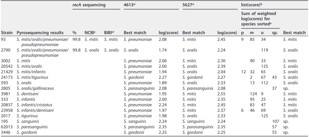
Examples of the identification results for nonpneumococcal
S. mitis
species group
strains are shown in Table 2. All the identification results obtained by MALDI Biotyper
for nonpneumococcal
S. mitis
species group strains are shown in Table S2, including the
log(score) ranking lists and the list(scores). When the earlier database version DB_4613
and classical log(score) algorithm were used, 66 of 101 nonpneumococcal
S. mitis
species group strains were misidentified as
S. pneumoniae
(Table 1). When database
version DB_5627 and the classical log(score) calculation were used, only one isolate
(62680), putatively identified as
S. infantis
by pyrosequencing, was incorrectly identified
as
S. pneumoniae
, with a log(score) of 2.03 (Table S2). When the new algorithm based
on weighted list(scores) was employed in addition to database version DB_5627, none
of the nonpneumococcal
S. mitis
species group strains tested were misidentified as
S.
pneumoniae
(Table 1).
Interestingly, even when the earlier database DB_4613 with the classical log(score)
algorithm was used, only one of the strains putatively identified as members of the
S.
sanguinis
species group (
S. gordonii
,
S. parasanguinis
, or
S. sanguinis
) by pyrosequencing
was misidentified as
S. pneumoniae
. With the updated version DB_5627 and the
classical log(score) algorithm, none of these strains were misidentified as
S. pneumoniae
and 19 of the 22 strains could be identified as members of this species group using the
classical log(score) algorithm (Table S2). All of the remaining three strains putatively
identified by pyrosequencing as belonging to the
S. sanguinis
group were identified by
MALDI-TOF as
S. oralis
. Therefore, a separate list(score) calculation for these three
species provided no further benefit.
[image:3.585.45.544.92.182.2]The species identification of
S. pneumoniae
and
S. oralis
appeared very clear when
using the list(scores). All 10 positions in the ranking list were almost always occupied
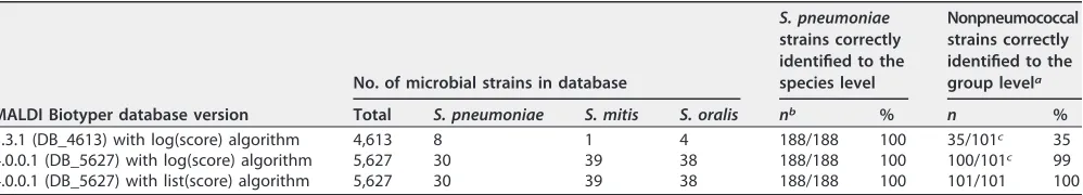
TABLE 1Differences in the database contents and in identification results between different MALDI Biotyper database versions tested in this study
MALDI Biotyper database version
No. of microbial strains in database
S. pneumoniae strains correctly identified to the species level
Nonpneumococcal strains correctly identified to the group levela
Total S. pneumoniae S. mitis S. oralis nb % n %
3.3.1 (DB_4613) with log(score) algorithm 4,613 8 1 4 188/188 100 35/101c 35
4.0.0.1 (DB_5627) with log(score) algorithm 5,627 30 39 38 188/188 100 100/101c 99
4.0.0.1 (DB_5627) with list(score) algorithm 5,627 30 39 38 188/188 100 101/101 100
aAs nonpneumococcal species in theS. mitisspecies group. bn, number of strains.
cAll misidentified strains were misidentified asStreptococcus pneumoniae.
on May 16, 2020 by guest
http://jcm.asm.org/
by the respective species only. Identification of the isolates putatively identified as
S.
mitis
by pyrosequencing delivered in most cases a much more diverse mixture of all
three species (
S. mitis
,
S. oralis
,
S. pneumoniae
). Nevertheless, the list(score) showed the
highest value for
S. mitis
in all cases (Table S2). For all isolates identified as
S. tigurinus
by pyrosequencing, the list(score) showed the highest value for
S. oralis
, as shown in
Table S2. For other nonpneumococcal
S. mitis
species group isolates, the highest valued
match given by the list(score) algorithm was either
S. mitis
,
S. oralis
, or other
S. mitis
species group strains but never
S. pneumoniae
(Table S2).
None of the 24 strains identified only ambiguously by pyrosequencing as
S.
pneu-moniae/S. pseudopneupneu-moniae/S. mitis/S. oralis
were most closely matched to
S.
pneu-moniae
or
S. pseudopneumoniae
by
recA
sequencing, whereas all were most closely
matched to either
S. mitis
or
S. oralis
(Table S2). Seven of these strains were
unambig-uously identified as
S. oralis
and 10 as
S. mitis
both by
recA
sequencing and by using
MALDI Biotyper. For two strains, the closest
recA
sequence match was to
S. mitis
, but
MALDI Biotyper identified them as
S. oralis
. For four strains, the closest
recA
sequence
match was to
S. mitis
, but MALDI Biotyper identifications were ambiguous (
S. mitis/S.
oralis
). One strain was identified as
S. mitis
both by
recA
sequencing and by the first two
matches using the standard MALDI Biotyper database but gave ambiguous
identifica-tion results (
S. mitis/S. oralis
) when the list(score) algorithm was used.
DISCUSSION
Using the classical interpretation rules of the MALDI Biotyper, the database
update from version DB_4613 to DB_5627 was already a considerable improvement
in
S. mitis
species group identification. The addition of more
S. pneumoniae
,
S. mitis
,
and
S. oralis
strains in the database led to much better “classical” identification rates
using log(scores) and ranking lists. The rate at which nonpneumococcal
S. mitis
species group streptococci were misidentified as pneumococci was reduced from
66 of 101 to 1 of 101 within this group in this study. The only misidentification as
S. pneumoniae
when the DB_5627 version of MALDI Biotyper was used with the
classical log(score) algorithm was one
S. infantis
isolate that was misidentified as
S.
[image:4.585.40.545.82.306.2]pneumoniae
. The new list(score) algorithm together with MALDI Biotyper version
TABLE 2Examples of identification of nonpneumococcalS. mitisgroup isolates
Strain Pyrosequencing results
recAsequencing 4613a 5627a list(score)b
% NCBIc BIBId Best match log(score) Best match log(score)
Sum of weighted log(scores) for species sortede
Best match p m o sp.
93 S. mitis/oralis/pneumoniae/
pseudopneumoniae
99.8 S. mitis S. mitis S. pneumoniae 2.08 S. mitis 2.45 9 85 34 S. mitis
2790 S. mitis/oralis/pneumoniae/
pseudopneumoniae
99.8 S. oralis S. oralis S. oralis 1.74 S. oralis 2.24 119 S. oralis
3002 S. mitis S. pneumoniae 2.06 S. mitis 2.30 90 33 S. mitis
20542 S. mitis/oralis S. pneumoniae 2.00 S. oralis 2.39 125 S. oralis
21429 S. mitis/infantis S. pneumoniae 1.94 S. oralis 2.04 12 32 65 S. oralis
24173 S. mitis/tigurinus S. gordonii 2.27 S. gordonii 2.27 2 67 43 S. oralis
593 S. oralis S. pneumoniae 1.89 S. oralis 2.33 13 112 S. oralis
2805 S. oralis/gallinaceus S. parasanguinis 2.08 S. parasanguinis 2.08 37 sp.
3981 S. dentisani S. pneumoniae 1.95 S. mitis 2.55 124 9 S. mitis
333 S. infantis S. pneumoniae 2.00 S. mitis 2.35 95 25 S. mitis
20837 S. infantis/cristatus S. pneumoniae 2.24 S. mitis 2.45 83 47 S. mitis
23958 S. infantis/dentisani S. pneumoniae 1.97 S. mitis 2.37 6 46 69 S. oralis
2017 S. tigurinus S. pneumoniae 1.98 S. oralis 2.33 125 S. oralis
195 S. sanguinis S. sanguinis 2.24 S. sanguinis 2.24 107 sp.
62013 S. parasanguinis S. parasanguinis 2.35 S. parasanguinis 2.35 57 sp.
3446 S. gordonii S. gordonii 2.25 S. gordonii 2.25 55 sp.
a4613, MALDI Biotyper database version 4613; 5627, MALDI Biotyper database version 5627. bThe list(score) is defined as the sum of weighted log(scores) for each single species (Fig. 2). cNCBI, species identification using therecAgene and the NCBI database.
dBIBI, species identification using therecAgene and the BIBI database.
ep,S. pneumoniae; m,S. mitis; o,S. oralis; sp., any other species in the Biotyper database.
on May 16, 2020 by guest
http://jcm.asm.org/
DB_5627 resulted in an even better capacity to distinguish between pneumococci
and other
S. mitis
species group strains, with no false identifications of
S.
pneu-moniae
for any of the nonpneumococcal isolates. The paucity of misidentifications
of
S. gordonii
,
S. parasanguinis
, and
S. sanguinis
as
S. pneumoniae
compared to other
nonpneumococcal
S. mitis
species group strains seems to be in line with the views
of those taxonomists who have separated
S. gordonii
,
S. parasanguinis
, and
S.
sanguinis
from
S. mitis
species group streptococci and grouped them in the
Streptococcus sanguinis
group (2).
The other nonpneumococcal isolates in the
S. mitis
species group could
gener-ally not be reliably identified to the species level even when MALDI Biotyper version
DB_5627 was used. This is not surprising, considering that many of the
nonpneu-mococcal isolates could not be identified to the species level even by
pyrosequenc-ing, and even the clearer species identifications provided by pyrosequencing
should be considered only putative identifications. Nevertheless, for clinical
labo-ratories, the exact identification to the species level among nonpneumococcal
S.
mitis
species group strains is by far less important than their discrimination from
S.
pneumoniae
. However, reliable identification of other viridans group streptococci to
the species level would enable researchers to gain new insights into their
patho-genic properties and antimicrobial resistance patterns.
As a final conclusion, it can be stated that adding further
S. mitis
species group
isolates (
S. mitis
,
S. oralis
, and
S. pneumoniae
) to the MALDI Biotyper database and using
the new list(score) algorithm to interpret MALDI Biotyper results led to significantly
improved differentiation of pneumococci from other species of the
S. mitis
species
group. MALDI Biotyper version DB_5627 together with the new algorithm might thus
provide routine clinical microbiological laboratories with a rapid and affordable method
for accurately distinguishing between these bacteria. Bruker Daltonics is currently
planning to introduce the list(score) result interpretation for users of the Research Use
Only (RUO) version of MALDI Biotyper software but not the In Vitro Diagnostics (IVD)
version.
Reliable identification to the species level within nonpneumococcal
S. mitis
species
group streptococci remains a challenge, as demonstrated recently for
S.
pseudopneu-moniae
(51). This is perhaps not surprising, given that exact species lineation within this
group is elusive even with the most sophisticated molecular techniques. For the newly
described species
S. tigurinus
and
S. dentisani
, the addition of spectra from reference
strains of these species to the MALDI Biotyper database might enable more accurate
species level identifications.
MATERIALS AND METHODS
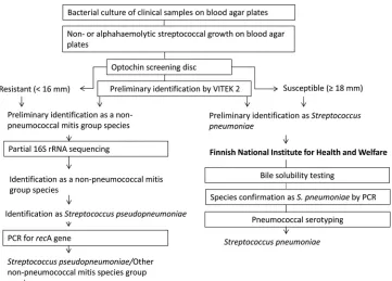
Bacterial strains.In this study, 188S. pneumoniaestrains and 101 strains belonging to other species of the S. mitisspecies group were analyzed. All the strains had been isolated from human clinical specimens at the Clinical Microbiology Laboratory of Turku University Hospital, Turku, Finland. The identification process for the strains is outlined in Fig. 1.
All S. pneumoniaestrains were identified as pneumococci by optochin sensitivity testing using optochin discs (optochin, 10g, Diatabs; ROSCO Diagnostica A/S, Taastrup, Denmark). AllS. pneumoniae
strains were subsequently serotyped and confirmed asS. pneumoniaeby optochin sensitivity and bile solubility testing at the National Institute for Health and Welfare (Helsinki, Finland). Also, a multiplex PCR with primers specific forS. pneumoniaewas performed at the National Institute for Health and Welfare for all the S. pneumoniaestrains as described by Siira et al. (52). The serotyping results for theS. pneumoniaestrains are shown in Table S2.
Fifty of theS. mitisspecies group strains other thanS. pneumoniaebelonged to the collection used in references 13 and 48. The remaining 51S. mitisspecies group strains were new isolates. The isolates had been presumptively identified as members of theS. mitisspecies group by the Vitek 2 instrument (bioMérieux, Grenoble, France) (13). An optochin resistance test (optochin, 10 g, Diatabs; ROSCO Diagnostica A/S, Taastrup, Denmark) was also performed. The strains were then identified by pyrose-quencing of the 16S rRNA gene at the Finnish National Institute for Health and Welfare as described by Haanperä et al. (13). Of theS. mitisspecies group strains tested, there were 24 strains for which the pyrosequencing results of the 16S rRNA differed by the same number of nucleotides (1 to 3 nucleotides) from the 16S rRNA sequences of the type strains ofS. pneumoniae,S. pseudopneumoniae,S. mitis, andS. oralis. In order to confirm whether these isolates belonged to the speciesS. pseudopneumoniae,recA
sequencing according to reference 32 was performed.
on May 16, 2020 by guest
http://jcm.asm.org/
Cultivation and storage.All isolates were stored in a mixture of skim milk and glycerol at⫺70°C before they were initially cultured on BD BBL Trypticase soy agar with 5% sheep blood (Becton Dickinson, Franklin Lakes, NJ, USA). Pure cultures were inoculated on the same plates from these initial culture plates. The resulting pure cultures were incubated at 35°C with 5% CO2for 17 to 24 h before analysis by
mass spectrometry.
Sample preparation.MALDI-TOF sample preparation of all strains was carried out as described in reference 47. Direct application was first carried out to generate MALDI-TOF MS spectra, and if it did not result in a score value ofⱖ2.0 (designated good identification to the species level by MALDI Biotyper software version 3.0 [RUO]), ethanol/formic acid extraction was performed. All strains were FIG 1Identification process forStreptococcus pneumoniaestrains and theS. mitisspecies group strains used in this study.
FIG 2List(score) calculation. “Strain # xyz” represents the spectrum of an unknown isolate; Spn,S. pneumoniae; Smi,S. mitis; Sor,S. oralis; Ssp,Streptococcusspp. In the first step, each log(score) of the first 10 positions of a ranking list is multiplied by a factor of 10 (1st position), a factor of 9 (2nd position), . . ., a factor of 2 (9th position), and a factor of 1 for the 10th position to calculate weighted log(scores). In the second step, the list(scores) are calculated by totaling weighted log(scores) for each single species appearing in the ranking list. List(scores) can be compared between all species appearing in the ranking list. In the example above, the highest list(score), 101.6, was observed forS. pneumoniae.
on May 16, 2020 by guest
http://jcm.asm.org/
[image:6.585.42.403.69.328.2] [image:6.585.47.364.498.671.2]measured on a Microflex LT instrument (Bruker Daltonics, Bremen, Germany) using Flex Control 3.0 software.
Data analysis.Spectra were analyzed using MALDI Biotyper software version 3.0 (RUO) for both database versions 3.3.1 (4,613 reference entries) and 4.0.0.1 (5,627 reference entries). The differences in these databases are summarized in Table 1. For result interpretation, the standard data interpretation rules [using log(scores) for the best and second best matches of the ranking list] were applied to both database versions.
MALDI Biotyper database version 3.3.1 (4,613 entries; RUO) contained 1, 4, and 8S. mitis,S. oralis, and S. pneumoniaestrains, respectively. For the updated database version 4.0.0.1 (5,627 entries; RUO), the spectra of several hundred well characterized strains ofS. mitis,S. oralis, andS. pneumoniae
from reference centers and routine laboratories covering strains from a broad variety of geograph-ical and other origins were analyzed, and several strains of each of these species were added in order to improve or optimize the species resolution between these three species. In DB_5627 as well as in the newer MALDI Biotyper database version DB_5989, the three species were represented by 39, 38, and 30 isolates, respectively (Table 1). When version DB_5627 was used, the first 10 positions of the ranking lists were also used to calculate a new supplementary list(score). The process of calculating the list(scores) is outlined in Fig. 2. At the first step, each log(score) of the first 10 positions of a ranking list is multiplied by a factor of 10 (1st position), a factor of 9 (2nd position), . . ., a factor of 2 (9th position), and a factor of 1 for the 10th position to calculate weighted log(scores). In the second step, each weighted log(score) is summarized for each single species appearing in the ranking list. List(scores) were compared between all species appearing in the ranking list. For example, if onlyS. pneumoniae
appears in the ranking list, the final list(score) is a sum of all 10 weighted log(scores). If only the first position of a ranking list reportsS. pneumoniaeand the remaining positions report onlyS. oralis, the influence of the first position is much lower in the overall summary. The use of such list(scores) as an additional tool to differentiate closely related microorganisms requires a database content preferably of more than 10 strains per species. The most abundant species in the ranking list is finally emphasized.
SUPPLEMENTAL MATERIAL
Supplemental material for this article may be found at
https://doi.org/10.1128/
JCM.01990-16
.
TEXT S1
, PDF file, 0.9 MB.
ACKNOWLEDGMENTS
This work was supported by the Research Fund of Hospital District of Southwest
Finland. Inka Harju has also received travel grants from the American Society for
Microbiology and from the University of Helsinki in order to present preliminary results
of this work as a poster at the General Meeting of American Society for Microbiology.
Inka Harju, Kaisu Rantakokko-Jalava, and Marjo Haanperä declare they have no
conflicts of interest. Cristoph Lange, Markus Kostrzewa, and Thomas Maier are
em-ployed by Bruker Daltonik GmbH.
REFERENCES
1. Spellerberg B, Brandt C. 2011. Streptococcus, p 331–349.InVersalovic J, Carroll KC, Funke G, Jorgensen JH, Landry ML, Warnock DW (ed), Manual of clinical microbiology, 10th ed. ASM Press, Washington, DC. 2. Doern C, Burnham C. 2010. It’s not easy being green: the viridans group
streptococci with a focus on pediatric clinical manifestations. J Clin Microbiol 48:3829 –3835.https://doi.org/10.1128/JCM.01563-10. 3. Krzys´ciak W, Pluskwa KK, Jurczak A, Kos´cielniak D. 2013. The
pathoge-nicity of theStreptococcus genus. Eur J Clin Microbiol Infect Dis 32: 1361–1376.https://doi.org/10.1007/s10096-013-1914-9.
4. Mitchell TJ. 2003. The pathogenesis of streptococcal infections: from tooth decay to meningitis. Nat Rev Microbiol 1:219 –230.https://doi.org/ 10.1038/nrmicro771.
5. Facklam R. 2002. What happened to the streptococci: overview of taxonomic and nomenclature changes. Clin Microbiol Rev 15:613– 630.
https://doi.org/10.1128/CMR.15.4.613-630.2002.
6. Arbique JC, Poyart C, Trieu-Cuot P, Quesne G, Carvalho Mda G, Steiger-walt AG, Morey RE, Jackson D, Davidson RJ, Facklam RR. 2004. Accuracy of phenotypic and genotypic testing for identification ofStreptococcus pneumoniae and description of Streptococcus pseudopneumoniae sp. nov. J Clin Microbiol 42:4686 – 4696. https://doi.org/10.1128/ JCM.42.10.4686-4696.2004.
7. Kawamura Y, Hou X-G, Sultana F, Miura H, Ezaki T. 1995. Determination of 16S rRNA sequences ofStreptococcus mitisand phylogenetic
relation-ships among members of the genusStreptococcus. Int J Syst Bacteriol 45:406 – 408.https://doi.org/10.1099/00207713-45-2-406.
8. Camelo-Castillo A, Benitez-Paez A, Belda-Ferre P, Cabrera-Rubio R, Mira A. 2014. Streptococcus dentisani sp. nov., a novel member of the mitis species group. Int J Syst Evol Microbiol 64:60 – 65.https://doi.org/ 10.1099/ijs.0.054098-0.
9. Zbinden A, Mueller NJ, Tarr PE, Sproer C, Keller PM, Bloemberg GV. 2012. Streptococcus tigurinus sp. nov., isolated from blood of patients with endocarditis, meningitis and spondylodiscitis. Int J Syst Evol Microbiol 62:2941–2945.https://doi.org/10.1099/ijs.0.038299-0.
10. Wessels E, Schelfaut JJ, Bernards AT, Claas EC. 2012. Evaluation of several biochemical and molecular techniques for identification ofStreptococcus pneumoniaeandStreptococcus pseudopneumoniaeand their detection in respiratory samples. J Clin Microbiol 50:1171–1177. https://doi.org/ 10.1128/JCM.06609-11.
11. Ikryannikova LN, Lapin KN, Malakhova MV, Filimonova AV, Ilina EN, Dubovickaya VA, Sidorenko SV, Govorun VM. 2011. Misidentification of alpha-hemolytic streptococci by routine tests in clinical practice. Infect Genet Evol 11:1709 –1715.https://doi.org/10.1016/j.meegid.2011.07.010. 12. Bascomb S, Manafi M. 1998. Use of enzyme tests in characterization and identification of aerobic and facultatively anaerobic gram-positive cocci. Clin Microbiol Rev 11:318 –340.
13. Haanperä M, Jalava J, Huovinen P, Meurman O, Rantakokko-Jalava K.
on May 16, 2020 by guest
http://jcm.asm.org/
2007. Identification of alpha-hemolytic streptococci by pyrosequencing the 16S rRNA gene and by use of VITEK 2. J Clin Microbiol 45:762–770.
https://doi.org/10.1128/JCM.01342-06.
14. Summanen PH, Rowlinson MC, Wooton J, Finegold SM. 2009. Evaluation of genotypic and phenotypic methods for differentiation of the mem-bers of the Anginosus group streptococci. Eur J Clin Microbiol Infect Dis 28:1123–1128.https://doi.org/10.1007/s10096-009-0758-9.
15. Chatzigeorgiou KS, Sergentanis TN, Tsiodras S, Hamodrakas SJ, Bagos PG. 2011. Phoenix 100 versus Vitek 2 in the identification of gram-positive and gram-negative bacteria: a comprehensive meta-analysis. J Clin Microbiol 49:3284 –3291.https://doi.org/10.1128/JCM.00182-11. 16. Teles C, Smith A, Ramage G, Lang S. 2011. Identification of clinically
relevant viridans group streptococci by phenotypic and genotypic anal-ysis. Eur J Clin Microbiol Infect Dis 30:243–250.https://doi.org/10.1007/ s10096-010-1076-y.
17. Kawamura Y, Whiley RA, Shu SE, Ezaki T, Hardie JM. 1999. Genetic approaches to the identification of the mitis group within the genus
Streptococcus. Microbiology 145:2605–2613. https://doi.org/10.1099/ 00221287-145-9-2605.
18. Whatmore AM, Efstratiou AP, Pickerill AP, Broughton K, Woodard G, Sturgeon D, George R, Dowson CG. 2000. Genetic relationships between clinical isolates ofStreptococcus pneumoniae,Streptococcus oralis, and
Streptococcus mitis: characterization of “atypical” pneumococci and or-ganisms allied toS. mitis harboringS. pneumoniae virulence factor-encoding genes. Infect Immun 68:1374 –1382.
19. Hoshino T, Fujiwara T, Kilian M. 2005. Use of phylogenetic and pheno-typic analyses to identify nonhemolytic streptococci isolated from bac-teremic patients. J Clin Microbiol 43:6073– 6085.https://doi.org/10.1128/ JCM.43.12.6073-6085.2005.
20. Chi F, Nolte O, Bergmann C, Ip M, Hakenbeck R. 2007. Crossing the barrier: evolution and spread of a major class of mosaic pbp2x in
Streptococcus pneumoniae, S. mitis, and S. oralis. Int J Med Microbiol 297:503–512.https://doi.org/10.1016/j.ijmm.2007.02.009.
21. Kilian M, Poulsen K, Blomqvist T, Håvarstein L, Bek-Thomsen M, Tettelin H, Sørensen UBS. 2008. Evolution ofStreptococcus pneumoniaeand its close commensal relatives. PLoS One 3:e2683.https://doi.org/10.1371/ journal.pone.0002683.
22. Do T, Jolley KA, Maiden MC, Gilbert SC, Clark D, Wade WG, Beighton D. 2009. Population structure ofStreptococcus oralis. Microbiology 155: 2593–2602.https://doi.org/10.1099/mic.0.027284-0.
23. Garnier F, Gerbaud G, Courvalin P, Galimand M. 1997. Identification of clinically relevant viridans group streptococci to the species level by PCR. J Clin Microbiol 35:2337–2341.
24. Teng LJ, Hsueh PR, Tsai JC, Chen PW, Hsu JC, Lai HC, Lee CN, Ho SW. 2002.groESLsequence determination, phylogenetic analysis, and spe-cies differentiation for viridans group streptococci. J Clin Microbiol 40:3172–3178.https://doi.org/10.1128/JCM.40.9.3172-3178.2002. 25. Täpp J, Thollesson M, Herrmann B. 2003. Phylogenetic relationships and
genotyping of the genusStreptococcusby sequence determination of the RNase P RNA gene,rnpB. Int J Syst Evol Microbiol 53:1861–1871.
https://doi.org/10.1099/ijs.0.02639-0.
26. Innings Å Krabbe M, Ullberg M, Herrmann B. 2005. Identification of 43
Streptococcusspecies by pyrosequencing analysis of thernpBgene. J Clin Microbiol 43:5983–5991. https://doi.org/10.1128/JCM.43.12.5983 -5991.2005.
27. Isaksson J, Rasmussen M, Nilson B, Svensson Stadler L, Kurland S, Olaison L, Ek E, Herrmann B. 2015. Comparison of species identification of endocarditis associated viridans streptococci usingrnpBgenotyping and 2 MALDI-TOF systems. Diagn Microbiol Infect Dis 81:240 –245.https:// doi.org/10.1016/j.diagmicrobio.2014.12.007.
28. Drancourt M, Roux V, Fournier PE, Raoult D. 2004.rpoBgene sequence-based identification of aerobic gram-positive cocci of the genera Strep-tococcus, Enterococcus,Gemella,Abiotropha, andGranulicatella. J Clin Microbiol 42:497–504.https://doi.org/10.1128/JCM.42.2.497-504.2004. 29. Poyart C, Quesne G, Coulon S, Berche P, Trieu-Cuot P. 1998. Identification
of streptococci to species level by sequencing the gene encoding the manganese-dependent superoxide dismutase. J Clin Microbiol 36: 41– 47.
30. Poyart C, Quesne G, Trieu-Cuot P. 2002. Taxonomic dissection of the Streptococcus bovis group by analysis of manganese-dependent super-oxide dismutase gene (sodA) sequences: reclassification of ‘Streptococ-cus infantarius subsp. coli’ as Streptococ‘Streptococ-cus lutetiensis sp. nov. and of Streptococcus bovis biotype 11.2 as Streptococcus pasteurianus sp. nov.
Int J Syst Evol Microbiol 52:1247–1255. https://doi.org/10.1099/ 00207713-52-4-1247.
31. Picard FJ, Ke D, Boudreau DK, Boissinot M, Huletsky A, Richard D, Ouellette M, Roy PH, Bergeron MG. 2004. Use of tufsequences for genus-specific PCR detection and phylogenetic analysis of 28 strepto-coccal species. J Clin Microbiol 42:3686 –3695.https://doi.org/10.1128/ JCM.42.8.3686-3695.2004.
32. Sistek V, Boissinot M, Boudreau DK, Huletsky A, Picard FJ, Bergeron MG. 2012. Development of a real-time PCR assay for the specific detection and identification of Streptococcus pseudopneumoniae using the recA gene: identification of S. pseudopneumoniae by real-time PCR. Clin Microb Infect 18:1089 –1096. https://doi.org/10.1111/j.1469 -0691.2011.03684.x.
33. Seng P, Drancourt M, Gouriet F, La Scola B, Fournier PE, Rolain JM, Raoult D. 2009. Ongoing revolution in bacteriology: routine identification of bacteria by matrix-assisted laser desorption ionization time-of-flight mass spectrometry. Clin Infect Dis 49:543–551.https://doi.org/10.1086/ 600885.
34. Bizzini A, Durussel C, Bille J, Greub G, Prod’hom G. 2010. Performance of matrix-assisted laser desorption ionization-time of flight mass spectrom-etry for identification of bacterial strains routinely isolated in a clinical microbiology laboratory. J Clin Microbiol 48:1549 –1554.https://doi.org/ 10.1128/JCM.01794-09.
35. Neville SA, Lecordier A, Ziochos H, Chater MJ, Gosbell IB, Maley MW, van Hal SJ. 2011. Utility of matrix-assisted laser desorption ionization-time of flight mass spectrometry following introduction for routine laboratory bacterial identification. J Clin Microbiol 49:2980 –2984.https://doi.org/ 10.1128/JCM.00431-11.
36. Friedrichs C, Rodloff AC, Chhatwal GS, Schellenberger W, Eschrich K. 2007. Rapid identification of viridans streptococci by mass spectrometric discrimination. J Clin Microbiol 45:2392–2397.https://doi.org/10.1128/ JCM.00556-07.
37. van Veen SQ, Claas EC, Kuijper EJ. 2010. High-throughput identification of bacteria and yeast by matrix-assisted laser desorption ionization-time of flight mass spectrometry in conventional medical microbiology lab-oratories. J Clin Microbiol 48:900 –907. https://doi.org/10.1128/ JCM.02071-09.
38. De Bel A, Wybo I, Pierard D, Lauwers S. 2010. Correct implementation of matrix-assisted laser desorption ionization-time of flight mass spectrom-etry in routine clinical microbiology. J Clin Microbiol 48:1991. (Reply, 48:1991–1992.)https://doi.org/10.1128/JCM.00403-10.
39. Scholz CF, Poulsen K, Kilian M. 2012. Novel molecular method for identification ofStreptococcus pneumoniaeapplicable to clinical micro-biology and 16S rRNA sequence-based microbiome studies. J Clin Mi-crobiol 50:1968 –1973.https://doi.org/10.1128/JCM.00365-12.
40. Martiny D, Busson L, Wybo I, El Haj RA, Dediste A, Vandenberg O. 2012. Comparison of the Microflex LT and Vitek MS systems for routine identification of bacteria by matrix-assisted laser desorption ionization-time of flight mass spectrometry. J Clin Microbiol 50:1313–1325.https:// doi.org/10.1128/JCM.05971-11.
41. Werno AM, Christner M, Anderson TP, Murdoch DR. 2012. Differentiation ofStreptococcus pneumoniaefrom nonpneumococcal streptococci of the
Streptococcus mitisgroup by matrix-assisted laser desorption ionization-time of flight mass spectrometry. J Clin Microbiol 50:2863–2867.https:// doi.org/10.1128/JCM.00508-12.
42. Davies AP, Reid M, Hadfield SJ, Johnston S, Mikhail J, Harris LG, Jenkinson HF, Berry N, Lewis AM, El-Bouri K, Mack D. 2012. Identification of clinical isolates of alpha-hemolytic streptococci by 16S rRNA gene sequencing, matrix-assisted laser desorption ionization-time of flight mass spectrom-etry using MALDI Biotyper, and conventional phenotypic methods: a comparison. J Clin Microbiol 50:4087– 4090. https://doi.org/10.1128/ JCM.02387-12.
43. Roa P, Sanchez Carrillo C, Marin M, Romero F, Cercenado E, Bouza E. 2013. Value of matrix-assisted laser desorption ionization-time of flight for routine identification of viridans group streptococci causing blood-stream infections. Clin Microbiol Infect 19:438 – 444. https://doi.org/ 10.1111/j.1469-0691.2012.03837.x.
44. Ikryannikova LN, Filimonova AV, Malakhova MV, Savinova T, Filimonova O, Ilina EN, Dubovickaya VA, Sidorenko SV, Govorun VM. 2013. Discrim-ination between Streptococcus pneumoniae and Streptococcus mitis based on sorting of their MALDI mass spectra. Clin Microbiol Infect 19:1066 –1071.https://doi.org/10.1111/1469-0691.12113.
45. Dubois D, Segonds C, Prere MF, Marty N, Oswald E. 2013. Identification of clinicalStreptococcus pneumoniaeisolates among other alpha and
on May 16, 2020 by guest
http://jcm.asm.org/
nonhemolytic streptococci by use of the Vitek MS matrix-assisted laser desorption ionization-time of flight mass spectrometry system. J Clin Microbiol 51:1861–1867.https://doi.org/10.1128/JCM.03069-12. 46. Branda JA, Markham RP, Garner CD, Rychert JA, Ferraro MJ. 2013.
Performance of the Vitek MS v2.0 system in distinguishingStreptococcus pneumoniaefrom nonpneumococcal species of theStreptococcus mitis
group. J Clin Microbiol 51:3079 –3082. https://doi.org/10.1128/ JCM.00824-13.
47. Woods K, Beighton D, Klein JL. 2014. Identification of the ‘Streptococcus anginosus group’ by matrix-assisted laser desorption ionization–time-of-flight mass spectrometry. J Med Microbiol 63:1143–1147. https:// doi.org/10.1099/jmm.0.076653-0.
48. Kärpänoja P, Harju I, Rantakokko-Jalava K, Haanpera M, Sarkkinen H. 2014. Evaluation of two matrix-assisted laser desorption ionization-time of flight mass spectrometry systems for identification of viridans group streptococci. Eur J Clin Microbiol Infect Dis 33:779 –788.https://doi.org/ 10.1007/s10096-013-2012-8.
49. Angeletti S, Dicuonozo G, Avola A, Crea F, Dedej E, Vailati F, Farina C, De
Florio L. 2015. Viridans group streptococci clinical isolates: MALDI-TOF mass spectrometry versus gene sequence-based identification. PLoS One 10:e0120502.https://doi.org/10.1371/journal.pone.0120502. 50. Rychert J, Burnham CA, Bythrow M, Garner OB, Ginocchio CC,
Jenne-mann R, Lewinski MA, Manji R, Mochon AB, Procop GW, Richter SS, Sercia L, Westblade LF, Ferraro MJ, Branda JA. 2013. Multicenter evaluation of the Vitek MS matrix-assisted laser desorption ionization-time of flight mass spectrometry system for identification of Gram-positive aerobic bacteria. J Clin Microbiol 51:2225–2231. https://doi.org/10.1128/ JCM.00682-13.
51. Van Preen J, van Veen S, Schelfaut JJG, Wessels E. 2016. MALDI-TOF mass spectrometry for differentiation betweenStreptococcus pneumoniaeand
Streptococcus pseudopneumoniae. Diagn Microb Infect Dis 85:9 –11.
https://doi.org/10.1016/j.diagmicrobio.2016.01.012.
52. Siira L, Kaijalainen T, Lambertsen L, Nahm MH, Toropainen M, Virolainen A. 2012. From quellung to multiplex PCR, and back when needed, in pneumococcal serotyping. 2012. J Clin Microbiol 50:2727–2731.https:// doi.org/10.1128/JCM.00689-12.