Typing and Clustering of
Yersinia pseudotuberculosis
Isolates by
Restriction Fragment Length Polymorphism Analysis Using Insertion
Sequences
E. Voskresenskaya,a,bC. Savin,aA. Leclercq,aG. Tseneva,bE. Carniela
YersiniaResearch Unit, National Reference Laboratory, WHO Collaborating Center forYersinia, Institut Pasteur, Paris, Francea
; Laboratory of Bacterial Respiratory Infections, Institut Pasteur, Saint Petersburg, Russiab
Yersinia pseudotuberculosisis an enteropathogen that has an animal reservoir and causes human infections, mostly in temperate
and cold countries. Most of the methods previously used to subdivideY. pseudotuberculosiswere performed on small numbers
of isolates from a specific geographical area. One aim of this study was to evaluate the typing efficiency of restriction fragment length polymorphism of insertion sequence hybridization patterns (IS-RFLP) compared to other typing methods, such as
sero-typing, ribosero-typing, and multilocus sequence typing (MLST), on the same set of 80 strains ofY. pseudotuberculosisof global
ori-gin. We found that IS100was not adequate for IS-RFLP but that both IS285and IS1541efficiently subtypedY.
pseudotuberculo-sis. The discriminatory index (DI) of IS1541-RFLP (0.980) was superior to those of IS285-RFLP (0.939), ribotyping (0.944), MLST
(0.861), and serotyping (0.857). The combination of the two IS (2IS-RFLP) further increased the DI to 0.998. Thus, IS-RFLP is a
powerful tool for the molecular typing ofY. pseudotuberculosisand has the advantage of exhibiting well-resolved banding
pat-terns that allow for a reliable comparison of strains of worldwide origin. The other aim of this study was to assess the clustering power of IS-RFLP. We found that 2IS-RFLP had a remarkable capacity to group strains with similar genotypic and phenotypic
markers, thus identifying robust populations withinY. pseudotuberculosis. Our study thus demonstrates that 2IS- and even
IS1541-RFLP alone might be valuable tools for the molecular typing of global isolates ofY. pseudotuberculosisand for the
analy-sis of the population structure of this species.
Y
ersinia pseudotuberculosisis an enteropathogenic speciesbe-longing to the genusYersinia. This species is transmitted by the fecal-oral route and has a wide range of animal reservoirs (1). In humans, the main clinical manifestations of pseudotuberculo-sis are abdominal pain (mimicking appendicitis), fever, and some-times diarrhea (2). Dissemination to deeper tissues and the blood-stream are frequent in older patients with underlying conditions (3). In some specific areas (e.g., Russia and Japan),Y.
pseudotu-berculosismay also cause a specific disease known as Far East
scar-let-like fever (4–6), characterized by a strong inflammatory syn-drome accompanying intestinal disorders. Although humanY.
pseudotuberculosisinfections are less frequent than those caused
byYersinia enterocoliticaworldwide, human pseudotuberculosis
outbreaks are reported in various parts of the world, such as Japan (7,8), Russia (9,10), France (3,11), and Scandinavia (12–15).
Phenotypically,Y. pseudotuberculosisis subdivided into 15 se-rotypes and several subsese-rotypes (16). The most common sero-types are O:1 to O:5, while the others are restricted to geographical areas in Asia. Because most strains isolated from human cases are of the major serotypes O:1 and O:3, the discriminatory power of serotyping is limited. However, sinceY. pseudotuberculosis exhib-its a certain degree of genetic polymorphisms (17), molecular techniques represent valuable alternatives for subtyping this spe-cies. One of the most commonly used molecular typing methods is the analysis of the genomic restriction fragment length poly-morphism (RFLP) obtained by pulsed-field gel electrophoresis (PFGE) (18–21). This technique is valuable for investigating an outbreak to find a common source of contamination. Typing ofY.
pseudotuberculosisby multilocus enzyme electrophoresis (22–24),
RFLP of the pYV virulence plasmid (25), or multilocus variable-number tandem-repeat analysis (MLVA) (26) have also been
de-scribed. These techniques have been applied to a small number of
Y. pseudotuberculosisstrains or to isolates from a given
geograph-ical area during outbreaks. Recently, larger sets ofY.
pseudotuber-culosisstrains of global origin have been subjected to other typing
methods. Ribotyping of 80Y. pseudotuberculosisstrains isolated from various countries and different hosts demonstrated that this technique allowed the subdivision of the strains into 27 ribotypes (27). However, the small number of hybridizing bands and the poor resolution of some profiles led to the conclusion that this technique has some intrinsic limitations (27). The analysis of mul-tilocus sequence typing (MLST) was also recently used to study the diversity ofY. pseudotuberculosis. In one work, 79 strains from various countries were analyzed (28), of which 11 were subse-quently found to beYersinia similisand 68 were trueY.
pseudotu-berculosis. Among these 68 Y. pseudotuberculosisisolates, 54
se-quence types (STs) were identified. In another work involving MLST, 417 strains belonging to theY. pseudotuberculosiscomplex and isolated from all continents were typed (29). The 386 trueY.
pseudotuberculosisstrains were divided into 76 STs. The
perfor-mance of IS-RFLP for typingY. pseudotuberculosisstrains has also been investigated in two studies. In one of them, IS1541-RFLP
Received12 February 2014 Returned for modification11 March 2014 Accepted20 March 2014
Published ahead of print26 March 2014
Editor:B. A. Forbes
Address correspondence to E. Carniel, [email protected].
Copyright © 2014, American Society for Microbiology. All Rights Reserved.
doi:10.1128/JCM.00397-14
on May 16, 2020 by guest
http://jcm.asm.org/
typing of 20Y. pseudotuberculosisisolates indicated that its dis-criminatory power was similar to that of PFGE (21). In another study, IS100- and IS285-RFLP typing of 27 isolates showed that IS-RFLP distinguished 24 IS types (30). Therefore, different dis-criminatory powers were observed for different methods and dif-ferent sets of strains.
An analysis of the genetic polymorphisms of a species may also be used to study subpopulations within this species and some-times to identify epidemiological links between geographical ori-gins or sources and specific genetic clusters. MLST analysis of a large set ofY. pseudotuberculosisstrains of global origins did not identify strong patterns of specificity for geographical origin, host type, or serotype (29). IS-RFLP typing using two or three different insertion sequences (IS100, IS285, and IS1541) has been success-fully applied to the molecular analysis of a collection of diverse strains ofYersinia pestis(31) and recently to the epidemiological study of a plague outbreak (32). The 3IS-RFLP dendrogram dem-onstrated a clear clustering ofY. pestisstrains, not only according to their biovars but also according to their geographical origin. Whether IS-RFLP can efficiently identify genetic subgroups ofY.
pseudotuberculosishas never been determined.
The aims of this study were to evaluate the discriminatory power of IS-RFLP compared to those of serotyping, ribotyping, and MLST on the same set of strains of global origin and to esti-mate the potential for IS-RFLP to delineate populations within the speciesY. pseudotuberculosis.
MATERIALS AND METHODS
Bacterial strains and growth conditions.The 80Y. pseudotuberculosis
strains used in this study were taken from the strain collection of the
YersiniaResearch Unit and the National Reference Laboratory at the In-stitut Pasteur. Their main characteristics are described inTable 1. The serotypes of the strains were determined by genoserotyping according to Bogdanovich et al. (33), with one modification: the O:8 genoserotype was redefined as O:4c since the strains agglutinated with the anti-O:4 antise-rum and their PCR profile were a combination of the O:4a and O:4b banding patterns. The ribotypes were determined in a previous study (27). The STs were already established for 66 strains (29). We determined the STs of the remaining strains, except for strain IP32921, which had lost its viability. Bacterial suspensions were prepared from stock cultures kept at
⫺80°C and streaked onto Luria-Bertani agar plates or grown in peptone broth with shaking for 24 h to 48 h at 28°C.
DNA extraction, restriction, and transfer to nylon membranes.The extraction of genomic DNA from eachY. pseudotuberculosisstrain was performed as described previously (34). Five micrograms of each sample was digested overnight at 37°C with EcoRI or EcoRV before being loaded onto 0.8% agarose gels and subjected to electrophoresis (50 V in 1⫻ Tris-borate-EDTA buffer) for 24 h. The DNA ofY. pestisstrain IP304 was systematically loaded on each gel to serve in intergel normalization. DNA bands were stained with ethidium bromide. Alkaline denaturation, neu-tralization, and transfer of total DNA onto nylon filters (Hybond-N⫹; Amersham, England) with a VacuGene apparatus (Pharmacia LKB Bio-technology, Uppsala, Sweden) were performed as previously described (35).
Preparation of the IS probes and hybridization.The primer pairs used to amplify a portion of the insertions sequences were IS100-F (5=-A AAACGTTCGAAGAGTATGA-3=) and IS100-R (5=-GATGAGCAGGCG GGGGGCCA-3=) (255 bp), IS1541-F (5=-AAAGCTTTCAGCTTTGGGT C-3=) and IS1541-R (5=-TCTTTCCCTTCAGGTACCCC-3=) (319 bp), and IS285-F (5=-AGCTTACCGAACACCTCGGG-3=) and IS285-R (5=-G TTGATGCCCAGCGCTAGGA-3=) (406 bp) (31). PCR amplification re-actions were performed on the genomic DNA ofY. pestisstrain CO92 as a template, as previously described (31). The IS probes were peroxidase
labeled with the enhanced chemiluminescence (ECL) direct nucleic acid labeling and detection system (Amersham). Hybridization was performed overnight at 42°C.
Bioinformatic analysis of the IS-RFLP patterns.The hybridization patterns obtained with each IS were scanned, and the computerized data were analyzed using the BioNumerics software version 6.6 (Applied Maths, Kortrijk, Belgium). Bands automatically assigned by the computer were checked by eyes and corrected manually when necessary. A position tolerance of 1.8 was selected for each IS. Cluster analysis of the individual or combined IS-RFLP patterns was done by the unweighted-pair group method using average linkages (UPGMA), using the Dice coefficient to analyze the similarities of the banding patterns. The discriminatory power of each IS-RFLP was determined by calculating the discrimination index (DI) based on the Simpson’s index of diversity (36). The DI depends on the number of types defined by the test method and the relative frequen-cies of these types, using the equation DI⫽1⫺{[1/(N⫻[N⫺1])]⫻ (⌺nj[nj⫺1])}, whereNis the total number of unrelated isolates andnjis the number of strains that belong to thejth type. An algorithm minimal spanning tree (MST) based on serotype, ribotype, ST, and 2IS type of each strain was constructed using BioNumerics version 6.6.
RESULTS
Setup of the IS-RFLP conditions.The 80Y. pseudotuberculosis
strains used to evaluate the IS-RFLP method were chosen based on their variety of geographical origins, sources, serotypes (O:1 to O:6), and on the fact that their ribotypes (27) and STs (29) were already determined for most of them, thus allowing for a compar-ison of the three molecular typing methods on the same set of diverse strains.
The IS100element was previously reported to be either missing or in low copy numbers (ⱕ5) in variousY. pseudotuberculosis
strains (30,37–39). Using a set of 11 strains of various serotypes, no IS100hybridizing fragments were detected in seven of them, and only one or two fragments were detected in three other strains (data not shown). Therefore, because of the low discriminatory power of IS100-RFLP, we decided not to use this IS further for IS-RFLP typing ofY. pseudotuberculosis. In contrast, IS1541and IS285were reported to be systematically present and in several copies in the genome ofY. pseudotuberculosisisolates previously tested (4,21,30,40). We therefore decided to estimate the perfor-mance of a typing method forY. pseudotuberculosisbased on the polymorphisms of individual and combined IS1541and IS285
fingerprints.
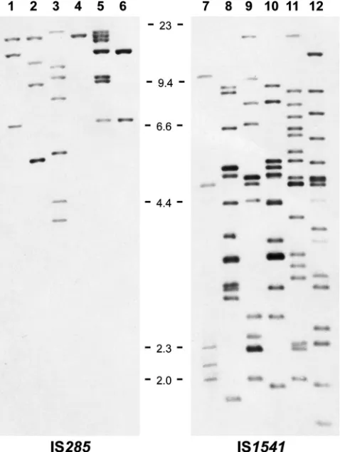
To optimize the resolution of the banding patterns, the genomic DNA of a few strains ofY. pseudotuberculosiswas di-gested with different restriction enzymes and hybridized with the IS1541and IS285probes. The hybridization profiles that gave the best resolution were obtained after EcoRI digestion for IS285and after EcoRV digestion for IS1541. An example of the banding pat-terns is provided inFig. 1. These two enzymes were therefore selected for further use.
To ensure that the hybridization profiles were correct, we also compared the sizes and numbers of fragments expected from the genome analysis of the sequenced strain IP32953 with those visu-alized after digestion of the DNA of this strain and IS1541or IS285
hybridization. Bands with the expected sizes were obtained. We also noted the absence of IS1541or IS285copies on the IP32953 pYV virulence plasmid, indicating that the loss of this replicon would not change the hybridization profile of the strain.
IS285-RFLP patterns. Upon hybridization with the IS285
probe of the DNA of the 80Y. pseudotuberculosisstrains, the num-ber of bands detected varied from 1 to 11 (Fig. 2), with 60% of the
on May 16, 2020 by guest
http://jcm.asm.org/
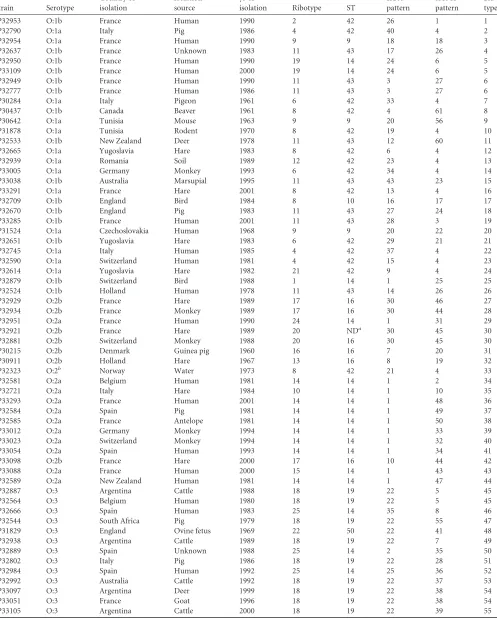
TABLE 1Characteristics of the 80 strains ofY. pseudotuberculosisused for IS-RFLP typing
Strain Serotype
Country of isolation
Isolation source
yr of
isolation Ribotype ST
IS285
pattern
IS1541
pattern
2IS type
IP32953 O:1b France Human 1990 2 42 26 1 1
IP32790 O:1a Italy Pig 1986 4 42 40 4 2
IP32954 O:1a France Human 1990 9 9 18 18 3
IP32637 O:1b France Unknown 1983 11 43 17 26 4
IP32950 O:1b France Human 1990 19 14 24 6 5
IP33109 O:1b France Human 2000 19 14 24 6 5
IP32949 O:1b France Human 1990 11 43 3 27 6
IP32777 O:1b France Human 1986 11 43 3 27 6
IP30284 O:1a Italy Pigeon 1961 6 42 33 4 7
IP30437 O:1b Canada Beaver 1961 8 42 4 61 8
IP30642 O:1a Tunisia Mouse 1963 9 9 20 56 9
IP31878 O:1a Tunisia Rodent 1970 8 42 19 4 10
IP32533 O:1b New Zealand Deer 1978 11 43 12 60 11
IP32665 O:1a Yugoslavia Hare 1983 8 42 6 4 12
IP32939 O:1a Romania Soil 1989 12 42 23 4 13
IP33005 O:1a Germany Monkey 1993 6 42 34 4 14
IP33038 O:1b Australia Marsupial 1995 11 43 43 23 15
IP33291 O:1a France Hare 2001 8 42 13 4 16
IP32709 O:1b England Bird 1984 8 10 16 17 17
IP32670 O:1b England Pig 1983 11 43 27 24 18
IP33285 O:1b France Human 2001 11 43 28 3 19
IP31524 O:1a Czechoslovakia Human 1968 9 9 20 22 20 IP32651 O:1b Yugoslavia Hare 1983 6 42 29 21 21
IP32745 O:1a Italy Human 1985 4 42 37 4 22
IP32590 O:1a Switzerland Human 1981 4 42 15 4 23 IP32614 O:1a Yugoslavia Hare 1982 21 42 9 4 24 IP32879 O:1b Switzerland Bird 1988 1 14 1 25 25 IP32524 O:1b Holland Human 1978 11 43 14 26 26
IP32929 O:2b France Hare 1989 17 16 30 46 27
IP32934 O:2b France Monkey 1989 17 16 30 44 28
IP32951 O:2a France Human 1990 24 14 1 31 29
IP32921 O:2b France Hare 1989 20 NDa 30 45 30
IP32881 O:2b Switzerland Monkey 1988 20 16 30 45 30 IP30215 O:2b Denmark Guinea pig 1960 16 16 7 20 31
IP30911 O:2b Holland Hare 1967 13 16 8 19 32
IP32323 O:2b Norway Water 1973 8 42 21 4 33
IP32581 O:2a Belgium Human 1981 14 14 1 2 34
IP32721 O:2a Italy Hare 1984 10 14 1 10 35
IP33293 O:2a France Human 2001 14 14 1 48 36
IP32584 O:2a Spain Pig 1981 14 14 1 49 37
IP32585 O:2a France Antelope 1981 14 14 1 50 38 IP33012 O:2a Germany Monkey 1994 14 14 1 33 39 IP33023 O:2a Switzerland Monkey 1994 14 14 1 32 40
IP33054 O:2a Spain Human 1993 14 14 1 34 41
IP33098 O:2b France Hare 2000 17 16 10 44 42
IP33088 O:2a France Human 2000 15 14 1 43 43
IP32589 O:2a New Zealand Human 1981 14 14 1 47 44 IP32887 O:3 Argentina Cattle 1988 18 19 22 5 45
IP32564 O:3 Belgium Human 1980 18 19 22 5 45
IP32666 O:3 Spain Human 1983 25 14 35 8 46
IP32544 O:3 South Africa Pig 1979 18 19 22 55 47 IP31829 O:3 England Ovine fetus 1969 22 50 22 41 48 IP32938 O:3 Argentina Cattle 1989 18 19 22 7 49
IP32889 O:3 Spain Unknown 1988 25 14 2 35 50
IP32802 O:3 Italy Pig 1986 18 19 22 28 51
IP32984 O:3 Spain Human 1992 25 14 25 36 52
IP32992 O:3 Australia Cattle 1992 18 19 22 37 53 IP33097 O:3 Argentina Deer 1999 18 19 22 38 54
IP33051 O:3 France Goat 1996 18 19 22 38 54
IP33105 O:3 Argentina Cattle 2000 18 19 22 39 55
(Continued on following page)
Voskresenskaya et al.
on May 16, 2020 by guest
http://jcm.asm.org/
strains displaying one to four copies of IS285. No hybridizing frag-ments common to all patterns were observed, but a band of⬇20.5 kb was found in 64 strains (80%), and another one of⬇7 kb was found in 41 strains (51%) (Fig. 2).
The 80 strains were separated into 43 distinct IS285profiles, while serotyping subdivided them into 10 groups, ribotyping into 27 groups, and MLST into 14 groups (Table 1). The dominant IS285profiles were: IS285profile/pattern 1 (285#1) (15 strains), 285#22 (11 strains), and 285#30 (8 strains). Together they repre-sented almost half of the strains (42.5%). Six profiles (285#3, 285#10, 285#20, 285#24, 285#31, and 285#41) were shared by two strains, and 34 profiles were unique. The discrimination index (DI) for IS285-RFLP was 0.939, compared to 0.857 for serotyping, 0.944 for ribotyping, and 0.861 for MLST. Therefore, IS285-RFLP had a discriminatory power higher than those of serotyping and MLST but lower than that of ribotyping.
We then examined whether a correlation could be observed between some epidemiological, phenotypic, or genotypic charac-teristics of the strains and their IS285types. The IS285profiles were not associated with specific geographical origins or sources, but it is worth noting that all four strains from Argentina belonged to 285#22, and all three strains from Russia belonged to 285#1 (Table 1). Furthermore, the majority of the 285#22 strains (9/11) were isolated from animal sources.
[image:4.585.45.542.77.305.2]No strict association between serotypes and IS285profiles was observed; nevertheless, the major IS285profiles were associated with a limited number of serotypes: 285#1 with serotypes O:1b and O:2a, 285#22 with serotype O:3, and 285#30 with serotypes O:2b and O:5a (Table 1). Similarly, some ribotypes correlated with specific IS285 profiles: all eight ribotype 14 (R14) and all three R27 strains were 285#1, the eight R17 strains were 285#10 or 285#30, and all 10 R18 strains were 285#22. Associations between the ST and IS285profiles were also noted in some instances: all 15 285#1 strains were from ST14 or ST2, 10 of the 11 285#22 strains
TABLE 1(Continued)
Strain Serotype
Country of isolation
Isolation source
yr of
isolation Ribotype ST
IS285
pattern
IS1541
pattern
2IS type IP33108 O:3 Bulgaria Human 1999 18 19 22 40 56
IP31411 O:4c Denmark Hare 1961 7 48 36 9 57
IP31833 O:4c England Sheep 1969 3 48 11 29 58
IP30151 O:4a Sweden Otter 1960 23 23 39 42 59
IP31830 O:4c England Human 1969 25 14 42 54 60 IP32687 O:4c France Wild species 1983 26 14 5 11 61
IP32817 O:5b Japan Hare 1986 19 73 38 52 62
IP32821 O:5a France Human 1986 17 16 30 14 63
IP32816 O:5b Japan Hare 1986 19 7 32 51 64
IP32952 O:5a France Human 1990 17 16 10 30 65
IP32463 O:5a Switzerland Guinea pig 1977 17 16 30 13 66 IP33061 O:5a Germany Monkey 1997 17 16 30 12 67 IP32699 O:5a France Wild species 1983 17 16 30 53 68 IP31553 O:6 Japan Guinea pig 1969 5 27 31 16 69 IP31554 O:6 Japan Guinea pig 1969 5 27 31 15 70
IP33156 O:1b Russia Human 2000 27 2 1 58 71
IP33157 O:1b Russia Human 2000 27 2 1 59 72
IP33158 O:1b Russia Human 2000 27 2 1 59 72
IP33162 O:1b Ukraine Human 1999 9 42 41 57 73
IP33161 O:1b Ukraine Rodent 1999 9 42 41 57 73
aND, not determined because the strain was no longer viable.
b
O:2: this strain agglutinated with the O:2 antiserum but had an unclassifiable genoserotype, different from O:2.
FIG 1Examples of IS-RFLP profiles obtained after EcoRI or EcoRV digestion of the genomic DNA of variousY. pseudotuberculosisstrains and hybridization with the IS285and IS1541probes, respectively. Lane 1, IP32879; lane 2, IP30911; lane 3, IP32614; lane 4, IP32938; lane 5, IP32984; lane 6, IP31554; lane 7, IP32953; lane 8, IP32581; lane 9, IP31411; lane 10, IP32721; lane 11, IP33051; lane 12, IP32817. The tick marks between the two panels indicate the size of the molecular mass markers (lambda DNA-HindIII digest) in kb.
on May 16, 2020 by guest
http://jcm.asm.org/
[image:4.585.44.283.339.656.2]FIG 2Dendrogram generated from the IS285-RFLP patterns of the 80Y. pseudotuberculosisstrains studied, using the UPGMA clustering analysis with the BioNumerics software. A position tolerance of 1.8% was chosen. Stars indicate hybridizing fragments of ca. 20.5 kb and 7 kb that were present in the majority of strains. Rectangles indicate clusters of strains of interest.
on May 16, 2020 by guest
http://jcm.asm.org/
[image:5.585.62.525.36.674.2]were from ST19, and seven 285#30 strains were from ST16 (Table 1). Therefore, a certain degree of association between some geno-types and IS285profiles was observed.
To further delineate the IS285genetic groups and their associ-ation with other phenotypic and genetic traits, a clustering analy-sis of the IS285profiles was performed. The resulting UPGMA dendrogram displayed two main branches (A and B) and one more distant branch corresponding to a single strain (IP30151, Fig. 2). The strains in branch A exhibited multiple IS285 -hybrid-izing fragments (Fig. 2). Most of these strains had a unique IS285
profile (10 among 12 strains), but they were all of serotype O:1b or O:4c, and they also belonged to a small number of MLST types (ST14, ST42, and ST43) and ribotypes (R9, R11, R25, and R26). Within branch B, at least four subclusters (B1 to B4) were delin-eated (Fig. 2). Subbranch B1, like branch A, contained strains with high copy numbers of IS285and with mostly unique IS285 pro-files. There was, however, a strong association with ST42 (14/21 strains), and to a lesser extent with R9, R8, and R4 (12/21 strains). Most of them were of serotype O:1a or O:1b (17/21). Subbranches B2, B3, and B4 had lower IS285copy numbers (Fig. 2). All strains in B2 had the same 285#1 profile, which was associated with sero-types O:1b and O:2a, ST14 (12/15 strains), and to a lesser extent with R14 (8/15 strains). B3 comprised strains exhibiting five dif-ferent IS285 profiles with a predominance of 285#30 (8/13 strains). Most strains in this cluster were of serotypes O:2b and O:5a (12/13 strains), ST16 (11/13 strains), and R17 (8/13 strains). B4 was composed of strains having the 285#22 profile, which was characterized by a single hybridizing fragment corresponding to the most conserved ⬇20.5-kb band (Fig. 2). All strains in this cluster were of serotype O:3, and all but one were ST19 and R18. The clustering ofY. pseudotuberculosisstrains based on their IS285
profiles thus showed a good, although not perfect, association with other phenotypic and genotypic traits.
IS1541-RFLP patterns.The polymorphisms of the IS1541
pro-files were greater than that of IS285, with 61 different RFLP pat-terns among 80Y. pseudotuberculosisstrains (Table 1). This higher number of profiles might be linked to an overall higher copy num-ber of IS1541, which varied from one to 33 copies per strain (Fig. 3). The most frequent IS1541copy number was two (13 strains), followed by seven (10 strains). No band common to all profiles was observed; however, a⬇8.5-kb hybridizing fragment was pres-ent in 40 strains (50%), and a⬇4.7-kb band was present in 44 strains (55%) (Fig. 3).
The most frequent IS1541profile was 1541#4, which was found in 11 strains (Table 1). The strains with this pattern were not those that had the most common IS285profiles. Of note, all but one strain with the 1541#4 pattern were of serotype O:1a, and all were of ST42. However, they had five different ribotypes, indicating that in that case, ribotyping was more discriminatory than IS1541 -RFLP. Except for 1541#4, no more than two strains shared a sim-ilar IS1541profile, and 51 of them had a unique IS1541profile. The discrimination index for IS1541-RFLP was 0.98 and therefore higher than those of all other compared methods (serotyping, MLST, ribotyping, and IS285-RFLP).
Because of this high degree of diversity, it was hardly possible to establish a link between individual IS1541profiles and specific countries of origin, serotypes, ribotypes, or STs. However, this relationship was examined in the UPGMA dendrogram generated by the clustering analysis. Similarly to IS285, the IS1541 dendro-grams displayed two main branches (A and B) and an additional
branch that, like in the IS285dendrogram, corresponded to strain IP30151 (Fig. 3). Branch A was characterized by a low number of IS1541copies and branch B by intermediate to high numbers of IS1541copies (Fig. 3). Branch A contained all strains with the 1541#4 pattern (10/11 of serotype O:1a) plus four other strains with distinct patterns, but all from serotype O:1b. There was a strong association with ST42 (14/15 strains) and to a lesser extent with R8, R6, and R4 (12/15 strains). Branch B was divided into six subbranches (B1 to B6) in which a robust association with specific serotypes, STs, and ribotypes was observed (Fig. 3). All strains in B1 were from O:2a and ST14, and most of them (4/6 strains) were R14. All 10 strains in B2 were O:3, ST19, and R18. This subbranch contained a large number of strains (6/10) of non-European ori-gin (from Argentina, Australia, and South Africa) that were iso-lated from animals (8/10 strains). Subbranch B3 was slightly more heterogeneous, with two serotypes (O:3 and O:4c), but all strains were ST14, and most of them (4/5) were R25. Most strains (5/6) in B4 were O:2a, all were ST14, and a majority (4/6) were R14. All 8 strains in B5 were O:1b, ST43, and R11. Finally, subbranch B6 was composed of strains of serotype O:2b or O:5a that all belonged to ST16 and most of which (8/12) belonged to R17. This subbranch was composed of European strains only, and most of them (10/12) had an animal source.
Additional strains that were not included in the above clusters were nevertheless grouped according to their epidemiological and genotypic characteristics (Fig. 3). Group 1 (G1) contained two strains isolated from hares in Japan that were both serotype O:5b and R19. G2 contained three strains that were O:1a, R9, and ST9. In G3, the two strains were from O:4c and ST48. Finally, within groups G4 to G7, the strains had identical serotypes, ribotypes, and STs, as well as the same geographical origin and sometimes the same source (guinea pigs in Japan, humans in France, and humans in Russia). Therefore, almost all strains that were grouped by IS1541-RFLP shared several common phenotypic and genotypic traits and sometimes even geographical origins or sources.
2IS-RFLP typing.We then wondered whether combining the
IS1541 and IS285 profiles would increase the discriminatory power and clustering potential of IS-RFLP. This combination yielded 73 different patterns (designated 2IS types), with no more than two strains sharing the same 2IS type (Table 1). The discrim-inatory power of 2IS typing (DI⫽0.998) was thus higher than that of IS1541-RFLP alone.
Remarkably, the seven pairs of strains that had the same 2IS type also had the same serotype, ribotype, and ST (Table 1), sug-gestive of a clonal origin of the strains composing each pair. Four of these pairs contained isolates from the same country: 2IS#5 and 2IS#6 from France, 2IS#72 from Russia, and 2IS#73 from Ukraine. This might indicate the local spread of a specific strain. However, strains from France that had the same 2IS type were isolated from distant regions, with a time interval of 4 years (2IS#6) or 10 years (2IS#5), indicating a geographical and temporal spread. Three other pairs were composed of strains isolated from different coun-tries (2IS#30 from France and Switzerland), and even from differ-ent contindiffer-ents (2IS#45 from Belgium and Argdiffer-entina and 2IS#54 from France and Argentina), further demonstrating the geo-graphical spread ofY. pseudotuberculosisclones, not only locally but also globally.
The 2IS-RFLP clustering analysis also identified two main branches (A and B) and, not unexpectedly, an outgroup branch corresponding to strain IP30151 (Fig. 4). Five clusters (B1 to B5)
on May 16, 2020 by guest
http://jcm.asm.org/
FIG 3Dendrogram generated from the IS1541-RFLP patterns of the 80Y. pseudotuberculosisstrains studied, using the UPGMA clustering analysis with the BioNumerics software. A position tolerance of 1.8% was chosen. Stars indicate hybridizing fragments of ca. 8.5 kb and 4.7 kb that were present in the majority of strains. Rectangles indicate clusters of strains of interest, and braces indicate groups of two or three strains with common characteristics.
Voskresenskaya et al.
on May 16, 2020 by guest
http://jcm.asm.org/
[image:7.585.43.539.65.682.2]were identified in branch B. Some similarities between the clus-tering obtained with 2IS- and IS285-RFLP were noted, but they were moderate. In contrast, despite having a different organiza-tion, the 2IS and IS1541dendrograms displayed almost identical clusters. Branch A1 in the 2IS dendrogram corresponded to sub-branch B5 in the IS1541dendrogram. The 2IS subbranch B1 was equivalent to IS1541branch A, 2IS B2 was equivalent to IS1541
branch B6, 2IS B4 grouped with IS1541B1 and B4, and 2IS B5 corresponded to IS1541B3 (Fig. 3and4). The only minor differ-ence was 2IS B3, which was very close to IS1541B2 but contained an additional strain (IP31829) that differed from all other strains in the cluster by a different ST (ST50) and ribotype (R22). Fur-thermore, six groups of strains outside these clusters that shared identical characteristics in the IS1541dendrogram (G2 to G7) were also identified in the 2IS dendrogram (Fig. 3and4).
The different branches, subbranches, and groups delineated in the 2IS dendrogram were defined by grouping strains that ap-peared to share some common genotypes or serotypes, but this grouping may have been somewhat arbitrary. To evaluate the so-lidity of this clustering, we constructed a minimal spanning tree (MST) based on the combination of the characteristics of each strain (serotype, ribotype, ST, and 2IS type). Remarkably, the MST formed clusters (Fig. 5) that corresponded to the subgroups identified in the 2IS dendrogram, demonstrating the robustness of the 2IS-RFLP clustering. These MST populations also corre-sponded to the IS1541clusters. However, the IS1541clusters B1 and B4, which formed distinct and distant branches in the den-drogram (Fig. 3), formed a single branch in the 2IS denden-drogram (Fig. 4) and also a single population in the MST (Fig. 5), indicating that 2IS-RFLP is slightly more potent than IS1541-RFLP for de-finingY. pseudotuberculosispopulations. Overall, our data indi-cate that IS-RFLP might be a useful and valuable tool for studying the population structure of the speciesY. pseudotuberculosis.
DISCUSSION
Different methods have been used to subtype the speciesY.
pseu-dotuberculosis. One of the most classical and simple techniques is
serotyping. However, this method has a poor discriminatory power, especially since most of the strains isolated from clinical cases belong to the major serotypes O:1 and O:3. Another com-monly used and more modern typing method is PFGE (18–21). This technique has a high discriminatory power and is therefore valuable for investigating the source of an outbreak. However, because of the complexity of its banding patterns, this technique is hardly applicable to the study of global isolates. A more recently developed method that may facilitate interstrain comparison is MLVA. This technique was recently and successfully applied to the typing ofY. pseudotuberculosis isolates from Finland (26). However, out of 63 strains studied from serotype O:1, 52 had the same MLVA profile, suggesting that the power of this method to differentiate strains within a given geographical area may be lim-ited. Ribotyping and MLST have also been used to subtypeY.
pseudotuberculosis(27–29). Both methods exhibited a higher
dis-criminatory power than serotyping and a lower complexity than PFGE, but which of these two methods has the higher capacity to differentiate isolates is unknown. In this study, the comparison of ribotypes and STs on the same set ofY. pseudotuberculosisstrains showed that ribotyping had a stronger discriminatory power (DI⫽0.944) than MLST (DI⫽0.861).
Previous works suggested that IS-RFLP might be a valuable
tool for subtyping the speciesY. pseudotuberculosis(21,30). We thus decided to evaluate the subtyping efficiencies of individual and combined IS-RFLP on a larger panel of strains of global origin and to compare them with those of ribotyping and MLST. The speciesY. pestishas previously been successfully subtyped by IS-RFLP using a combination of three IS (IS100, IS285, and IS1541) (31). However, we found that IS100was frequently in low copy number or absent from the Y. pseudotuberculosisgenome and therefore that this insertion element was not usable for the iden-tification of subgroups within this species. In contrast, IS285and IS1541proved to be systematically present, and often in several copy numbers on theY. pseudotuberculosischromosome; there-fore, they were utilizable for IS-RFLP. IS285-RFLP was slightly less discriminatory (DI⫽0.939) than ribotyping (DI⫽0.944) despite a higher number of profiles, because almost half of the strains belonged to three main IS285profiles. IS1541had a much stronger discriminatory power (DI⫽0.98) and yielded 61 different IS1541
patterns among the 80Y. pseudotuberculosisstrains studied. The higher subtyping efficiency of IS1541-RFLP over IS285-RFLP is most likely due to the higher copy number of IS1541in the ge-nome ofY. pseudotuberculosis. A combination of the two IS pro-files further increased the discriminatory power, allowing the sub-division of the isolates into 73 different types and yielding a discrimination index as high as 0.998. 2IS-RFLP is thus an ex-tremely powerful tool for the molecular typing ofY.
pseudotuber-culosis. It has the advantage over PFGE of exhibiting a better
res-olution of the banding patterns, thus permitting an easier and more reliable comparison of strains of worldwide origin.
Despite the high number of profiles obtained with IS285- and IS1541-RFLP, strains were nonetheless grouped based on the sim-ilarity of their banding patterns. The IS285and IS1541 dendro-grams generated were different, but several branches or sub-branches were relatively similar (although not identical) in the two dendrograms, suggesting that the location and copy number of these two IS are quite stable and that they reflect the existence of defined genetic groups withinY. pseudotuberculosis.
The clustering efficiency of IS1541-RFLP was more robust than that of IS285-RFLP. This was illustrated, for instance, by the fact that the two strains of serotype O:5 and ribotype R19 isolated from hares in Japan clustered together in the IS1541 dendrogram (group G1), while they were in different subbranches in the IS285
dendrogram. Similarly, the two strains that formed group G3 in the IS1541dendrogram and were from both O:4c and ST48 were more distantly related (although both in branch A) in the IS285
dendrogram. IS1541-RFLP also allowed a better distinction of ge-netic clusters, as the three strains from Russia that had identical sources, serotypes, ribotypes, and STs formed a specific cluster (G6) in the IS1541dendrogram, while they had an IS285pattern identical to that of strains with completely different epidemiolog-ical and genotypic characteristics (branch B2 of the IS285 dendro-gram). We then wondered whether combining the two IS would further enhance the clustering performance of IS-RFLP. When the IS1541 and 2IS dendrograms were compared, it appeared that their shapes were different, but they generated almost identical clusters and groups of strains. We noted that IS1541-RFLP was more efficient at grouping the two strains isolated from hares in Japan (G1) that were at distant positions in the 2IS dendrogram, but on the other hand, 2IS-RFLP grouped in cluster B4 strains that had several similar characteristics and that were in two distinct clusters (B1 and B4) in the IS1541dendrogram. Overall, our data
on May 16, 2020 by guest
http://jcm.asm.org/
FIG 42IS-RFLP dendrogram. This dendrogram was generated after combination of the two individual RFLP patterns (IS285and IS1541). Rectangles indicate clusters of strains of interest, and braces indicate groups of two or three strains with common characteristics.
on May 16, 2020 by guest
http://jcm.asm.org/
[image:9.585.71.514.35.675.2]indicate that IS1541-RFLP and 2IS-RFLP have similar efficiencies for grouping genetically related strains.
We observed a remarkable association between ST and 2IS patterns. Except for one strain in cluster B3, all six 2IS clusters and all six 2IS groups contained a single ST. Although less robust, there was also a clear association between serotypes and 2IS clusters, with no more than two different serotypes per cluster of strains. The ribotypes within each 2IS cluster were more diverse but still in limited numbers; this is also indicative of an association between these two genetic traits. The higher discriminatory power of 2IS-RFLP over MLST or ribotyping reflects a higher plasticity of the patterns, most likely due to IS-mediated chromosomal recombi-nation. It is of interest to analyze the 2IS-RFLP patterns ofY.
pseudotuberculosis strains isolated during one or repeated
out-breaks in the same area to evaluate the rate of 2IS-type modifica-tion under natural condimodifica-tions. Although we did not have such isolates in this study, it is still possible that in several instances, strains isolated the same year from the same sources in a specific country clustered together, as exemplified by those forming G4, G6, and G7. InY. pestis, which contains a very high IS copy num-ber (⬎100), IS-RFLP still robustly clustered strains according to their phylogenetic group and geographical origin (31). Since the IS copy number is 10 times lower in the chromosome ofY.
pseu-dotuberculosis, the rate of IS recombination is expected to be lower
than inY. pestis, and therefore, the IS patterns are expected to be even more stable.
Our previous MLST study suggested a lack of clear population structure inY. pseudotuberculosisdue to mutations and
intraspe-cific recombination events leading to diffuse groups of sequence types (29). In contrast, this IS-RFLP analysis demonstrates the existence of clusters of strains that share identical or nearly iden-tical characteristics. The robustness of these clusters was con-firmed when a minimal spanning tree that incorporated the geno-type and serogeno-type of each strain was constructed. Discrete populations that matched well with the 2IS clusters were identified in the MST. Since the characteristics analyzed correspond to com-pletely different markers (position and number of IS elements in the genome, sequences of different housekeeping genes, restric-tion sites flanking the rRNA genes, and composirestric-tion of the O antigen), the fact that they were tightly associated within each cluster strongly argues for the existence of solid genetic groups in the speciesY. pseudotuberculosis.
One of these clusters (subbranch B3 in the 2IS dendrogram) was also identified as forming a monophyletic group by MLST and being composed of melibiose nonfermenting strains (29). This cluster contains all four strains from Argentina as well as isolates from Europe, Australia, and South Africa. This and the fact that these strains were isolated mostly from farming animals suggest that this clade spread globally via livestock transportation. Strengthening this hypothesis is the previous observation that these isolates do not circulate among wild animals (41). We also noted that some 2IS clusters (B2, B4, and B5) exclusively con-tained strains from Europe, suggesting a regional spread of these groups of strains. However, since strains of European origin pre-dominated in our samples (65/80), this may be due to a geograph-ical bias. Similarly, some clusters (B1 to B3) contained mostly
FIG 5Minimal spanning tree of the 80Y. pseudotuberculosisstrains colored to delineate the different branches (A1, B1 to B5) and groups (G2 to G5) identified in the 2IS-RFLP dendrogram. The tree was constructed using the combination of the serotype, ribotype, ST, and 2IS type for each strain. The thickness of the lines between circles reflects the closeness of the strains, with thicker lines representing a closer relationship.
on May 16, 2020 by guest
http://jcm.asm.org/
[image:10.585.119.470.66.353.2]strains of animal origin, suggesting a predominant circulation of these strains in their animal reservoirs, but then again, most of the studied strains were of animal origin, with only 28/80 being hu-man strains. Therefore, a much larger number of strains of diverse geographical origins and sources are needed to establish solid ep-idemiological links with 2IS clusters.
One strain (IP30151) had a unique serotype, ribotype, and ST and formed an outgroup in both the IS285and IS1541 dendro-grams, as it carried the lowest number of IS copies in its genome (one copy of IS285and two copies of IS1541). Likewise, this strain was previously found to form an outgroup in the ribotype den-drogram (27). The MLST analysis also placed it in a specific cluster designated the Korean group, which was distinct from theY.
pseu-dotuberculosisclusters (29). Although this strain has been isolated
in Sweden, it was shown to carry several genes found in Russian isolates that cause a specific and severe disease known as Far East scarlet-like fever (4). We initially thought that this strain was aY.
pseudotuberculosisisolate; however, recent phenotypic and genetic
characterization of Korean group strains indicate that this group forms a new and potentially pathogenic species ofYersinianamed
Yersinia wautersii (42). Our results thus suggest that IS-RFLP
might also have the power to differentiate closely related but dis-tinct species within theY. pseudotuberculosiscomplex. An analysis of additional strains belonging to other species (Y. wautersiiandY.
similis) within this complex is necessary to confirm this
observa-tion.
In conclusion, we show here that 2IS-RFLP is a powerful tool for the molecular typing ofY. pseudotuberculosis. The superior resolution of the banding patterns, compared to PFGE or ribotyp-ing, should facilitate intra- and interlaboratory comparisons of isolates. 2IS-RFLP is also a useful means to delineate and study the structures ofY. pseudotuberculosispopulations. To simplify the method, it is also possible to use IS1541-RFLP alone, with a min-imal loss in sensitivity and grouping efficiency. Since IS-RFLP allows an efficient and reliable analysis ofY. pseudotuberculosis
isolates of diverse geographical origins, sources, and times of iso-lation, the incorporation of a large number of new strains in the existing database might help uncover epidemiological links that were not known until now.
ACKNOWLEDGMENTS
This work was funded in part by the Action Concertées des Instituts Pas-teur et Instituts Associés (ACIP) and by the Institut de Veille Sanitaire (InVS, Saint-Maurice, France).
REFERENCES
1.Fukushima H, Gomyoda M, Kaneko S.1991. Wild animals as the source of infection withYersinia pseudotuberculosisin Shimane Prefecture, Japan. Contrib. Microbiol. Immunol.12:1– 4.
2.Mollaret HH.1965. Les formes cliniques de l’infection humaine à bacille de Malassez et Vignal. Pathol. Biol.13:554 –556. (In French.)
3.Vincent P, Leclercq A, Martin L,YersiniaSurveillance Network, Duez JM, Simonet M, Carniel E.2008. Sudden onset of pseudotuberculosis in humans, France, 2004 – 05. Emerg. Infect. Dis.14:1119 –1122.http://dx .doi.org/10.3201/eid1407.071339.
4.Eppinger M, Rosovitz MJ, Fricke WF, Rasko DA, Kokorina G, Fayolle C, Lindler LE, Carniel E, Ravel J.2007. The complete genome sequence ofYersinia pseudotuberculosisIP31758, the causative agent of Far East scarlet-like fever. PLoS Genet.3:e142.http://dx.doi.org/10.1371/journal .pgen.0030142.
5.Mollaret HH, Carniel E, Guilvout I. 1990. La fièvre scarlatiniforme d’Extrême Orient. Med. Mal. Infec. 20:519 –529.http://dx.doi.org/10 .1016/S0399-077X(05)80007-1.
6.Sato K, Ouchi K, Taki M.1983.Yersinia pseudotuberculosisinfection in children, resembling Izumi fever and Kawasaki syndrome. Pediatr. Infect. Dis.2:123–126.http://dx.doi.org/10.1097/00006454-198303000-00011. 7.Toyokawa Y, Ohtomo Y, Akiyama T, Masuda K, Kasai M, Kaneko S,
Maruyama T.1993. Large scale outbreak ofYersinia pseudotuberculosis
serotype 5a infection at Noheji-machi in Aomori Prefecture. Kansen-shogaku Zasshi67:36 – 44.
8.Inoue M, Nakashima H, Ueba O, Ishida T, Date H, Kobashi S, Takagi K, Nishu T, Tsubokura M.1984. Community outbreak ofYersinia pseu-dotuberculosis. Microbiol. Immunol. 28:883– 891. http://dx.doi.org/10 .1111/j.1348-0421.1984.tb00744.x.
9.Markov IS, Tkachenko VI, Silin DD.1989. Outbreaks of pseudotuber-culosis and intestinal yersiniosis among Soviet specialists and members of their families in the Mongolian People’s Republic. Zh. Mikrobiol. Epide-miol. Immunobiol.8:43– 49. (In Russian.)
10. Tseneva GY, Chesnokova MV, Timofeevich KV, Aleksandrovna VE, Burgasova OA, Sayapina LV, Aleksandrovna TK, Karimova TV.2012. Pseudotuberculosis in the Russian federation. Adv. Exp. Med. Biol.954:
63– 68.http://dx.doi.org/10.1007/978-1-4614-3561-7_9.
11. De Smet P, Van Ussel E.1966. Toxi-infection alimentaire collective par
Pasteurella pseudotuberculosis. Acta Gastroenterol. Belg.29:341–350. (In French.)
12. Jalava K, Hakkinen M, Valkonen M, Nakari UM, Palo T, Hallanvuo S, Ollgren J, Siitonen A, Nuorti JP.2006. An outbreak of gastrointestinal illness and erythema nodosum from grated carrots contaminated with
Yersinia pseudotuberculosis. J. Infect. Dis.194:1209 –1216.http://dx.doi .org/10.1086/508191.
13. Jalava K, Hallanvuo S, Nakari UM, Ruutu P, Kela E, Heinasmaki T, Siitonen A, Nuorti JP.2004. Multiple outbreaks ofYersinia pseudotuber-culosisinfections in Finland. J. Clin. Microbiol.42:2789 –2791.http://dx .doi.org/10.1128/JCM.42.6.2789-2791.2004.
14. Tertti R, Granfors K, Lehtonen OP, Mertsola J, Mäkelä AL, Välimäki I, Hänninen P, Toivanen A.1984. An outbreak ofYersinia pseudotubercu-losis infection. J. Infect. Dis. 149:245–250. http://dx.doi.org/10.1093 /infdis/149.2.245.
15. Rimhanen-Finne R, Niskanen T, Hallanvuo S, Makary P, Haukka K, Pajunen S, Siitonen A, Ristolainen R, Pöyry H, Ollgren J, Kuusi M.
2009.Yersinia pseudotuberculosiscausing a large outbreak associated with carrots in Finland, 2006. Epidemiol. Infect.137:342–347.http://dx.doi .org/10.1017/S0950268807000155.
16. Tsubokura M, Aleksic´ S.1995. A simplified antigenic scheme for sero-typing ofYersinia pseudotuberculosis: phenotypic characterization of ref-erence strains and preparation of O and H factor sera. Contrib. Microbiol. Immunol.13:99 –105.
17. Achtman M, Zurth K, Morelli C, Torrea G, Guiyoule A, Carniel E.1999.
Yersinia pestis, the cause of plague, is a recently emerged clone ofYersinia pseudotuberculosis. Proc. Natl. Acad. Sci. U. S. A.96:14043–14048.http: //dx.doi.org/10.1073/pnas.96.24.14043.
18. Iteman I, Najdenski H, Carniel E.1995. High genomic polymorphism in
Yersinia pseudotuberculosis, p 106 –111.InRavagnan G, Chiesa C (ed), Yersiniosis: present and future, vol 13. Karger, Postfach, Basel, Switzer-land.
19. Niskanen T, Fredriksson-Ahomaa M, Korkeala H.2002.Yersinia pseu-dotuberculosiswith limited genetic diversity is a common finding in tonsils of fattening pigs. J. Food Prot.65:540 –545.
20. Nuorti JP, Niskanen T, Hallanvuo S, Mikkola J, Kela E, Hatakka M, Fredriksson-Ahomaa M, Lyytikainen O, Siitonen A, Korkeala H, Ruutu P.2004. A widespread outbreak ofYersinia pseudotuberculosisO:3 infec-tion from iceberg lettuce. J. Infect. Dis.189:766 –774.http://dx.doi.org/10 .1086/381766.
21. Odaert M, Berche P, Simonet M. 1996. Molecular typing ofYersinia pseudotuberculosisby using an IS200-like element. J. Clin. Microbiol.34:
2231–2235.
22. Dolina M, Peduzzi R.1993. Population genetics of human, animal, and environmentalYersiniastrains. Appl. Environ. Microbiol.59:442– 450. 23. Goullet P, Picard B. 1984. Distinctive electrophoretic and isoelectric
focusing patterns of esterases fromYersinia enterocoliticaandYersinia pseudotuberculosis. J. Gen. Microbiol.130:1471–1480.
24. Goullet P, Picard B.1988. Characterization ofYersinia enterocolitica,
Yersinia intermedia,Yersinia aldovae,Yersinia frederiksenii,Yersinia kris-tenseniiandYersinia pseudotuberculosisby electrophoretic polymorphism of acid phosphatase, esterases, and glutamate and malate dehydrogenases. J. Gen. Microbiol.134:317–325.
Voskresenskaya et al.
on May 16, 2020 by guest
http://jcm.asm.org/
25. Fukushima H, Gomyoda M, Kaneko S, Tsubokura M, Takeda N, Hongo T, Shubin FN.1994. Restriction endonuclease analysis of viru-lence plasmids for molecular epidemiology ofYersinia pseudotuberculosis
infections. J. Clin. Microbiol.32:1410 –1413.
26. Halkilahti J, Haukka K, Siitonen A. 2013. Genotyping of outbreak-associated and sporadicYersinia pseudotuberculosisstrains by novel mul-tilocus variable-number tandem repeat analysis (MLVA). J. Microbiol. Methods95:245–250.http://dx.doi.org/10.1016/j.mimet.2013.09.007. 27. Voskressenskaya E, Leclercq A, Tseneva G, Carniel E.2005. Evaluation
of ribotyping as a tool for molecular typing ofYersinia pseudotuberculosis
strains of worldwide origin. J. Clin. Microbiol.43:6155– 6160.http://dx .doi.org/10.1128/JCM.43.12.6155-6160.2005.
28. Ch’ng SL, Octavia S, Xia Q, Duong A, Tanaka MM, Fukushima H, Lan R.2011. Population structure and evolution of pathogenicity ofYersinia pseudotuberculosis. Appl. Environ. Microbiol.77:768 –775.http://dx.doi .org/10.1128/AEM.01993-10.
29. Laukkanen-Ninios R, Didelot X, Jolley KA, Morelli G, Sangal V, Kristo P, Brehony C, Imori PF, Fukushima H, Siitonen A, Tseneva G, Voskressenskaya E, Falcao JP, Korkeala H, Maiden MC, Mazzoni C, Carniel E, Skurnik M, Achtman M.2011. Population structure of theYersinia pseudotuberculosiscomplex according to multilocus sequence typing. Environ. Microbiol. 13:3114 –3127. http://dx.doi.org/10.1111/j .1462-2920.2011.02588.x.
30. Bobrov AG, Filippov AA.1997. Prevalence of IS285 and IS100 inYersinia pestisandYersinia pseudotuberculosisgenomes. Mol. Gen. Mikrobiol. Vi-rusol. (2):36 – 40. (In Russian.)
31. Torrea G, Chenal-Francisque V, Leclercq A, Carniel E.2006. Efficient tracing of global isolates ofYersinia pestisby restriction fragment length polymorphism analysis using three insertion sequences as probes. J. Clin. Microbiol.44:2084 –2092.http://dx.doi.org/10.1128/JCM.02618-05. 32. Cabanel N, Leclercq A, Chenal-Francisque V, Annajar B, Rajerison M,
Bekkhoucha S, Bertherat E, Carniel E.2013. Plague outbreak in Libya, 2009, unrelated to plague in Algeria. Emerg. Infect. Dis.19:230 –236.http: //dx.doi.org/10.3201/eid1902.121031.
33. Bogdanovich T, Carniel E, Fukushima H, Skurnik M. 2003. Use of antigen gene cluster-specific PCRs for the identification and O-genotyping ofYersinia pseudotuberculosisandYersinia pestis. J. Clin. Mi-crobiol. 41:5103–5112. http://dx.doi.org/10.1128/JCM.41.11.5103-5112 .2003.
34. Carniel E, Mercereau-Puijalon O, Bonnefoy S.1989. The gene coding for
the 190,000-dalton iron-regulated protein ofYersiniaspecies is present only in the highly pathogenic strains. Infect. Immun.57:1211–1217. 35. Guiyoule A, Grimont F, Iteman I, Grimont PAD, Lefèvre M, Carniel E.
1994. Plague pandemics investigated by ribotyping ofYersinia pestis
strains. J. Clin. Microbiol.32:634 – 641.
36. Hunter PR, Gaston MA.1988. Numerical index of the discriminatory ability of typing systems: an application of Simpson’s index of diversity. J. Clin. Microbiol.26:2465–2466.
37. Podladchikova ON, Dikhanov GG, Rakin AV, Heesemann J. 1994. Nucleotide sequence and structural organization ofYersinia pestis inser-tion sequence IS100. FEMS Microbiol. Lett.121:269 –274.http://dx.doi .org/10.1111/j.1574-6968.1994.tb07111.x.
38. Buchrieser C, Brosch R, Bach S, Guiyoule A, Carniel E. 1998. The high-pathogenicity island ofYersinia pseudotuberculosiscan be inserted into any of the three chromosomalasntRNA genes. Mol. Microbiol.30:
965–978.http://dx.doi.org/10.1046/j.1365-2958.1998.01124.x.
39. McDonough KA, Hare JM.1997. Homology with a repeatedYersinia pestisDNA sequence IS100 correlates with pesticin sensitivity inYersinia pseudotuberculosis. J. Bacteriol.179:2081–2085.
40. Chain PS, Carniel E, Larimer FW, Lamerdin J, Stoutland PO, Regala WM, Georgescu AM, Vergez LM, Land ML, Motin VL, Brubaker RR, Fowler J, Hinnebusch J, Marceau M, Medigue C, Simonet M, Chenal-Francisque V, Souza B, Dacheux D, Elliott JM, Derbise A, Hauser LJ, Garcia E.2004. Insights into the evolution ofYersinia pestisthrough whole-genome comparison withYersinia pseudotuberculosis. Proc. Natl. Acad. Sci. U. S. A. 101:13826 –13831. http://dx.doi.org/10.1073/pnas .0404012101.
41. Fukushima H, Matsuda Y, Seki R, Tsubokura M, Takeda N, Shubin FN, Paik IK, Zheng XB.2001. Geographical heterogeneity between Far East-ern and WestEast-ern countries in prevalence of the virulence plasmid, the superantigenYersinia pseudotuberculosis-derived mitogen, and the high-pathogenicity island among Yersinia pseudotuberculosis strains. J. Clin. Microbiol. 39:3541–3547. http://dx.doi.org/10.1128/JCM.39.10 .3541-3547.2001.
42. Savin C, Martin L, Bouchier C, Filali S, Chenau J, Zhou Z, Becher F, Fukushima H, Thomson NR, Scholz HC, Carniel E.2014. TheYersinia pseudotuberculosiscomplex: characterization and delineation of a new species,Yersinia wautersii. Int. J. Med. Microbiol.304:452– 463.http://dx .doi.org/10.1016/j.ijmm.2014.02.002.