Microarray and Next-Generation Sequencing
Nicholas A. Be,aJonathan E. Allen,bTrevor S. Brown,c,dShea N. Gardner,bKevin S. McLoughlin,bJonathan A. Forsberg,c,d Benjamin C. Kirkup,e,fBrett A. Chromy,gPaul A. Luciw,g,hEric A. Elster,c,d,eCrystal J. Jainga
Physical and Life Sciences Directorate, Lawrence Livermore National Laboratory, Livermore, California, USAa
; Computation/Global Security Directorates, Lawrence Livermore National Laboratory, Livermore, California, USAb
; Regenerative Medicine Department, Naval Medical Research Center, Silver Spring, Maryland, USAc ; Surgical Critical Care Initiative, Uniformed Services University of the Health Sciences, Bethesda, Maryland, USAd
; Norman M. Rich Department of Surgery, F. Edward Hébert School of Medicine, Uniformed Services University of the Health Sciences, Bethesda, Maryland, USAe
; Department of Wound Infections, Walter Reed Army Institute of Research, Silver Spring, Maryland, USAf
; Department of Pathology and Laboratory Medicine, School of Medicine, University of California at Davis, Davis, California, USAg ; Center for Comparative Medicine, University of California at Davis, Davis, California, USAh
Combat wound healing and resolution are highly affected by the resident microbial flora. We therefore sought to achieve comprehensive detection of microbial populations in wounds using novel genomic technologies and bioinformatics analy-ses. We employed a microarray capable of detecting all sequenced pathogens for interrogation of 124 wound samples from extremity injuries in combat-injured U.S. service members. A subset of samples was also processed via next-generation sequencing and metagenomic analysis. Array analysis detected microbial targets in 51% of all wound samples, with Acin-etobacter baumanniibeing the most frequently detected species. MultiplePseudomonasspecies were also detected in tissue bi-opsy specimens. Detection of theAcinetobacterplasmid pRAY correlated significantly with wound failure, while detection of enteric-associated bacteria was associated significantly with successful healing. Whole-genome sequencing revealed broad mi-crobial biodiversity between samples. The total wound bioburden did not associate significantly with wound outcome, although temporal shifts were observed over the course of treatment. Given that standard microbiological methods do not detect the full range of microbes in each wound, these data emphasize the importance of supplementation with molecular techniques for thor-ough characterization of wound-associated microbes. Future application of genomic protocols for assessing microbial content could allow application of specialized care through early and rapid identification and management of critical patterns in wound bioburden.
M
odern combat environments create an array of difficulties relevant to the medical treatment of injured warfighters. Im-provised explosive devices, increasing number and severity of in-juries per casualty, and longer periods of time spent by the patient in transport represent unique challenges and necessitate a reas-sessment of our approach to wound management (1–3). The se-verity of blasts creates massive zones of injury that involve tissue, bone, and the neurovasculature. Since such wounds require serial debridements prior to definitive closure, surgeons must deter-mine the optimal time for closure to reduce morbidity (4, 5). Although it has been shown that both infection and subsequent inflammatory pathology play an important role in wound pro-gression, objective criteria for assessing and accurately estimating the likelihood of successful wound healing have yet to be clearly established (4,6,7).Previous studies of wound infection have focused on a rel-atively small subset of well-characterized pathogens (8,9). Re-cent studies have shown, however, that standard techniques overestimate the contribution of easily cultivated bacteria to the overall impact of the wound microbiota (10). Focusing only on cultured organisms reduces assessed diversity, and subse-quent selection of single bacterial colonies applies a fur-ther bottleneck to downstream characterization. Analyses of chronic wounds have shown that the wound microflora is com-posed of a spatially structured (11–13) community of organ-isms that impacts healing either directly or indirectly through
host immune and inflammatory responses (6, 14). Many of
these organisms may be difficult or impossible to culture under
standard conditions, and their role in colonization of acutely wounded tissue is not well understood. In acute wounds, it is possible that organisms undetected by conventional tech-niques may impact the inflammatory response and play a sig-nificant role in the wound healing process.
Modern molecular techniques allow for comprehensive as-sessment of the microbial flora unique to each wound. These protocols provide superior reproducibility, precision, shorter assay duration, and lower overall costs for the acquired infor-mation compared to standard culture. These analyses could allow for personalized care based on the unique microbial flora of individual wounds rather than standardized treatment mo-dalities directed toward a narrow range of microbes. These data could further be paired with assessment of the host inflamma-tory response to better estimate the likelihood of
wound-spe-Received4 March 2014Returned for modification31 March 2014
Accepted5 May 2014
Published ahead of print14 May 2014
Editor:R. Patel
Address correspondence to Nicholas A. Be, [email protected], or Crystal J. Jaing, [email protected].
Supplemental material for this article may be found athttp://dx.doi.org/10.1128
/JCM.00556-14.
Copyright © 2014, American Society for Microbiology. All Rights Reserved.
doi:10.1128/JCM.00556-14
on May 16, 2020 by guest
http://jcm.asm.org/
cific complications. We believe that this type of comprehensive approach is needed to more completely understand the inter-acting roles of microbial communities and host response mechanisms in acute wounds. Samples obtained from the most complex of wounds, many of which are due to blasts, provide the ideal samples for these analyses.
We applied the Lawrence Livermore microbial detection array (LLMDA) for microbial analysis of 124 extremity wound samples (both tissue biopsy specimens and effluent from negative-pressure wound therapy [NPWT]) representing combat-injured U.S. service members. The LLMDA contains DNA probes capable of detecting all sequenced microbial spe-cies (15). This technology represents a cost-effective, high-throughput platform for analysis of wound infections, capable of detecting fastidious or unculturable organisms. We also em-ployed whole-genome next-generation sequencing for high-resolution analysis of the complete wound microbiome in se-lect samples. We applied a whole-genome methodology instead of targeted 16S rRNA gene sequencing, since a whole-genome approach facilitates deeper taxonomic resolution (16). Addi-tionally, methods based on 16S rRNA genes will not identify plasmids or sequence derived from viruses or fungi, which could be relevant to the wound healing process. Although the unbiased whole-genome approach applied in this study may limit depth of individual species analysis, these methods are expected to more accurately represent bacterial abundance as indicated by sequence coverage.
By employing unbiased genomic technologies, we sought to examine microbial detection in the context of wound healing suc-cess or failure for identification of associations with clinical out-come. In combination with pathology and host response data, such information would be critically informative to combat wound management.
MATERIALS AND METHODS
Sample collection.Study participants were recruited from wounded U.S. service members evacuated from Iraq and Afghanistan to the National Capital Area. Samples were collected between September 2007 and January 2012. The study was approved by the Walter Reed National Military Medical Center Institutional Review Board (proto-col number 352334, titled “The use of the vacuum assisted wound closure device in treating extremity wounds”) in compliance with all federal regulations governing the protection of human subjects and informed consent. This study was a prospective, observational study and thus was exempt from registration.
Serial debridement of wounds was performed every 48 to 72 h with utilization of negative-pressure wound therapy (NPWT) application until surgical closure (4). Tissue and effluent samples were collected from 124 debridement procedures representing 61 wounds in 44 combat-injured patients. NPWT comprises attachment of a vacuum pump to the wound under an airtight, adhesive covering, which creates controlled negative pressure, causing effluent to flow from the wound bed into a sterile col-lection reservoir (without a gel pack; Kinetic Concepts, Inc., San Antonio, TX). A 1-cm3wound tissue specimen was obtained from the center of the wound bed at each debridement and divided, as described elsewhere (4,6).
The tissue fraction for nucleic acid processing was immediately stored in RNAlater solution (Ambion, Austin, TX) at 4°C for 24 h. RNAlater was then removed, and the sample was stored at⫺80°C until analysis. The tissue fraction for quantitative bacteriology was placed in a sterile 15-ml conical vial and stored at 4°C until analysis, which typically occurred within 4 days. Samples were obtained from wounds that either healed
successfully or failed to heal. Wound failure was defined by reoperation for persistent drainage, progressive erythema, frank dehiscence following closure, or less than 90% engraftment of a split-thickness skin graft (4,17). Each sample was assigned an identifier with three components: patient identification (ID) number, wound number, and sample type (e.g., 1-1-WA). Tissue biopsy specimens were designated “W,” and effluent was designated “E.” Samples were obtained from up to three procedures per wound (designated A, B, and C). The annotation “ON” indicates that an effluent sample was collected overnight starting the evening before de-bridement, while “2” indicates that the effluent was collected for 2 h fol-lowing the procedure.
Quantitative bacterial culture from wound samples.Wound sam-ples were examined for bacterial colonization using quantitative culture as previously described (6). Briefly, tissue biopsy specimens were fully ho-mogenized with a sterile, disposable tissue grinder and diluted 1:10 (wt/ vol) in fastidious broth (0.1 g tissue per ml). Dilute homogenates or wound effluents were inoculated (1l and 10l) on sheep’s blood agar and MacConkey plates in triplicate prior to overnight incubation at 37°C. Following incubation, colonies were counted and the number of CFU per gram of tissue or ml of effluent was determined. Phenotypic identification of colonies was accomplished using the Phoenix automated bacterial identification system (Becton, Dickinson, Sparks, MD).
Nucleic acid processing.Wound tissue and effluent samples were ho-mogenized and DNA was extracted using the QiaAMP DNA minikit and QIAamp cador pathogen minikit (Qiagen, Valencia, CA) according to the manufacturer’s recommendations. Tissue samples of 15 to 20 mg were cut into small pieces and digested via incubation in proteinase K. Samples were disrupted via high-speed vortexing with 0.1-mm zirconia beads to facilitate disruption of Gram-positive bacteria. Purification of nucleic acid from sample homogenate was performed using QiaAMP spin col-umns. Fluorescent labeling of DNA was performed using the Nimblegen One-Color DNA labeling kit (Roche, Indianapolis, IN) according to the manufacturer’s instructions (15).
Microarray processing and analysis.The LLMDA contains probes designed to detect all currently sequenced microbial pathogens. This study employed the 12-plex format of this array (version 5), which is restricted to pathogens associated with vertebrate infection, including 1,856 viral, 1,398 bacterial, 123 archaean, 48 fungal, and 94 protozoan species (current as of December 2011). Strategies for probe design and quality control have been previously published (15).
DNA samples were prepared for hybridization using the Nimblegen hybridization kit LS (Roche, Indianapolis, IN). Ten micrograms labeled DNA was hybridized to each array, followed by incubation for 45 to 60 h at 42°C. Arrays were washed using the Nimblegen wash buffer kit (Roche, Indianapolis, IN) and scanned using the Nimblegen MS200 microarray scanner. Microbial targets were identified as present or absent using com-posite likelihood maximization (CLiMax), a previously described web-integrated data analysis algorithm (15). Probe intensity thresholds were set at the 99th or 95th percentile of negative control probe intensities, as indicated.
Next-generation sequencing and analysis. Sequencing was per-formed by the Vincent J. Coates Genomics Sequencing Laboratory at the University of California, Berkeley. Overnight effluent samples (annotated E_ON) were prioritized for sequencing in order to maximize the quantity of bacterial sequence data relative to human background sequence. Sam-ples were processed on the Illumina Hiseq 2000 platform using 100-bp paired-end reads, multiplexing three samples in each flow cell lane. Re-sultant data were processed using the Livermore Metagenomics Analysis Toolkit (LMAT), a software platform developed at Lawrence Livermore National Laboratory (LLNL) for scalable metagenomic taxonomy classi-fication using a unique reference genome database (18).
Statistical analysis.Associations between microbial detection and wound healing outcome were assessed using Fisher’s exact test. The Phy-loseq package (version 1.6.0) in the R (version 3.0.2) software environ-ment was applied for data interpretation and graphical representation
on May 16, 2020 by guest
http://jcm.asm.org/
(19). Heat maps were built using h clustering in the gplots package (ver-sion 2.12.1) (20) or nonmetric multidimensional scaling (NMDS)-based ordering using a Phyloseq implementation of the NeatMap approach (21).
RESULTS
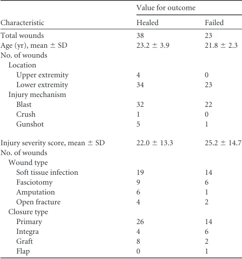
Patient demographics.Tissue biopsy and effluent samples were obtained from 124 debridement procedures representing 61 wounds in 44 patients with extremity war wounds. Wound loca-tions, characteristics, and closure methods are given inTable 1. All patients were free of comorbid conditions, such as immune or connective tissue disorders, that could potentially confound re-sults.
Microorganisms detected in wound samples by microarray. The LLMDA was applied for analysis of 124 wound samples, ob-tained from 44 patients. Microbial species detected in samples derived from each patient by both culture and LLMDA are shown inTable 2. A patient is indicated as positive for a given microor-ganism if any corresponding sample tested positive for that target. In cases where individual strains were detected, these were col-lapsed to the parent species. Complete microbial status and indi-vidual strain detection for all samples are shown in Data Set S1 in the supplemental material. At least one microbial target was de-tected by the LLMDA in 63 samples (51%). Wound samples were also interrogated for bacterial presence via quantitative culture. Of the 79 samples for which no bacterial growth was observed on culture, 27 (34% of culture-negative samples) were indicated to contain at least one microbial species. It was also observed in 22 samples (18% of total) that a species detected by culture was not identified by the corresponding microarray analysis. The most commonly observed microorganism in wound samples was Acin-etobacter baumannii, represented in 28 samples (23% of total) (Fig. 1).
Association of microbial detection with wound resolution. Fifty-four of the 124 total samples (44%) were derived from wounds that failed to heal. Of the 61 total wounds examined, 22 (36%) failed to heal. Of the 44 patients from whom samples were derived, 16 individuals (36%) exhibited one or more failed wounds, with some patients demonstrating multiple wounds with different outcomes. LLMDA detection status for each individual sample is shown inFig. 2, along with clinical outcome of the cor-responding wound. Individual samples inFig. 2are clustered ac-cording to all the microbial targets identified by LLMDA in each sample. Samples derived from wounds with the same outcome did not cluster together in all cases, indicate multifactorial causes of wound healing failure.Table 3shows wound failure rates seg-mented at the sample and wound levels. A wound was classified as positive for a microbial target if one or more derived samples tested positive. No association was observed between culture sta-tus and wound failure. When only effluent samples were consid-ered,A. baumanniiwas detected by LLMDA in 14% of samples from healed wounds, compared to 27% for wounds that under-went dehiscence. Although this association with wound failure was not statistically significant (P⫽0.150), a larger sample set could provide additional statistical power.
MultiplePseudomonasspecies were detected in wound sam-ples, includingP. aeruginosa, P. entomophila,P. putida, and P. stutzeri. When considering effluent samples only, the LLMDA de-tectedPseudomonasin 3% of samples from wounds that healed successfully, compared to 17% of samples from wounds that failed to heal (P⫽0.059).Pseudomonaswere detected at the wound level in 3% of healed wounds and 23% of wounds that failed to heal (P⫽0.020). Paradoxically, an inverse correlation was observed with detection of bacterial species associated with the gastrointes-tinal system (Bacteroides fragilis,Bacteroides plebeius,Enterobacter cloacae, Enterobacter sp., Enterococcus faecium, Escherichia coli, Salmonella enterica, andSalmonella entericaserovar Enteritidis). These organisms were detected most commonly in tissue samples, in which 30% of samples from healed wounds contained one or more of these species, compared to only 4% in those from failed wounds (P⫽0.013).
In addition to the more commonly detected microorganisms, detection events in which species possibly relevant to wound heal-ing were observed at low frequency also occurred. Examples in-cluded detection of threeStaphylococcusspecies in one sample (S. aureus,S. epidermidis, andS. lugdunensis) and detection of the opportunistic pathogenMycobacterium abscessus.
Association of Acinetobacter sequence-specific detection with wound resolution.One of the strengths of the microarray platform is the ability to detect sequence from plasmid DNA targets. TheAcinetobacterplasmid pRAY was the most commonly detected plasmid sequence in wound samples. Within the effluent samples studied, only 3% of healed samples contained pRAY, compared to 23% of samples that underwent dehiscence (P⫽ 0.012) (Table 3). In tissue samples, pRAY was detected in 24% of healed samples and 42% of failed wound samples (P⫽0.134). When examining detection status by wound, pRAY was detected in 15% of healed wounds and 41% of failed wounds (P⫽0.029). Microbial diversity identified by next-generation
sequenc-ing.DNA extracted from a subset of 21 wound samples was
[image:3.585.40.286.77.340.2]subjected to whole-genome sequencing. All data were analyzed using the Livermore Metagenomics Analysis Toolkit (LMAT) (18). Due to elevated human content in these samples and our TABLE 1Patient demographics and wound characteristics
Characteristic
Value for outcome
Healed Failed
Total wounds 38 23
Age (yr), mean⫾SD 23.2⫾3.9 21.8⫾2.3 No. of wounds
Location
Upper extremity 4 0
Lower extremity 34 23
Injury mechanism
Blast 32 22
Crush 1 0
Gunshot 5 1
Injury severity score, mean⫾SD 22.0⫾13.3 25.2⫾14.7 No. of wounds
Wound type
Soft tissue infection 19 14
Fasciotomy 9 6
Amputation 6 1
Open fracture 4 2
Closure type
Primary 26 14
Integra 4 6
Graft 8 2
Flap 0 1
on May 16, 2020 by guest
http://jcm.asm.org/
whole-genome approach, the majority of total reads aligned to the human genome. Results revealed a broad range in mapped microbial sequence data between individual wound samples. Microbial sequence abundance was lowest for wound samples which tested negative both by culture and LLMDA (Fig. 3), suggesting that the extent of coverage for microbial sequence data derived from wounds is reflective of microbial status.
No-tably, the two samples with the highest microbial sequence abundance (26-1-ECON and 27-2-EBON) both tested negative for microbial presence via culture but positive by LLMDA. This may indicate that the relevant species successfully proliferated within the wound but, because of possible strain-specific fac-tors, did not multiply under standard culture conditions and thus remained undetectable via standard microbiology. De-TABLE 2Microorganisms detected by culture and LLMDA for each patienta
Patient Culture result LLMDA detectionb Patient Culture result LLMDA detectionb
1 Pseudomonas stutzeri Mycobacterium abscessusⴱ 24 Acinetobacter baumannii Acinetobacter baumannii
Citrobacter freundii Salmonella entericaⴱ Human parvovirus
Klebsiella pneumoniaeⴱ HPV71ⴱ
HPV71ⴱ Salmonella entericaⴱ
Acinetobactersp. Borrelia afzeliiⴱ
2 NG None detected Mycobacterium abscessusⴱ
3 NG Acinetobactersp. 25 NG None detected
4 NG None detected 26 Staphylococcus capitis Acinetobacter baumannii
5 NG None detected Achromobactersp. Pseudomonas putida
6 NG None detected Pseudomonas entomophila
7 NG None detected Ralstonia solanacearumⴱ
8 NG None detected Mycobacterium abscessusⴱ
9 Acinetobacter baumannii Acinetobacter baumannii Borrelia afzeliiⴱ
Corynebacterium bovis 27 Acinetobactersp. Acinetobacter baumannii
Klebsiella pneumoniaeⴱ Unidentified Shigella boydii
10 Acinetobacter baumannii None detected Shigella sonnei
Enterococcus faecium Escherichia coliⴱ
11 Acinetobacter baumannii Acinetobacter baumannii 28 NG Pseudomonassp.ⴱ
Salmonella enterica 29 NG None detected
12 Acinetobacter baumannii Acinetobactersp. 30 NG None detected
13 NG None detected 31 NG Bacteroides plebeius
14 Escherichia coli Bacteroides fragilisⴱ Bacteroides fragilis
HPV57ⴱ Enterococcus faeciumⴱ
15 NG HHV-6 32 Acinetobacter baumannii Acinetobacter baumannii
Pseudomonassp.ⴱ HPV57
Ralstonia solanacearumⴱ 33 Acinetobacter baumannii Acinetobacter baumannii
16 Stenotrophomonas maltophilia Acinetobacter baumannii Achromobactersp. Borrelia afzeliiⴱ
Achromobactersp. Achromobacter xylosoxidans HPV57ⴱ
Moraxellasp. Pseudomonas aeruginosa 34 NG None detected
Staphylococcus epidermidis 35 NG None detected
Staphylococcus lugdunensis 36 Acinetobacter baumannii Acinetobacter baumannii
Enterobacter cloacae 37 NG None detected
Escherichia coli 38 NG None detected
Bordetella avium 39 ND None detected
Borrelia afzeliiⴱ 40 Acinetobacter baumannii Acinetobacter baumannii
Staphylococcus aureusⴱ Escherichia coli
Klebsiella pneumoniaeⴱ Klebsiella pneumoniae
17 Enterococcus faecium Klebsiella pneumoniaeⴱ Salmonella enteritidis
Salmonella entericaⴱ 41 NG None detected
HPV57ⴱ 42 NG None detected
HPV71ⴱ 43 Enterococcus faecium Bacteroides fragilis
18 Enterococcus faecium HPV57ⴱ Streptomyces avermitilisⴱ
19 Bacillus cereus Bacillus cereus HPV71ⴱ
Acinetobactersp. Acinetobactersp. Salmonella entericaⴱ
Alloiococcus otitidis Human parvovirus Borrelia afzeliiⴱ
Streptococcus agalactiae Pseudomonas stutzeriⴱ 44 Acinetobacter baumannii Acinetobacter baumannii
20 NG None detected Enterobacter cloacae Enterobacter cloacae
21 NG Roseburia hominisⴱ Escherichia coli
22 NG Pasteurella multocidaⴱ Uncultured bacterium
23 Acinetobacter baumannii Acinetobacter baumannii HPV71ⴱ
Enterococcus faecium Human parvovirus
aDetection in all wounds and samples was collapsed at the patient level. Strain detection was collapsed at the species level. Complete detection data for all samples and strains are
available in Data Set S2 in the supplemental material. NG, no growth; ND, culture not done; HPV, human papillomavirus. bⴱ, LLMDA detection at lower-intensity threshold (0.95).
on May 16, 2020 by guest
http://jcm.asm.org/
[image:4.585.48.544.79.602.2]tected bacterial species (via both LMAT and LLMDA) are shown for each sequenced sample in Data Set S2 in the supple-mental material.
In the sample set examined, wound-derived samples did not group exclusively according to successful or failed healing based on microbial community composition. This is observed inFig. 4A, in which samples were ordered according to their corresponding microbial profiles, and samples from wounds with similar outcomes were not observed to group together definitively (Fig. 4A).Figure 4Bshows a heat map representing a subset of high-abundance microorganisms detected by LMAT analysis of sequence data. Again, samples did not group according to outcome. Further, samples derived from the same patient did not group as closely in this analysis as when all detected organisms were considered (Fig. 4A), indicating that the more comprehensive assessment of bioburden may yield a higher degree of clinical relevance. In one particular case in which multiple sampling time points were available (patient 26), samples obtained from the same wound at different times were observed to be compositionally distinct (Fig. 4C). In this wound, a population ofAcinetobacterwas present during the first procedure (sample EA2) and expanded during the following (sample EBON), while the latter procedures (samples EB2 and ECON) displayed prominentPseudomonasdetection. It should be noted that bacterial sequence coverage was higher in samples de-rived from sample ECON (Fig. 3), suggesting that colonization by Pseudomonaswas characterized by greater bacterial abundance thanAcinetobacter.
The relatively high sequence coverage forA. baumannii in some samples raised the possibility of identifying the strain associated with infection. Sequence alignment and unique sin-gle nucleotide polymorphism (SNP)-based identification ap-proaches (22,23) were therefore applied for strain resolution. These analyses revealed some clustering according to patient
and outcome; however, coverage and sample size were not suf-ficient to confidently assign a clinical correlation. These data and methods are detailed in the supplemental material (see Appendix S1).
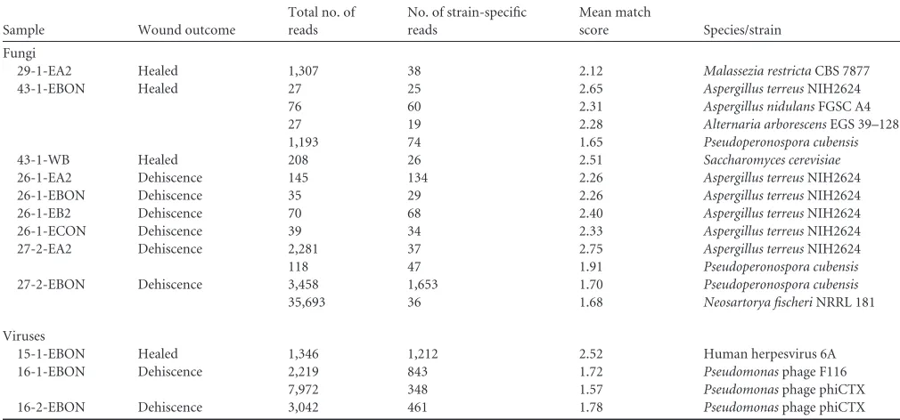
Fungal and viral groups identified in wounds by next-gener-ation sequencing.LMAT was also applied for examination of fun-gal and viral sequence data. For each identified funfun-gal strain, total mapped reads and strain-specific mapped reads are shown, as well as the mean match score per read for each detected species/strain (Table 4). These scores were calculated as the ratio of the summed match score for all strain-specific mapped reads to the absolute number of strain-specific reads. Only those species with mean match scores exceeding 1.5, greater than 25 total mapped reads, and greater than 10 strain-specific mapped reads are shown. Reads corresponding to detected species were verified via BLAST analy-sis. Fungal species identified in wound samples above the set threshold includedAspergillus terreus,Aspergillus nidulans, Alter-naria arborescens, Saccharomyces cerevisiae, Malassezia restricta, Pseudoperonospora cubensis, andNeosartorya fischeri.Aspergillus was detected in all samples derived from patient 26, whose wounds failed to heal.Aspergilluswas also observed in one other sample derived from a failed wound and one from a healed wound.
Due to low observed microbial coverage and a relatively small genome size, viral identification could be reliably verified in only three samples. Identical filtering cutoffs were applied as described above (Table 4).Pseudomonasphage was observed in samples ob-tained from both wounds for patient 16, and both wounds failed to heal. This observation corresponded with the detection of mul-tiplePseudomonas species in these samples via sequencing and LLMDA. Human herpesvirus 6 (HHV-6) was detected in one sample derived from patient 15, in agreement with LLMDA detec-tion.
FIG 1Microbial species detected by LLMDA in combat wound tissue and effluent samples. Nucleic acid was extracted from combat wound samples and hybridized to a microbial detection microarray. Tissue biopsy and effluent samples were analyzed independently. The number of positive detection events for each microbial target is shown. For samples in which a non-species-specific microbial target was detected (e.g.,Acinetobacterplasmid) and no other species-specific target was observed in that sample, the sample was classified according to genus (e.g.,Acinetobactersp.).
on May 16, 2020 by guest
http://jcm.asm.org/
[image:5.585.113.474.66.291.2]DISCUSSION
Studies of microbial colonization in traumatic wounds have been performed for combat injuries ranging from the Vietnam era (24) to modern environments (6, 25,26) and in burn and chronic wounds from civilian populations (9,10,27). Although quantita-tive culture techniques were applied in this study, current stan-dard practices for combat wound clinical microbiology involve only qualitative culture using surface swabs and phenotypic bac-terial identification. These traditional culture methods result in an overestimation of the relative presence of easily cultured and iden-tifiable microbes (10). Further, these techniques do not yield de-tailed genomic information, with the possible exception of drug resistance testing, the results of which can be slow and inconclu-sive.
This study shows that results from currently applied culture techniques underestimate the wound bioburden and do not cor-relate significantly with wound outcome. These methods should therefore be supplemented with molecular techniques. Microar-ray-based detection ofAcinetobacter andPseudomonastrended
toward association with wound failure, although this correlation was not statistically significant at aPvalue of⬍0.05. It is likely, however, that a larger sample size would provide additional sta-tistical power toward establishing this connection. This is sup-ported by the fact that theAcinetobacterplasmid pRAY was signif-icantly associated with wound failure. This plasmid has been implicated in multiple drug resistance, which may partially ex-plain its association with wound failure (28).Staphylococcuswas detected in only two wounds by microarray despite relatively fre-quent colonization of chronic and burn wounds by these bac-teria. This observation may be owing to the unique conditions under which these wounds were acquired and treated, which are highly distinct from those of civilian injuries. It is therefore possible that incidence of this pathogen and amenability to colonization in these severe wounds are different from what might be observed elsewhere. The relatively low incidence of Staphylococcusis in agreement with previous studies of combat wound colonization (6).
One of the unique and statistically significant findings for these FIG 2Clustering of samples from healed and failed combat wounds according to microbial species detected by microarray. Wound samples were ordered by hierarchical clustering. Samples were clustered according to their detected microbial profile, as determined by microarray detection. Individual patient samples are shown in columns and are labeled along the bottom horizontal axis. Patient samples are labeled according to the following scheme: patient number-wound number-extraction method (e.g., 9-1-WA). Sample extraction types are detailed in Materials and Methods. Detected microbial species are shown in rows and are labeled along the right vertical axis. As in the previous figure, when a non-species-specific microbial target was detected and no other species-specific target was observed in that sample, the sample was classified according to genus. Positive microbial detection is shown in light blue, and negative microbial detection is shown in dark blue. Wound outcome is indicated in a horizontal bar above the plot. Samples obtained from healed wounds are indicated in green, and samples from failed wounds are in red.
on May 16, 2020 by guest
http://jcm.asm.org/
[image:6.585.39.530.68.415.2]samples was the observation that enteric bacterial species associ-ated more strongly with healed wounds. This seemingly paradox-ical observation is in agreement with findings for chronic diabetic foot ulcers, in which ulcer duration correlated negatively with Staphylococcusabundance (29). This may reflect changes in im-munoregulation and remodeling of the wound, where a microen-vironment that is progressing toward healing is an amenable niche for distinct classes of bacteria from an environment that is immu-nologically distant from successful resolution. Detection of these microbial targets may provide a useful metric that is reflective of wound status. These results support the use of microarrays as a detection technology that could fill an important diagnostic niche, capable of delivering comprehensive microbial detection ap-proaching the capacity of sequencing, but with monetary and time costs closer to those of PCR.
While microarray analysis revealed numerous instances where microbial presence was not detected by culture, cases in which a cultured organism was not detected by microarray analysis were also observed. Several factors may have contrib-uted to this observation. Due to inherent differences in avail-able sequence data between target organisms, probe coverage and detection capacity may differ between sequence targets, with the array exhibiting reduced power for identifying some organisms depending on reference genome quality and anno-tation. Addressing this issue is an active area of interest, and subsequent versions of the LLMDA are undergoing continuous
optimization. Also, while the Gram-positive bacteria Enterococ-cus faeciumandAlloiococcus otitidiswere detected by culture,A. otitidiswas never identified via microarray and E. faeciumwas detected only once. While extensive bead beating was performed to facilitate Gram-positive lysis, it is possible that further optimi-zation would improve extraction efficiency and detection capac-ity. Finally, the culture and molecular analyses were performed with different aliquots of tissue or effluent obtained from a given wound sample. It is possible that the microbes within these ali-quots may not have been homogenously distributed. Discrepan-cies between culture-based and molecular techniques have simi-larly been observed in chronic wound studies, where results derived from distinct detection modalities depended on the bac-terial group in question (30). These observations reinforce the difficulty in exclusive use of a single diagnostic tool and indicate that complementary approaches may be useful for yielding fully comprehensive results.
[image:7.585.42.549.78.373.2]To further evaluate total wound bioburden, a subset of samples was selected for metagenomic analysis. Clear distinctions were not observed in overall community structure between successfully healed wounds and those that failed to heal. Failed wounds, how-ever, did trend toward higher overall bacterial sequence coverage, and observations from one patient revealed that microbial status may shift over time. While the complete microbiome profile did not associate with clinical status, our observed microarray data TABLE 3Correlation of target detection by microarray with wound resolution statusa
Category
All samples Individual wounds
No. (%) healed No. (%) failed Pvalue No. (%) healed No. (%) failed Pvalue
All 39 (63.9) 22 (36.1)
Effluent 37 (55.2) 30 (44.8) Tissue 33 (57.9) 24 (42.1)
Culture 14 (35.9) 11 (50.0) 0.210
Effluent 7 (18.9) 8 (26.7) 0.321
Tissue 16 (48.5) 11 (45.8) 0.528
LLMDA 19 (48.7) 13 (59.1) 0.305
Effluent 13 (35.1) 12 (40.0) 0.437
Tissue 23 (69.7) 15 (62.5) 0.386
A. baumannii 10 (25.6) 9 (40.9) 0.171
Effluent 5 (13.5) 8 (26.7) 0.149
Tissue 9 (27.3) 6 (25.0) 0.548
AcinetobacterpRAY 6 (15.4) 9 (40.9) 0.029
Effluent 1 (2.7) 7 (23.3) 0.012
Tissue 8 (24.2) 10 (41.7) 0.134
Pseudomonasb 1 (2.6) 5 (22.7) 0.020
Effluent 1 (2.7) 5 (16.7) 0.059
Tissue 0 (0.0) 0 (0.0)
Enteric bacteriac 9 (23.1) 2 (9.1) 0.155
Effluent 2 (5.4) 1 (3.3) 0.579
Tissue 10 (30.3) 1 (4.2) 0.013
aHealing and failure rates are shown by individual sample and wound. Wounds were considered positive for a microbial target if one or more derived samples tested positive. AllP values were calculated using Fisher’s exact test.
bPseudomonas:P. aeruginosa,P. entomophila,P. putida,P. stutzeri, andPseudomonassp. c
Enteric bacteria:Bacteroides fragilis,Bacteroides plebeius,Enterobacter cloacae,Enterobactersp.,Enterococcus faecium,Escherichia coli,Salmonella enterica, andSalmonella enterica
serovar Enteritidis.
on May 16, 2020 by guest
http://jcm.asm.org/
suggest that detection of specific microbial sequence targets does demonstrate association with wound outcome.
In some cases, sequencing analysis of wound samples revealed much larger numbers of taxonomic targets than were observed via LLMDA or identified organisms in samples with no targets de-tected by LLMDA. This was observed primarily in samples where less than approximately 2,000 reads were mapped to a given spe-cies/strain. This likely reflects the approximate limit of detection for the microarray platform, which is expected to be less sensitive than sequencing analysis (31). In other samples (e.g., 16-1-EBON), the total number of taxonomic targets was inflated by identification of many individual substrains, again due in part to the high sensitivity of sequencing, which could potentially have been collapsed in the sequencing analysis. It is likely that identifi-cation of these substrains is reflective of only a few actual coloniz-ing strains.
In addition to species-specific detection, strain identity could also be relevant to wound care. This is particularly true in the case ofA. baumannii, which is included in the ESKAPE pathogen group (Enterococcus faecium, Staphylococcus aureus, Klebsiella pneumoniae, Acinetobacter baumannii, Pseudomonas aeruginosa
andEnterobactersp.), containing organisms especially capable of evading clinically applied microbicidal efforts (32). It has been shown previously that different species and strains within the Acinetobactergenus demonstrate distinct tolerances for pH and antimicrobial pressure (33). Further, separate A. baumannii strains derived from the same patient have been shown to be phe-notypically distinct in a mouse model, as well as demonstrating different morphologies and propensities for catheter adherence (34). It has also been shown that differences in the infecting strain ofA. baumanniican impact the resultant pathology and the cyto-kine response in a mouse model of pulmonary infection (35). TheA. baumanniigenotyping analysis outlined in Appendix S1 in the supplemental material did not attribute a specific response to the nearest-neighbor strains or a subset of unique SNP markers and thus was not a central focus of this study. However, these data do raise the possibility of classifying wound samples according to the most likely colonizing strain, and our application of alignment and SNP-based approaches demonstrates the potential of these tools for rapid genotyping ofA. baumannii.
Although less common than bacterial colonization, fungal in-fections, in particular infection withAspergillus, have been associ-FIG 3Quantity of next-generation sequence data mapped to microbial species by LMAT. Nucleic acid extracted from wound samples was processed via next-generation sequencing. Resultant reads were aligned to bacterial reference genomes using the Livermore Metagenomics Analysis Toolkit (LMAT). The abundance of reads mapped to microbial reference genomes is shown for each sample. Samples are shown along the horizontal axis, listed in numerical order by patient. The patient and wound from which each sample was derived are shown, along with sample extraction type (detailed in Materials and Methods). In some cases, multiple wounds from the same patient were analyzed. Metrics shown below the axis for each sample include wound clinical outcome (H, healed; D, dehiscence), LLMDA microbial detection status, and microbial culture status.
on May 16, 2020 by guest
http://jcm.asm.org/
[image:8.585.139.452.62.414.2]FIG 4Microbial profiles in combat wound samples as determined by LMAT analysis of next-generation sequence data. Sequence data from wound samples were analyzed to determine the total quantity of reads mapping to each microbial species identified as present by LMAT. Microbial abundance within each sample, as measured by mapped reads, was used to order samples within a heat map using NMDS ordination by Phyloseq. Individual wound samples are given in columns. Labels below the horizontal axis show patient number, wound number, and sample extraction type (detailed in Materials and Methods). In some cases, more than one wound from the same patient was analyzed. Wound outcome is also indicated (H, healed; D, dehiscence). Microbial species are represented in rows and are shown along the vertical axis. (A) Heat map showing all microbial species detected by LMAT analysis. Individual species are not labeled due to the high number of total targets. (B) Heat map showing only those microbial species to which 1,000 or more total reads were assigned across all samples. (C) Heat map showing only those samples derived from patient 26, with samples ordered according to temporal collection point. Only those microbial species to which 1,000 or more total reads were mapped across all species are shown.
on May 16, 2020 by guest
http://jcm.asm.org/
[image:9.585.44.543.62.621.2]ated with traumatic combat injury and can significantly compli-cate wound care (36,37). One possible source of fungal material could be incorporation of contaminating organic matter at the time of injury (38). This is supported by detection of the fungal phytopathogens Alternaria arborescens and Pseudoperonospora cubensis, which may have been embedded concurrently with plant matter at the point of trauma, possibly consistent with the mech-anism of injury from improvised explosive device (IED) blasts. Despite identification of fungal species via sequence data, fungi were not detected by the LLMDA, possibly due to minimal cover-age or low quality and annotation of draft fungal reference se-quences used for probe design.
Similarly, relatively few viral targets were identified with high confidence, and it is likely that elevated coverage would be required for reliable characterization. The majority of viral se-quence data were expected to be derived from bacteriophage, and indeed,Pseudomonasphage was detected in two samples de-rived from separate wounds in the same patient. Further assess-ment of wound bacteriophage communities could be relevant to future development of novel phage therapy for addressing drug-resistantA. baumanniiandP. aeruginosainfections (39,40).
Wound failure is a consequence not only of the microbial bioburden but also of local and systemic inflammatory status. Numerous studies have identified human mediators of wound healing and the tissue remodeling response (6,7,17,41). These studies have implicated a range of cytokines, chemokines, and matrix metalloproteinases, and support the notion that inflam-matory dysregulation is central to wound healing failure. An increasing number of novel human biomarkers are being iden-tified as predictive of wound healing progression (4,42). The combination of molecular assays for host protein markers of inflammation with the advanced microbial detection protocols in this study could greatly improve care and reduce the high morbidity associated with blast and otherwise combat-related wounds.
In summary, these data support the inclusion of integrated molecular techniques for detection of microbial species and plasmid- or strain-specific sequences. Clinical assessment of the microbial flora unique to each patient could provide clini-cians with invaluable information during the debridement process. More effective and timely assessments based on quan-tifiable metrics would reduce surgical morbidity, accelerate re-habilitation, and decrease the length of hospital stays. The po-tential for reduction in overall health care costs further supports the application of these molecular protocols as a pru-dent and cost-effective addition to the wound diagnostics ar-mamentarium. These techniques could represent an important step toward personalized assessment of individual patients and rational design of tailored treatment regimens.
ACKNOWLEDGMENTS
This work was sponsored by the U.S. Army Medical Research and Materiel Command (MIPR1EO89M1115). The U.S. Army Medical Research Ac-quisition Activity (Fort Detrick, MD) is the awarding and administering acquisition office. A portion of this effort was also supported by the U.S. Navy Bureau of Medicine and Surgery under the Medical Development Program and Office of Naval Research work unit (604771N.0933.001. A0604). This study was performed under the auspices of the U.S. Depart-ment of Energy by Lawrence Livermore National Laboratory under con-tract DE-AC52-07NA27344.
[image:10.585.39.544.78.314.2]This document was prepared as an account of work sponsored by an agency of the U.S. Government. Neither the U.S. Government nor Law-rence Livermore National Security, LLC, nor any of their employees makes any warranty, expressed or implied, or assumes any legal liabil-ity or responsibilliabil-ity for the accuracy, completeness, or usefulness of any information, apparatus, product, or process disclosed or repre-sents that its use would not infringe privately owned rights. Reference herein to any specific commercial product, process, or service by trade name, trademark, manufacturer, or otherwise does not necessarily constitute or imply its endorsement, recommendation, or favoring by the U.S. Government or Lawrence Livermore National Security, LLC. The views and opinions of authors expressed herein do not necessarily TABLE 4Fungi and viruses identified in wound samples through analysis of next-generation sequence data with LMATa
Sample Wound outcome
Total no. of reads
No. of strain-specific reads
Mean match
score Species/strain
Fungi
29-1-EA2 Healed 1,307 38 2.12 Malassezia restrictaCBS 7877
43-1-EBON Healed 27 25 2.65 Aspergillus terreusNIH2624
76 60 2.31 Aspergillus nidulansFGSC A4
27 19 2.28 Alternaria arborescensEGS 39–128
1,193 74 1.65 Pseudoperonospora cubensis
43-1-WB Healed 208 26 2.51 Saccharomyces cerevisiae
26-1-EA2 Dehiscence 145 134 2.26 Aspergillus terreusNIH2624 26-1-EBON Dehiscence 35 29 2.26 Aspergillus terreusNIH2624 26-1-EB2 Dehiscence 70 68 2.40 Aspergillus terreusNIH2624 26-1-ECON Dehiscence 39 34 2.33 Aspergillus terreusNIH2624 27-2-EA2 Dehiscence 2,281 37 2.75 Aspergillus terreusNIH2624
118 47 1.91 Pseudoperonospora cubensis
27-2-EBON Dehiscence 3,458 1,653 1.70 Pseudoperonospora cubensis
35,693 36 1.68 Neosartorya fischeriNRRL 181
Viruses
15-1-EBON Healed 1,346 1,212 2.52 Human herpesvirus 6A
16-1-EBON Dehiscence 2,219 843 1.72 Pseudomonasphage F116 7,972 348 1.57 Pseudomonasphage phiCTX 16-2-EBON Dehiscence 3,042 461 1.78 Pseudomonasphage phiCTX
a
Fungi and viruses with more than 25 total mapped reads, more than 10 strain-specific mapped reads, and mean match scores exceeding 1.5 are shown.
on May 16, 2020 by guest
http://jcm.asm.org/
state or reflect those of the U.S. Government or Lawrence Livermore National Security, LLC, and shall not be used for advertising or prod-uct endorsement purposes.
E.A.E., J.A.F., and B.C.K. are military service members and employees of the U.S. Government. This work was prepared as part of their official duties.
REFERENCES
1.Brethauer SA, Chao A, Chambers LW, Green DJ, Brown C, Rhee P, Bohman HR.2008. Invasion vs insurgency: US Navy/Marine Corps for-ward surgical care during Operation Iraqi Freedom. Arch. Surg.143:564 – 569.http://dx.doi.org/10.1001/archsurg.143.6.564.
2.Ritenour AE, Blackbourne LH, Kelly JF, McLaughlin DF, Pearse LA, Holcomb JB, Wade CE.2010. Incidence of primary blast injury in US military overseas contingency operations: a retrospective study. Ann. Surg.251:1140 –1144.http://dx.doi.org/10.1097/SLA.0b013e3181e01270. 3.Ramasamy A, Hill AM, Clasper JC.2009. Improvised explosive de-vices: pathophysiology, injury profiles and current medical manage-ment. J. R. Army Med. Corps.155:265–272.http://dx.doi.org/10.1136 /jramc-155-04-05.
4.Hawksworth JS, Stojadinovic A, Gage FA, Tadaki DK, Perdue PW, Forsberg J, Davis TA, Dunne JR, Denobile JW, Brown TS, Elster EA. 2009. Inflammatory biomarkers in combat wound healing. Ann. Surg. 250:1002–1007.http://dx.doi.org/10.1097/SLA.0b013e3181b248d9. 5.Forsberg JA, Elster EA, Andersen RC, Nylen E, Brown TS, Rose MW,
Stojadinovic A, Becker KL, McGuigan FX.2008. Correlation of procal-citonin and cytokine expression with dehiscence of wartime extremity wounds. J. Bone Joint Surg. Am.90:580 –588.http://dx.doi.org/10.2106 /JBJS.G.00265.
6.Brown TS, Hawksworth JS, Sheppard FR, Tadaki DK, Elster E.2011. Inflammatory response is associated with critical colonization in combat wounds. Surg. Infect. (Larchmt.)12:351–357.http://dx.doi.org/10.1089 /sur.2010.110.
7.Evans KN, Forsberg JA, Potter BK, Hawksworth JS, Brown TS, Ander-sen R, Dunne JR, Tadaki D, Elster EA.2012. Inflammatory cytokine and chemokine expression is associated with heterotopic ossification in high-energy penetrating war injuries. J. Orthop. Trauma26:e204 – e13.http: //dx.doi.org/10.1097/BOT.0b013e31825d60a5.
8.Bowler PG, Duerden BI, Armstrong DG.2001. Wound microbiology and associated approaches to wound management. Clin. Microbiol. Rev. 14:244 –269.http://dx.doi.org/10.1128/CMR.14.2.244-269.2001. 9.McGuckin M, Goldman R, Bolton L, Salcido R. 2003. The clinical
relevance of microbiology in acute and chronic wounds. Adv. Skin Wound Care16:12–23; quiz, 24 –25.http://dx.doi.org/10.1097/00129334 -200301000-00011.
10. Davies CE, Hill KE, Wilson MJ, Stephens P, Hill CM, Harding KG, Thomas DW.2004. Use of 16S ribosomal DNA PCR and denaturing gradient gel electrophoresis for analysis of the microfloras of healing and nonhealing chronic venous leg ulcers. J. Clin. Microbiol.42:3549 –3557.
http://dx.doi.org/10.1128/JCM.42.8.3549-3557.2004.
11. Davis SC, Ricotti C, Cazzaniga A, Welsh E, Eaglstein WH, Mertz PM. 2008. Microscopic and physiologic evidence for biofilm-associated wound colonization in vivo. Wound Repair Regen.16:23–29.http://dx.doi.org /10.1111/j.1524-475X.2007.00303.x.
12. Hurlow J, Bowler PG.2009. Clinical experience with wound biofilm and management: a case series. Ostomy Wound Manage.55:38 – 49. 13. Ryan TJ.2007. Infection following soft tissue injury: its role in wound
healing. Curr. Opin. Infect. Dis.20:124 –128.http://dx.doi.org/10.1097 /QCO.0b013e32801a3e7c.
14. Frankel YM, Melendez JH, Wang NY, Price LB, Zenilman JM, Lazarus GS.2009. Defining wound microbial flora: molecular microbiology open-ing new horizons. Arch. Dermatol.145:1193–1195.http://dx.doi.org/10 .1001/archdermatol.2009.246.
15. Gardner SN, Jaing CJ, McLoughlin KS, Slezak TR.2010. A microbial detection array (MDA) for viral and bacterial detection. BMC Genomics 11:668.http://dx.doi.org/10.1186/1471-2164-11-668.
16. Kuczynski J, Lauber CL, Walters WA, Parfrey LW, Clemente JC, Gevers D, Knight R.2012. Experimental and analytical tools for studying the human microbiome. Nat. Rev. Genet. 13:47–58. http://dx.doi.org/10 .1038/nrg3129.
17. Utz ER, Elster EA, Tadaki DK, Gage F, Perdue PW, Forsberg JA, Stojadinovic A, Hawksworth JS, Brown TS. 2010. Metalloproteinase
expression is associated with traumatic wound failure. J. Surg. Res.159: 633– 639.http://dx.doi.org/10.1016/j.jss.2009.08.021.
18. Ames SK, Hysom DA, Gardner SN, Lloyd GS, Gokhale MB, Allen JE. 2013. Scalable metagenomic taxonomy classification using a reference ge-nome database. Bioinformatics29:2253–2260.http://dx.doi.org/10.1093 /bioinformatics/btt389.
19. McMurdie PJ, Holmes S.2013. Phyloseq: an R package for reproducible interactive analysis and graphics of microbiome census data. PLoS One 8:e61217.http://dx.doi.org/10.1371/journal.pone.0061217.
20. Warnes GR, Bolker B, Bonebakker L, Gentleman R, Huber W, Liaw A, Lumley T, Maechler M, Magnusson A, Moeller S, Schwartz M, Venables B.2013. Gplots: various R programming tools for plotting data. R package version 2.12.1.
21. Rajaram S, Oono Y.2010. NeatMap—non-clustering heat map alterna-tives in R. BMC Bioinformatics11:45.http://dx.doi.org/10.1186/1471 -2105-11-45.
22. Gardner SN, Slezak T.2010. Scalable SNP analyses of 100⫹bacterial or viral genomes. J. Forensic Res. 1:107. http://dx.doi.org/10.4172/2157 -7145.1000107.
23. Gardner SN.2013. When whole-genome alignments just won’t work: kSNP v2 software for alignment-free SNP discovery and phylogenetics of hundreds of microbial genomes. PLoS One8:e81760.http://dx.doi.org/10 .1371/journal.pone.0081760.
24. Bendy RH, Jr, Nuccio PA, Wolfe E, Collins B, Tamburro C, Glass W, Martin CM.1964. Relationship of quantitative wound bacterial counts to healing of decubiti: effect of topical gentamicin. Antimicrob. Agents Che-mother.10:147–155.
25. Murray CK.2008. Infectious disease complications of combat-related injuries. Crit. Care Med.36:S358 –S364.http://dx.doi.org/10.1097/CCM .0b013e31817e2ffc.
26. Murray CK, Wilkins K, Molter NC, Li F, Yu L, Spott MA, Eastridge B, Blackbourne LH, Hospenthal DR.2011. Infections complicating the care of combat casualties during operations Iraqi Freedom and Enduring Freedom. J. Trauma71:S62–S73.http://dx.doi.org/10.1097 /TA.0b013e3182218c99.
27. Azzopardi EA, Azzopardi SM, Boyce DE, Dickson WA.2011. Emerging gram-negative infections in burn wounds. J. Burn Care Res.32:570 –576.
http://dx.doi.org/10.1097/BCR.0b013e31822ac7e6.
28. Hamidian M, Nigro SJ, Hall RM.2012. Variants of the gentamicin and tobramycin resistance plasmid pRAY are widely distributed in Acinetobac-ter. J. Antimicrob. Chemother.67:2833–2836.http://dx.doi.org/10.1093 /jac/dks318.
29. Gardner SE, Hillis SL, Heilmann K, Segre JA, Grice EA. 2013. The neuropathic diabetic foot ulcer microbiome is associated with clinical fac-tors. Diabetes62:923–930.http://dx.doi.org/10.2337/db12-0771. 30. Dowd SE, Sun Y, Secor PR, Rhoads DD, Wolcott BM, James GA,
Wolcott RD.2008. Survey of bacterial diversity in chronic wounds using pyrosequencing, DGGE, and full ribosome shotgun sequencing. BMC Mi-crobiol.8:43.http://dx.doi.org/10.1186/1471-2180-8-43.
31. Be NA, Thissen JB, Gardner SN, McLoughlin KS, Fofanov VY, Koshin-sky H, Ellingson SR, Brettin TS, Jackson PJ, Jaing CJ.2013. Detection of
Bacillus anthracisDNA in complex soil and air samples using next-generation sequencing. PLoS One 8:e73455.http://dx.doi.org/10.1371 /journal.pone.0073455.
32. Pendleton JN, Gorman SP, Gilmore BF.2013. Clinical relevance of the ESKAPE pathogens. Expert Rev. Anti Infect. Ther.11:297–308.http://dx .doi.org/10.1586/eri.13.12.
33. Peleg AY, de Breij A, Adams MD, Cerqueira GM, Mocali S, Galardini M, Nibbering PH, Earl AM, Ward DV, Paterson DL, Seifert H, Dijk-shoorn L.2012. The success ofAcinetobacterspecies; genetic, metabolic and virulence attributes. PLoS One7:e46984.http://dx.doi.org/10.1371 /journal.pone.0046984.
34. Kempf M, Eveillard M, Deshayes C, Ghamrawi S, Lefrancois C, Georgeault S, Bastiat G, Seifert H, Joly-Guillou ML.2012. Cell surface properties of two differently virulent strains ofAcinetobacter baumannii
isolated from a patient. Can. J. Microbiol.58:311–317.http://dx.doi.org /10.1139/w11-131.
35. de Breij A, Eveillard M, Dijkshoorn L, van den Broek PJ, Nibbering PH, Joly-Guillou ML.2012. Differences inAcinetobacter baumanniistrains and host innate immune response determine morbidity and mortality in experimental pneumonia. PLoS One7:e30673.http://dx.doi.org/10.1371 /journal.pone.0030673.
on May 16, 2020 by guest
http://jcm.asm.org/
36. Warkentien T, Rodriguez C, Lloyd B, Wells J, Weintrob A, Dunne JR, Ganesan A, Li P, Bradley W, Gaskins LJ, Seillier-Moiseiwitsch F, Mur-ray CK, Millar EV, Keenan B, Paolino K, Fleming M, Hospenthal DR, Wortmann GW, Landrum ML, Kortepeter MG, Tribble DR. 2012. Invasive mold infections following combat-related injuries. Clin. Infect. Dis.55:1441–1449.http://dx.doi.org/10.1093/cid/cis749.
37. Paolino KM, Henry JA, Hospenthal DR, Wortmann GW, Hartzell JD. 2012. Invasive fungal infections following combat-related injury. Mil. Med.177:681– 685.http://dx.doi.org/10.7205/MILMED-D-11-00364. 38. Hajdu S, Obradovic A, Presterl E, Vecsei V.2009. Invasive mycoses
following trauma. Injury40:548 –554.http://dx.doi.org/10.1016/j.injury .2008.03.034.
39. Krylov V, Shaburova O, Krylov S, Pleteneva E.2013. A genetic approach
to the development of new therapeutic phages to fightPseudomonas aeruginosa in wound infections. Viruses5:15–53.http://dx.doi.org/10 .3390/v5010015.
40. Mihu MR, Martinez LR.2011. Novel therapies for treatment of multi-drug resistantAcinetobacter baumanniiskin infections. Virulence2:97– 102.http://dx.doi.org/10.4161/viru.2.2.15061.
41. Hahm G, Glaser JJ, Elster EA.2011. Biomarkers to predict wound healing: the future of complex war wound management. Plast. Reconstr. Surg. 127(Suppl 1):21S–26S.http://dx.doi.org/10.1097/PRS.0b013e3181fbe291. 42. Chromy BA, Eldridge A, Forsberg J, Brown TS, Kirkup BC, Jaing C, Be
NA, Elster E, Luciw PA.2013. Wound outcome in combat injuries is associated with a unique set of protein biomarkers. J. Transl. Med.11:281.
http://dx.doi.org/10.1186/1479-5876-11-281.