www.impactjournals.com/oncotarget/
Oncotarget, Vol. 6, No. 14
Loss of Wave1 gene defines a subtype of lethal prostate cancer
Adam G. Sowalsky
1,5, Rebecca Sager
2, Rachel J. Schaefer
1, Gennady Bratslavsky
3,
Pier Paolo Pandolfi
1,4,5, Steven P. Balk
1,5, Leszek Kotula
2,31Department of Medicine, Beth Israel Deaconess Medical Center, Harvard Medical School, Boston, MA 02215, USA 2Department of Biochemistry and Molecular Biology, SUNY Upstate Medical University, Syracuse, NY 13210, USA 3Department of Urology, SUNY Upstate Medical University, Syracuse, NY 13210, USA
4Department of Pathology, Beth Israel Deaconess Medical Center, Harvard Medical School, Boston, MA 02215, USA 5Cancer Research Institute, Beth Israel Deaconess Cancer Center, Harvard Medical School, Boston, MA 02215, USA
Correspondence to:
Leszek Kotula, e-mail: [email protected]
Adam G. Sowalsky, e-mail: [email protected]
Keywords: WAVE, prostate cancer, genomics, androgen receptor, castration resistance
Received: January 09, 2015 Accepted: March 13, 2015 Published:March 31, 2015
ABSTRACT
Genetic alterations involving TMPRSS2-ERG alterations and deletion of key tumor suppressor genes are associated with development and progression of prostate cancer (PCa). However, less defined are early events that may contribute to the development of high-risk metastatic prostate cancer. Bioinformatic analysis of existing tumor genomic data from PCa patients revealed that WAVE complex gene alterations are associated with a greater likelihood of prostate cancer recurrence. Further analysis of primary vs. castration resistant prostate cancer indicate that disruption of WAVE complex gene expression, and particularly WAVE1 gene (WASF1) loss, is also associated with castration resistance, where WASF1 is frequently co-deleted with PTEN and resists androgen deprivation therapy (ADT). Hence, we propose that WASF1 status defines a subtype of ADT-resistant patients. Better understanding of the effects of WAVE pathway disruption will lead to development of better diagnostic and treatment modalities.
INTRODUCTION
As the most-common noncutaneous cancer in men
worldwide [1], mechanisms contributing to development
of prostate cancer at all stages of disease remain of high
interest for both diagnosis and treatment of
clinically-relevant disease. Consistent with the multi-hit hypothesis,
several genes that control critical growth, survival, and/
or apoptotic pathways [2] must be altered to lead to fully
penetrant prostate cancer [3–5]. A wide body of literature
has identified many complex genetic alterations involved
in neoplastic transformation, including TMPRSS2-ETS
(ERG) chromosomal translocations and deletion of tumor
suppressor genes (PTEN,
TP53,
RB1,
NKX3–1, and
CDKN1B) [3–10]. Of these, selection for the deletion
of
PTEN (phosphatase and tensin homolog) occurs in
approximately 30% of prostate cancers, as lowered
levels of PTEN increase the availability of
phosphatidyl-inositol 3, 4, 5 triphosphate (PIP3) for driving PI 3-kinase
(PI3K)-dependent signaling, such as cellular growth
pathways downstream of AKT (protein kinase B) [7, 11].
In addition, we previously implicated ABI1 (SSH3BP1) as
a putative tumor suppressor in prostate cancer [12] and
have demonstrated that disruption of Abi1 in the mouse
prostate leads to prostatic intraepithelial neoplasia (PIN),
but not to invasive prostate cancer [13]. We have therefore
sought to find additional genes that cooperate with
ABI1 in
prostate tumor progression.
In cells Abi1 is incorporated in the WAVE complex.
In mammalian cells, several distinct WAVE complexes can
form, depending on the isoform of proteins that is expressed
[14]. Each WAVE complex assembles from ubiquitously
expressed variants of 5 polypeptides: WAVE, Abelson inter
Nck-associated protein (Nap), and Brk1 (HSPC300)
[15–17]. Except for Brk1 [18], for which only one isoform
exists, each of these proteins is a member of a protein family
consisting of 2–3 genes in mammals including alternatively
spliced isoforms of 10 genes [14, 19]: (i)
WASF1, WAVE2
(WASF2), and WAVE3 (WASF3), (ii) ABI1,
ABI2, and
ABI3, (iii) Sra-1 and PIR121 (CYFIP1 and CYFIP2), and
(iv) Nap1 (NCKAP1) and Hem1 (NCKAP1L1). Functionally,
WAVE complexes are major actin cytoskeleton regulatory
factors that promote actin polymerization through an Arp2/3
dependent mechanism [20]. Other functions of WAVE
complex or its components involve binding to variety of
membrane receptors [21], intracellular signaling [22, 23],
and transcription [22, 24]. WAVE complexes are involved
in cell motility and migration, cellular adhesion, cell-to-cell
communication, cell division, and immunological responses
[17, 25–31]. Thus, WAVE complexes are involved numerous
cellular functions requiring actin cytoskeleton reorganization
and dynamics, processes with roles in tumor progression and
metastasis [32].
Consistent with this, dysregulation of WAVE
complex or its components is associated with human
cancer [33–35] including prostate cancer [12, 36–39], as
the WAVE pathway cross talks with the PI3K pathway. It
has been shown previously that PIP3 can bind directly to
WAVE protein [40], and we and others have demonstrated
that the p85 regulatory subunit of PI3K interacts with Abi1
[23, 41]. Importantly, we have also shown that genetic
inactivation of Abi1 led to down-regulation of WASF1
(and
WASF2 [42]) which in turn resulted in increased
phosphorylation of Akt in murine prostate tissue [13].
To this effect, altered levels of WAVE complex genes
would result in disruption of the WAVE complex’s ability
to regulate p85, and thus would phenocopy deletion of
PTEN in mediating cancer progression. Therefore we
hypothesize that prostate cancers select for cells with
lower levels of WAVE complex proteins.
In patients with metastatic PCa, standard treatments
involve either surgical or medical castration, which
effectively prevents testicular testosterone from driving
androgen receptor (AR) activation in the tumor cell [43].
The fact that most patients invariably relapse has led to
profound interest in identifying mechanisms contributing
to the development of castration-resistant prostate cancer
(CRPC) and commercial production of inhibitors of adrenal
sources of androgens and AR pathway inhibitors such
as galeterone, abiraterone and enzalutamide [44–46].
However, the fact that PTEN is deleted or down-regulated
in these cancers has intensified investigation of how
PI3K contributes to androgen independence via a
PTEN-dependent mechanism. Not surprisingly, simultaneous
inhibition of both AR and PI3K effectively blocked growth
of human PCa xenografts [47–49]. These data further
reinforce the need to identify causative mechanisms of
CRPC development, especially in the context of
PTEN
deletion.
Therefore, to assess whether prostate cancer
progression to castration resistance is mediated by changes
to WAVE complex, we performed bioinformatic meta
analyses on several published datasets, including mutation,
copy number, and gene expression data accumulated
as part of The Cancer Genome Atlas (TCGA), as well
as other publicly-available datasets deposited in GEO
from experiments performed at Beth Israel Deaconess
Medical Center (BIDMC), Memorial Sloan Kettering
Medical Center (MSKCC) and the University of Michigan
(UMICH) [7, 10, 50]. By comparing results across
independent cohorts, we observed shifts in the frequency
of WAVE complex gene alterations, which suggest that
WAVE complex disruption may be a putative driver of
prostate tumorigenesis. Importantly, we observed that
deletion of WASF1, the gene that codes for WAVE1,
occurs more frequently with
PTEN deletion in metastatic
lethal
vs. primary disease, suggesting that WASF1 loss
represents an aggressive of subtype of prostate cancer. It is
thus possible that patients who harbor tumors with WASF1
deletion may benefit from either earlier or more aggressive
intervention.
RESULTS AND DISCUSSION
We had previously assessed the status of ABI1
in a small cohort of primary prostate tissue [51], so we
expanded our interrogation of WAVE complex alterations
to The Cancer Genome Atlas (TCGA) and molecular
analyses performed on these samples. We hypothesized
that alterations to the WAVE complex may contribute to
increased tumorigenicity and therefore poorer long-term
outcome. When we interrogated the dataset for cases
with mutations, copy number alterations, or abnormal
expression, we found that alterations to WAVE complex
genes were significantly associated with a 13.43% increase
in the rate of biochemical recurrence (BCR) within
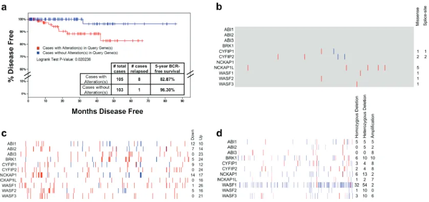
5 years (Figure 1a). Interrogating other datasets for which
biochemical recurrence rates were available, we observed
a similar trend in the MKSCC dataset, where alterations
to WAVE complex genes were associated with a 11.14%
increase in the fiveyear BCR rate (Supplementary
Figure 1). Strikingly, the most common alterations were
not mutations (Figure 1b) nor recurrent up- or
down-regulation (Figure 1c), but rather copy number variation
to WAVE complex genes, preeminently WAVE1 (
WASF1)
(Figure 1d), which was deleted hemizygously in 54 cases
(16.9%) and homozygously in 32 cases (10%).
These observations are consistent with several recent
reports that describe many recurrent translocations and
deletions in prostate cancer and surprisingly few recurrent
mutations. Indeed, mutations to SPOP, MED12 and FOXA1
comprise less than 10% (each) recurrence in primary
PCa exomes sequenced as part of TCGA and previous
studies [52], but large chromosomal events occur far more
involving TMPRSS2 and ETS-family transcription factors
(such as ERG
) in approximately 50% of cases, or tumor
suppressor deletions (PTEN, NKX3–1
) in approximately
30–50% of cases [7]. Our observation of
WASF1 deletion
was not as frequent (approximately 25%). Although these
deletions were identified, these same cases did not indicate
lower levels of WASF1 mRNA. While we cannot rule out
the possibility that cells can compensate by up-regulating
the remaining copy, recent evidence suggests that the WAVE
complex is regulated mostly at the protein level [42, 53].
Nonetheless, the increased frequency of biochemical
recurrence of tumors with WAVE complex gene alterations
and our observation of recurrent deletion of WASF1 in
primary prostate cancer therefore led us to ask whether
WAVE complex was similarly disrupted in advanced PCa,
in a castrationresistant (CRPC) setting. Examination
of gene expression microarrays from 131 primary PCa
(MSKCC dataset [7]) and 33 CRPC (BIDMC dataset
[50]) revealed marked down-regulation of ABI1, ABI2,
and NCKAP1 in the CRPC samples (Figure 2a). WASF1
expression displayed modest variation (within and
between sets), which is consistent with the observation
from TCGA (see Figure 1c) that WASF1 down-regulation
by itself is not a mechanism of progression in PCa,
although down-regulation of ABI1, ABI2, or NCKAP1 may
lead to destabilization of WAVE complex and thus lower
levels of WAVE1 protein.
Importantly, the WAVE complex acts as a mediator
PI-3 kinase-directed cell motility, and thus may also
contribute to overall tumorigenenicity when dysregulated
[54, 55]. Given its function downstream of PI3K, we
therefore sought to explore
WASF1 copy number status in
the context of
PTEN
deletion, a frequent genomic lesion
found in primary and advanced PCa [56–58]. To make
comparisons, we selected two published datasets (from
MSKCC [7] and UMICH [10]) in which copy number
analysis was performed simultaneously in both primary
PCa and CRPC. As anticipated, PTEN and WASF1 were
frequently deleted (hemizygously and homozygously) in
primary PCa from both datasets (Figures 2b and 2c). To
our surprise however, PTEN and WASF1 deletion were
mutually exclusive in primary PCa, but hemizygous
WASF1 deletion co-occurred with both hemizygous and
homozygous PTEN deletion in CRPC (P < 0.0001 for the
MSKCC set, P = 0.0006, for the UMICH set). As PTEN
and
WASF1
are on different chromosomes (10q23 and
6q21) these events are not physically linked, and because
[image:3.612.99.518.48.245.2]in CPRC nearly all WASF1 deletions were in cases that
Figure 1: Spectrum of alterations of WAVE complex in primary prostate cancer.
Data on prostate adenocarcinomaanalyses performed as part of The Cancer Genome Atlas (TCGA) as of the time of writing was downloaded from cBioPortal [64], the
NCBI TCGA data portal (https://tcgadata.nci.nih.gov) and CGHub [65].
a.
Kaplan-Meier survival distribution showing the time untilbiochemical recurrence for patients with alterations in WAVE complex genes (ABI1, ABI2, ABI3, BRK1, CYFIP1, CYFIP2, NCKAP1, NCKAP1L, WASF1, WASF2, and WASF3). Log rank P value: 0.020 suggesting a significant difference between groups.
b.
Distribution of nonsynonymous somatic mutations to WAVE complex genes from 493 cases of PCa whole exome sequencing data generated by the TCGA. Blue: splicesite mutation; red: missense mutation; gray: unchanged.c.
Gene expression changes in tumor samples relative to the mean of the sample set’s 75th percentile-normalized RSEM values (z-score/standard deviations, s.d.) from 493 cases of TCGA PCaRNAseq data [66]. Normalized RSEM values are given in Supplementary Table 1, and z-score values are given in Supplementary Table
2. Blue: ≥ 2 s.d. downregulated; Red: ≥ 2 s.d. upregulated. Note that due to variability in the purity of the tumor sample (Supplementary Table 3) as determined by ESTIMATE [67], some up or downregulation of WAVE complex genes may be due to stromal contamination.
d.
Somatic copy number analysis of WAVE complex genes from 319 cases of TCGA PCa Affymetrix 6.0 SNP arrays, where 1 (white) = two copies. Red: > 1.2, inferring amplification; Blue: < 0.8, inferring deletion. Homozygous deletion: copy number less than 0.6. Hemizygous deletion: copy number 0.6–0.8. Cases with tumor cell purity less than 90% are excluded from totals, but values for all cases are given inFigure 2: WAVE complex disruption in castration-resistant prostate cancer. a.
Heatmap of gene expression changes in WAVE complex genes relative to the mean of the sample. Microarray data for primary PCa (Affymetrix Human Exon 1.0 array, accession ID GSE21034) and CRPC (Affymetrix U133A array, accession ID GSE32269) were downloaded from the Gene Expression Omnibus (GEO) and normalized to the same scale using SCAN for Bioconductor [68]. Gene expression values in each row are displayed according to their z-score (number of standard deviations (s.d.) greater or lower than the mean for the sample set). Note that probes to BRK1 and ABI3 werenot present on the Affymetrix U133A microarray and thus are excluded from this analysis. Red: high expression (greater than 1 s.d. up regulated); blue: low expression (greater than 1 s.d. downregulated). Normalized intensity values are given in Supplementary Table 5 and z-scores are values are given in Supplementary Table 6.
b.
Somatic copy number depiction of PTEN and WASF1 of 157 primary PCa and28 CRPC from the MKSCC dataset [7] (Agilent 244A aCGH array, accession ID GSE21032). P < 0.0001 by Fisher’s exact test for the frequency of cooccurrence of hemizygous WASF1 deletion with combined frequencies of hemizygous and homozygous deletion of PTEN in CRPC vs. primary PCa. Numeric copy number calls are given in Supplementary Table 7.
c.
Somatic copy number depiction of PTEN and WASF1 of 59 primary PCa and 35 lethal CRPC from the UMICH dataset [10] (Agilent 44K aCGH array, accession ID GSE35988). P =0.0006 by Fisher’s exact test for the frequency of cooccurrence of hemizygous WASF1 deletion with combined frequencies of hemizygous
also harbored at least one copy of PTEN (but not vice
versa), these data suggest that WASF1 deletion is an earlier
event preceding tumor cells’ acquisition of
PTEN deletions
that likely occur later as tumors progress.
Importantly, as PTEN and WASF1 were rarely
deleted simultaneously in the same case in primary
PCa (see Figures 2b and 2c), selection for dual loss
may be associated with processes that mediate disease
progression. Specifically, the frequency of
WASF1 deletion
in lethal prostate cancer and its co-deletion with PTEN
raised the possibility that advanced PCa cells select for
PTEN deletion in cases with WASF1 already deleted in
order to drive PI 3-kinase signaling in castration-resistant
cases with stronger AR reactivation. Furthermore,
because we did not observe frequent homozygous
[image:5.612.127.496.46.510.2]deletion of WASF1, tumors with lower WAVE1 levels
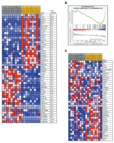
Figure 3:
WASF1
deletion may identify an aggressive subtype of prostate cancer. a.
Of the 36 CRPC samples in the BIDMC dataset (Affymetrix U133A array, accession ID GSE32269), the 9 samples with the highest quartile of WASF1 expression (gray, mean = 2.67) were compared against the 9 samples with the lowest quartile of WASF1 expression (yellow, mean = 1.41), and the genes contributing most to the distinguishing of these groups were identified with the ComparativeMarkerSelection [69] module in GenePattern [70] (fold change ≥ ± 2.4 × , P < 0.05 by Student’s ttest). Red: high expression; blue: low expression. Expression intensity values are given inSupplementary Table 9.
b.
Enrichment plot of WASF1-high vs. WASF1low gene expression and correlation with Androgen Receptor (AR) activity. Normalized enrichment score: –1.50; P = 0.038.c.
Heatmap of gene expression associated with AR activity positively correlated(rather than total loss of WAVE1) may similarly confer
a selective advantage at later stages of disease. Indeed,
WASF1
expression within the BIDMC dataset of CRPC
was varied to permit stratification of samples by the top
quartile of
WASF1
expression (
WASF1-high) vs. the lower
quartile (
WASF1-low) to predict the potential effects of
WASF1 genomic deletion (Figure 3a). Consistent with
our hypothesis that low WASF1
expression represents
an aggressive phenotype, we observed significant down
regulation of the putative tumor suppressors CHD1 and
CDH10 (cadherin-10) [59, 60]. Moreover, amongst the
genes most up-regulated in the WASF1-low group are
HSD17B4, which codes for an androgen inactivating
enzyme [61], and PIP (prolactin-induced protein),
whose expression was recently reported as a readout of
AR (Androgen Receptor) activity [62]. Therefore, to test
whether AR activity is indeed increased in the
WASF1-low cases, we performed Geneset Enrichment Analysis
(high vs
. low) using the expression values for all genes.
In the WASF1-low phenotype, we observed negative
enrichment for up-regulated genes derived from a dataset
in which LNCaP cells were treated with methyltrienolone/
R1881 (Figure 3b) [63]. Indeed, these genes include the
AR targets KLK3 (PSA), TMPRSS2, and FKBP5, which
are up-regulated in the WASF1-low group (Figure 3c),
suggesting that CRPC with lower levels of WASF1 have
increased tumorigenicity.
While metastatic CRPC circumvents androgen
deprivation by intratumoral testosterone synthesis
[50], increased expression of AR target genes suggests
higher stability of ligand-bound AR and thus higher
levels of androgen synthesis. Because the reactivation
of AR activity and down-regulation of additional tumor
suppressors in these cases (see Figure 3a) represents an
aggressive prostate cancer phenotype, deletion of WASF1
and lower levels of ABI1 and ABI2 likely cooperate
with other perturbations in these CRPC for overcoming
androgen deprivation, providing a selective advantage
for those tumors harboring these genetic changes. Thus,
the normal function of the WAVE complex may be to
serve as a tumor suppressor. Therefore, WASF1 deletion
co-occurring with PTEN in advanced prostate cancers
may result in even stronger PI3K signaling, having
removed inhibitory forces from p85 (
WASF1 deletion)
and increasing levels of PIP3 (PTEN deletion). Further
investigation is needed to determine the biological
consequences of
WASF1 deletion, the interactions between
WAVE pathway and the AR signaling axis, and the role
of WAVE pathway in establishing tumors with long-term
aggressive potential.
ACkNOWLEDgmENTS
This work was supported by the National Institutes
of Health (DF/HCCProstate Cancer SPORE P50
CA090381 to AGS, R01 CA161018 to LK) and the
Department of Defense Prostate Cancer Research Program
(Postdoctoral Training Award W81XWH13–10267 to
AGS). Portions of this research were conducted on the
Orchestra High Performance Compute Cluster at Harvard
Medical School. The results published here are in whole
or part based upon data generated by the TCGA Research
Network: http://cancergenome.nih.gov/.
FUNDINg SOURCES
National Cancer Institute, Department of Defense.
CONFLICTS OF INTEREST
The authors have no conflicts of interest to declare.
REFERENCES
1. Siegel R, Ma J, Zou Z, Jemal A. Cancer statistics. CA
Cancer J Clin. 2014; 64:9–29.
2. Hanahan D, Weinberg RA. Hallmarks of cancer: the next generation. Cell. 2011; 144:646–674.
3. Chen Z, Trotman LC, Shaffer D, Lin HK, Dotan ZA,
Niki M, Koutcher JA, Scher HI, Ludwig T, Gerald W,
CordonCardo C, Pandolfi PP. Crucial role of p53
dependent cellular senescence in suppression of
Pten-deficient tumorigenesis. Nature. 2005; 436:725–730.
4. Kim MJ, BhatiaGaur R, BanachPetrosky WA, Desai N, Wang Y, Hayward SW, Cunha GR, Cardiff RD, Shen MM, AbateShen C. Nkx3.1 mutant mice recapitulate early stages of prostate carcinogenesis. Cancer research. 2002; 62:2999–3004.
5. Di Cristofano A, De Acetis M, Koff A, CordonCardo C, Pandolfi PP. Pten and pKIP1 cooperate in prostate can
-cer tumor suppression in the mouse. Nat Genet. 2001; 27:222–224.
6. Tomlins SA, Rhodes DR, Perner S, Dhanasekaran SM, Mehra R, Sun XW, Varambally S, Cao X, Tchinda J,
Kuefer R, Lee C, Montie JE, Shah RB, Pienta KJ, Rubin MA, Chinnaiyan AM. Recurrent fusion of TMPRSS2 and ETS transcription factor genes in prostate cancer.
Science. New York, NY. 2005; 310:644–648.
7. Taylor BS, Schultz N, Hieronymus H, Gopalan A, Xiao Y,
Carver BS, Arora VK, Kaushik P, Cerami E, Reva B,
Antipin Y, Mitsiades N, Landers T, Dolgalev I, Major JE, Wilson M, et al. Integrative genomic profiling of human prostate cancer. Cancer Cell. 2010; 18:11–22.
8. Di Cristofano A, Pesce B, CordonCardo C, Pandolfi PP.
Pten is essential for embryonic development and tumour
suppression. Nat Genet. 1998; 19:348–355.
9. Hill R, Song Y, Cardiff RD, Van Dyke T. Heterogeneous
preclinical prostate cancer model. Cancer research. 2005; 65:10243–10254.
10. Grasso CS, Wu YM, Robinson DR, Cao X, Dhanasekaran SM, Khan AP, Quist MJ, Jing X, Lonigro RJ, Brenner JC, Asangani IA, Ateeq B, Chun SY, Siddiqui J,
Sam L, Anstett M, et al. The mutational landscape of
lethal castrationresistant prostate cancer. Nature. 2012; 487:239–243.
11. Hemmings BA, Restuccia DF. PI3KPKB/Akt pathway. Cold Spring Harb Perspect Biol. 2012; 4:a011189.
12. Macoska JA, Xu J, Ziemnicka D, Schwab TS, Rubin MA, Kotula L. Loss of expression of human spectrin src homol -ogy domain binding protein 1 is associated with 10p loss in human prostatic adenocarcinoma. Neoplasia. New York,
NY. 2001; 3:99–104.
13. Xiong X, Chorzalska A, Dubielecka PM, White JR,
Vedvyas Y, Hedvat CV, Haimovitz-Friedman A,
Koutcher JA, Reimand J, Bader GD, Sawicki JA, Kotula L. Disruption of Abi/Hssh3bp expression induces prostatic
intraepithelial neoplasia in the conditional Abi/Hssh3bp KO
mice. Oncogenesis. 2012; 1:e26.
14. Stovold CF, Millard TH, Machesky LM. Inclusion of Scar/
WAVE3 in a similar complex to Scar/WAVE1 and 2. BMC cell biology. 2005; 6:11.
15. Eden S, Rohatgi R, Podtelejnikov AV, Mann M,
Kirschner MW. Mechanism of regulation of
WAVE1-induced actin nucleation by Rac1 and Nck. Nature. 2002; 418:790–793.
16. Stradal TE, Rottner K, Disanza A, Confalonieri S,
Innocenti M, Scita G. Regulation of actin dynamics by WASP and WAVE family proteins. Trends in cell biology.
2004; 14:303–311.
17. Takenawa T, Suetsugu S. The WASP-WAVE protein
net-work: connecting the membrane to the cytoskeleton. Nature reviews. 2007; 8:37–48.
18. Derivery E, Fink J, Martin D, Houdusse A, Piel M, Stradal TE, Louvard D, Gautreau A. Free Brick1 is a
trimeric precursor in the assembly of a functional wave
complex. PloS one. 2008; 3:e2462.
19. Dubielecka PM, Cui P, Xiong X, Hossain S, Heck S, Angelov L, Kotula L. Differential regulation of macropinocy
-tosis by Abi1/Hssh3bp1 isoforms. PloS one. 2010; 5:e10430.
20. Derivery E, Gautreau A. Generation of branched actin networks: assembly and regulation of the NWASP and WAVE molecular machines. Bioessays. 2010; 32:119–131.
21. Chen B, Brinkmann K, Chen Z, Pak CW, Liao Y, Shi S, Henry L, Grishin NV, Bogdan S, Rosen MK. The WAVE
regulatory complex links diverse receptors to the actin cyto
-skeleton. Cell. 2014; 156:195–207.
22. Scita G, Nordstrom J, Carbone R, Tenca P, Giardina G,
Gutkind S, Bjarnegard M, Betsholtz C, Di Fiore PP. EPS8 and E3B1 transduce signals from Ras to Rac. Nature. 1999; 401:290–293.
23. Dubielecka PM, Machida K, Xiong X, Hossain S,
Ogiue-Ikeda M, Carrera AC, Mayer BJ, Kotula L. Abi1/
Hssh3bp1 pY213 links Abl kinase signaling to p85 regula -tory subunit of PI-3 kinase in regulation of
macropinocy-tosis in LNCaP cells. FEBS letters. 2010; 584:3279–3286.
24. Miyamoto K, Teperek M, Yusa K, Allen GE, Bradshaw CR,
Gurdon JB. Nuclear Wave1 is required for reprogram -ming transcription in oocytes and for normal development.
Science. New York, NY. 2013; 341:1002–1005.
25. Stradal TE, Scita G. Protein complexes regulating Arp2/3
mediated actin assembly. Current opinion in cell biology.
2006; 18:4–10.
26. Ladwein M, Rottner K. On the Rho’d: the regulation of
membrane protrusions by Rho-GTPases. FEBS letters.
2008; 582:2066–2074.
27. Zhuang C, Tang H, Dissanaike S, Cobos E, Tao Y, Dai Z. CDK1mediated phosphorylation of Abi1 attenuates Bcr
Abl-induced F-actin assembly and tyrosine phosphorylation
of WAVE complex during mitosis. The Journal of biologi
-cal chemistry. 2011; 286:38614–38626.
28. King JS, Veltman DM, Georgiou M, Baum B, Insall RH.
SCAR/WAVE is activated at mitosis and drives
myosin-independent cytokinesis. Journal of cell science. 2010; 123:2246–2255.
29. Suetsugu S, Yamazaki D, Kurisu S, Takenawa T. Differential roles of WAVE1 and WAVE2 in dorsal and peripheral ruffle formation for fibroblast cell migration. Developmental cell. 2003; 5:595–609.
30. Legg JA, Bompard G, Dawson J, Morris HL, Andrew N,
Cooper L, Johnston SA, Tramountanis G, Machesky LM.
NWASP involvement in dorsal ruffle formation in mouse embryonic fibroblasts. Molecular biology of the cell. 2007; 18:678–687.
31. Zipfel PA, Bunnell SC, Witherow DS, Gu JJ, Chislock EM,
Ring C, Pendergast AM. Role for the Abi/wave protein
complex in T cell receptormediated proliferation and cyto
-skeletal remodeling. Curr Biol. 2006; 16:35–46.
32. Campellone KG, Welch MD. A nucleator arms race: cel
-lular control of actin assembly. Nature reviews. 2010; 11:237–251.
33. Taki T, Shibuya N, Taniwaki M, Hanada R, Morishita K, Bessho F, Yanagisawa M, Hayashi Y. ABI-1, a human homolog to mouse Abl-interactor 1, fuses the MLL gene in
acute myeloid leukemia with t(10, 11)(p11.2;q23). Blood. 1998; 92:1125–1130.
34. Wang C, TranThanh D, Moreno JC, Cawthorn TR, Jacks LM, Wang DY, McCready DR, Done SJ. Expression of Abl interactor 1 and its prognostic significance in breast cancer: a tissuearraybased investigation. Breast Cancer Res Treat. 2011; 129:373–386.
36. Silva JM, Ezhkova E, Silva J, Heart S, Castillo M, Campos Y, Castro V, Bonilla F, Cordon-Cardo C,
Muthuswamy SK, Powers S, Fuchs E, Hannon GJ. Cyfip1
is a putative invasion suppressor in epithelial cancers. Cell.
2009; 137:1047–1061.
37. Fernando HS, Sanders AJ, Kynaston HG, Jiang WG. WAVE1 is associated with invasiveness and growth of
prostate cancer cells. J Urol. 2008; 180:1515–1521. 38. Fernando HS, Sanders AJ, Kynaston HG, Jiang WG.
WAVE3 is associated with invasiveness in prostate cancer
cells. Urol Oncol. 2010; 28:320–327.
39. Hossain S, Dubielecka PM, Sikorski AF, Birge RB, Kotula L. Crk and ABI1: binary molecular switches that
regulate abl tyrosine kinase and signaling to the
cytoskel-eton. Genes & cancer. 2012; 3:402–413.
40. Oikawa T, Yamaguchi H, Itoh T, Kato M, Ijuin T, Yamazaki D, Suetsugu S, Takenawa T. PtdIns(3, 5)P3 bind -ing is necessary for WAVE2-induced formation of
lamel-lipodia. Nature cell biology. 2004; 6:420–426.
41. Innocenti M, Frittoli E, Ponzanelli I, Falck JR,
Brachmann SM, Di Fiore PP, Scita G. Phosphoinositide 3kinase activates Rac by entering in a complex with Eps8, Abi1, and Sos1. The Journal of cell biology. 2003; 160:17–23.
42. Dubielecka PM, Ladwein KI, Xiong X, Migeotte I,
Chorzalska A, Anderson KV, Sawicki JA, Rottner K, Stradal TE, Kotula L. Essential role for Abi1 in embryonic
survival and WAVE2 complex integrity. Proceedings of
the National Academy of Sciences of the United States of
America. 2011; 108:7022–7027.
43. Yuan X, Cai C, Chen S, Yu Z, Balk SP. Androgen receptor
functions in castration-resistant prostate cancer and mecha-nisms of resistance to new agents targeting the androgen
axis. Oncogene. 2014; 33:2815–2825.
44. Chen E, Sowalsky AG, Gao S, Cai C, Voznesensky O,
Schaefer R, Loda M, True LD, Ye H, Troncoso P, Lis RT,
Kantoff P, Montgomery B, Nelson PS, Bubley GJ, Balk SP, et al. Abiraterone Treatment in Castration-Resistant Prostate Cancer Selects for Progesterone Responsive Mutant Androgen Receptors. Clin Cancer Res. 2014.
45. Yu Z, Cai C, Gao S, Simon NI, Shen HC, Balk SP. Galeterone prevents androgen receptor binding to chroma-tin and enhances degradation of mutant androgen receptor.
Clin Cancer Res. 2014; 20:4075–4085.
46. Beer TM, Tombal B. Enzalutamide in metastatic
pros-tate cancer before chemotherapy. N Engl J Med. 2014; 371:1755–1756.
47. Carver BS, Chapinski C, Wongvipat J, Hieronymus H, Chen Y, Chandarlapaty S, Arora VK, Le C, Koutcher J, Scher H, Scardino PT, Rosen N, Sawyers CL. Reciprocal feedback regulation of PI3K and androgen receptor signaling in
PTEN-deficient prostate cancer. Cancer Cell. 2011; 19:575–586. 48. Lin HK, Hu YC, Lee DK, Chang C. Regulation of andro
-gen receptor signaling by PTEN (phosphatase and tensin
homolog deleted on chromosome 10) tumor suppressor through distinct mechanisms in prostate cancer cells. Mol
Endocrinol. 2004; 18:2409–2423.
49. Nan B, Snabboon T, Unni E, Yuan XY, Whang YE,
Marcelli M. The PTEN tumor suppressor is a negative modulator of androgen receptor transcriptional activity. J
Mol Endocrinol. 2003; 31:169–183.
50. Stanbrough M, Bubley GJ, Ross K, Golub TR, Rubin MA,
Penning TM, Febbo PG, Balk SP. Increased expression
of genes converting adrenal androgens to testosterone in
androgenindependent prostate cancer. Cancer Res. 2006; 66:2815–2825.
51. Xiong X, Chorzalska A, Dubielecka PM, White JR,
Vedvyas Y, Hedvat CV, Haimovitz-Friedman A,
Koutcher JA, Reimand J, Bader GD, Sawicki JA, Kotula L. Disruption of Abi1/Hssh3bp1 expression induces prostatic
intraepithelial neoplasia in the conditional Abi1/Hssh3bp1
KO mice. Oncogenesis. 2012; 1:e26.
52. Barbieri CE, Baca SC, Lawrence MS, Demichelis F, Blattner M, Theurillat JP, White TA, Stojanov P, Van
Allen E, Stransky N, Nickerson E, Chae SS, Boysen G,
Auclair D, Onofrio RC, Park K, et al. Exome sequencing identifies recurrent SPOP, FOXA1 and MED12 mutations in prostate cancer. Nat Genet. 2012; 44:685–689.
53. Kunda P, Craig G, Dominguez V, Baum B. Abi, Sra1, and
Kette control the stability and localization of SCAR/WAVE to regulate the formation of actin-based protrusions. Curr
Biol. 2003; 13:1867–1875.
54. Kölsch V, Charest PG, Firtel RA. The regulation of cell
motility and chemotaxis by phospholipid signaling. J Cell Sci. 2008; 121:551–559.
55. Kotula L. Abi1, a critical molecule coordinating actin cytoskeleton reorganization with PI-3 kinase and growth
signaling. FEBS Lett. 2012; 586:2790–2794.
56. Schmitz M, Grignard G, Margue C, Dippel W, Capesius C,
Mossong J, Nathan M, Giacchi S, Scheiden R, Kieffer N.
Complete loss of PTEN expression as a possible early prog -nostic marker for prostate cancer metastasis. Int J Cancer.
2007; 120:1284–1292.
57. Feilotter HE, Nagai MA, Boag AH, Eng C, Mulligan LM.
Analysis of PTEN and the 10q23 region in primary prostate carcinomas. Oncogene. 1998; 16:1743–1748.
58. Bismar TA, Yoshimoto M, Vollmer RT, Duan Q, Firszt M, Corcos J, Squire JA. PTEN genomic deletion is an early
event associated with ERG gene rearrangements in prostate
cancer. BJU Int. 2011; 107:477–485.
59. Walker MM, Ellis SM, Auza MJ, Patel A, Clark P. The intercellular adhesion molecule, cadherin-10, is a marker for
human prostate luminal epithelial cells that is not expressed in prostate cancer. Mod Pathol. 2008; 21:85–95.
60. Huang S, Gulzar ZG, Salari K, Lapointe J, Brooks JD, Pollack JR. Recurrent deletion of CHD1 in prostate can
61. Rasiah KK, Gardiner-Garden M, Padilla EJ, Möller G,
Kench JG, Alles MC, Eggleton SA, Stricker PD, Adamski J, Sutherland RL, Henshall SM, Hayes VM. HSD17B4 over
-expression, an independent biomarker of poor patient outcome in prostate cancer. Mol Cell Endocrinol. 2009; 301:89–96.
62. Baniwal SK, Little GH, Chimge NO, Frenkel B. Runx2
controls a feed-forward loop between androgen and
prolactininduced protein (PIP) in stimulating T47D cell proliferation. J Cell Physiol. 2012; 227:2276–2282.
63. Nelson PS, Clegg N, Arnold H, Ferguson C, Bonham M, White J, Hood L, Lin B. The program of androgen- responsive genes in neoplastic prostate epithelium. Proc
Natl Acad Sci U S A. 2002; 99:11890–11895.
64. Cerami E, Gao J, Dogrusoz U, Gross BE, Sumer SO,
Aksoy BA, Jacobsen A, Byrne CJ, Heuer ML, Larsson E, Antipin Y, Reva B, Goldberg AP, Sander C, Schultz N. The
cBio cancer genomics portal: an open platform for explor
-ing multidimensional cancer genomics data. Cancer Discov. 2012; 2:401–404.
65. Wilks C, Cline MS, Weiler E, Diehkans M, Craft B, Martin C, Murphy D, Pierce H, Black J, Nelson D,
Litzinger B, Hatton T, Maltbie L, Ainsworth M, Allen P,
Rosewood L, et al. The Cancer Genomics Hub (CGHub):
overcoming cancer through the power of torrential data.
Database (Oxford). 2014; 2014.
66. Li B, Dewey CN. RSEM: accurate transcript quantification from RNASeq data with or without a reference genome. BMC Bioinformatics. 2011; 12:323.
67. Yoshihara K, Shahmoradgoli M, Martinez E, Vegesna R, Kim H, Torres-Garcia W, Trevino V, Shen H, Laird PW,
Levine DA, Carter SL, Getz G, StemkeHale K, Mills GB,
Verhaak RG. Inferring tumour purity and stromal and
immune cell admixture from expression data. Nat Commun. 2013; 4:2612.
68. Piccolo SR, Withers MR, Francis OE, Bild AH, Johnson WE. Multiplatform single-sample estimates of
transcriptional activation. Proc Natl Acad Sci U S A. 2013; 110:17778–17783.
69. Gould J, Getz G, Monti S, Reich M, Mesirov JP. Comparative gene marker selection suite. Bioinformatics.
2006; 22:1924–1925.
70. Reich M, Liefeld T, Gould J, Lerner J, Tamayo P,