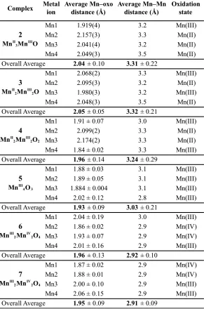
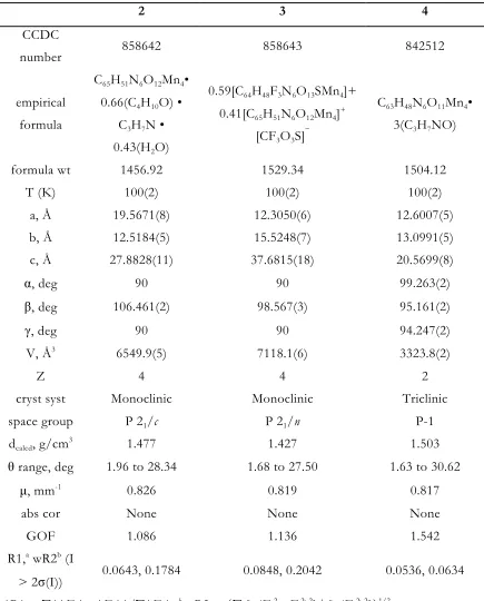
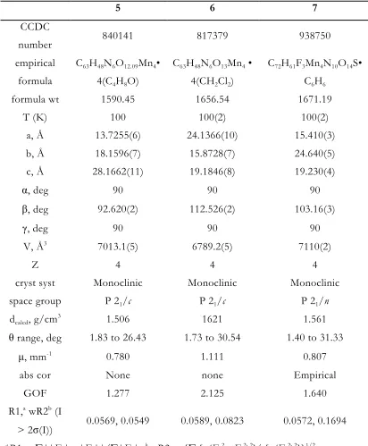
Models of the Oxygen-
Evolving Complex of Photosystem II
Thesis by Jacob Steven Kanady
In Partial Fulfillment of the Requirements for the Degree of Doctor of Philosophy
Division of Chemistry and Chemical Engineering
CALIFORNIA INSTITUTE OF TECHNOLOGY Pasadena, California
2015
Dedicated to Andi.
ACKNOWLEDGEMENTS
Over my five years at Caltech I have had the privilege to meet and work with a lot of great people while still still being able to spend time with my closest friends and family in Southern California. I would like to acknowledge up front that without my family, especially my wife Andi, there is no chance that I would have made it through. Now then:
I would first like to thank my advisor, Theodor Agapie. He has shown unflagging support of my scientific career, pushing me to apply for, and get with his helpl, the NSF, a trip to the Inorganic Chemistry G.R.C., and a trip to the Lindau Nobelauretes Meeting in Germany. And even when I was thinking about leaving and applying for jobs, although he disagreed with me, he supported me. He was also patient with me when science took longer than expected, took the time to talk to me about issues big and small, and always showed me respect. He also got me to experience the Eastern Sierras; without the group trips, I would never have gone on out there on my own. For all this, thank you.
Thanks to my committee members Jonas Peters, Mark Davis, Harry Gray, and Bob Grubbs. They gave excellent advice throughout my five years, and it was always evident that they cared about my progress and my future career. I would like to especially thank committee chair Jonas, for spending extra time with me after my props exam to discuss the beginnings of my independent career.
My graduate work was not done in a vacuum, and I would therefore like to acknowledge my fellow Agapie group members. Obviously, first and foremost, Dr. Emily Tsui. Much of the work in this thesis was done in collaboration with her. Her intensity and rigor made our science, and my scientific skill, much better. Thanks for the fun trips to Northern California and Mt. Whitney too!
Dr Zhiji Han, Dr. Siti Riduan, and Dr. Graham de Ruiter. Your lack of fear will suit this project well.
To rest of the group: Guy, you helped me through more stuff than you know. Thanks for being a good dude. Kyle, thanks for playing music that reminded me of home, and taking all the warmth into your legs so the rest of us could chill. Those days were always good. Justin, Thanks for humoring me with the glovebox trap situation; that…conversation taught me a lot, so thanks. Buss, thanks for getting me to go to the second pass and giving me something to chase up Mt. Whitney. To the Youngin’s Marcus and Jes: thanks for being fun and nice people to be around; keep it up! Finally, to the original four: thank you all for setting up the lab and giving us examples of how to do synthetic inorganic chemistry and how to (and how not to) get enough sleep. Maddy, thanks for trying to herd cats and keep the lab safe. Paul and Sibo: thanks for all the great music. I really appreciate all the music you both introduced me to.
I would like to mention my two mentees, Wei Jian Ong and Alessandro Maggi; I learned a lot from both of you about leadership and communication. Thank you for forcing me to step-up and mature some.
The Caltech staff were extremely helpful. Thanks to Agnes Tong for talking me through some bad times; Mona Shahgholi for keeping the ESI working, which was absolutely critical for all of my projects; Dave VanderVelde for keeping the NMRs running; Mike Day and Mike Takase for help with some really tough crystallography; and finally Larry Henling, who helped me at all hours to mount crystals and solve their structures. He was also a great outfielder during softball season.
work, our understanding of our compounds and their relevance in the community would be greatly diminished.
Before my time at Caltech, a few people really helped start my scientific career. My high school chemistry teacher Mr. Ray Cruickshank first planted the seed. My undergraduate advisor Prof. Chris Vanderwal supported me and really introduced me to the idea of graduate school as an option, and my mentor Dr. Grant Shibuya’s excellent lab technique set an great example when I began with research. I would also like to acknowledge Prof. Andy Borovik for getting me excited about bioinorganic chemistry, and Prof. Keith Woerpel for all of the life advice and honest friendship.
Finally, I would like to thank my friends and family. I have made some great friends here at Caltech. Davide and Mike, thanks for chilling with me the first few years. Drinking and golfing was always fun. Thanks to Grant, Naeem, Joey, Ryan, and Peter for pulling me through here at the end. I would also like to mention the Cp-Allstars; thanks for the good times and excuse to be outside. From home, I would like to thank James, my best man and best friend, for always giving me perspective on what is important in life and to always remember to chill out.
To my sister Jesica, you have always set the example for me and I had loved talking science and life with you. To my brother-in-law Nathan: thank you for being such a great friend and getting me to the top of Mt. Whitney. To Yen and Phiet, thank you for supporting me with love and food and advice. To my parents, thank you for rasing me to be curious and planting the desire to understand the world around me. Also for feeding me and clothing me and sending me to college.
Last, to my wife Andi: thank you for being the one thing in my life I am sure of, for loving me and listening to me, for taking me all over the world. I can’t imagine my life without you.
RESPECTIVE CONTRIBUTIONS
Much of the work described in this thesis is the result of collaborative efforts. Specific notes are included for compounds not synthesized by the author; some general comments are given here.
Many of the studies of multimetallic clusters were carried out in close collaboration with Dr. Emily Y. Tsui. She originally synthesized and characterized the triarylbenzene ligand framework used throughout this thesis. She synthesized the first metal complexes of the ligand (M = CuII, FeII, ZnII), of which magnetic susceptibility
data are presented in Chapter 2, which Dr. Tsui and I collected in collaboration. All magnetic data was fit using a Matlab program written by Dr. Tsui. Additionally, the PMe3 studies of the oxidized heterometallic cubane clusters (Chapter 5) were run in
collaboration with Dr. Tsui.
ABSTRACT
In the five chapters that follow, I delineate my efforts over the last five years to synthesize structurally and chemically relevant models of the Oxygen Evolving Complex (OEC) of Photosystem II. The OEC is nature’s only water oxidation catalyst, in that it forms the dioxygen in our atmosphere necessary for oxygenic life. Therefore understanding its structure and function is of deep fundamental interest and could provide design elements for artificial photosynthesis and manmade water oxidation catalysts. Synthetic endeavors towards OEC mimics have been an active area of research since the mid 1970s and have mutually evolved alongside biochemical and spectroscopic studies, affording ever-refined proposals for the structure of the OEC and the mechanism of water oxidation. This research has culminated in the most recent proposal: a low symmetry Mn4CaO5 cluster with a distorted Mn3CaO4 cubane bridged to
a fourth, dangling Mn. To give context for how my graduate work fits into this rich history of OEC research, Chapter 1 provides a historical timeline of proposals for OEC structure, emphasizing the role that synthetic Mn and MnCa clusters have played, and ending with our Mn3CaO4 heterometallic cubane complexes.
In Chapter 2, the triarylbenzene ligand framework used throughout my work is introduced, and trinuclear clusters of Mn, Co, and Ni are discussed. The ligand scaffold consistently coordinates three metals in close proximity while leaving coordination sites open for further modification through ancillary ligand binding. The ligands coordinated could be varied, with a range of carboxylates and some less coordinating anions studied. These complexes’ structures, magnetic behavior, and redox properties are discussed.
Chapter 3 explores the redox chemistry of the trimanganese system more thoroughly in the presence of a fourth Mn equivalent, finding a range of oxidation states and oxide incorporation dependent on oxidant, solvent, and Mn salt. Oxidation states from MnII
4 to MnIIIMnIV3 were observed, with 1-4 O2– ligands incorporated,
modeling the photoactivation of the OEC. These complexes were studied by X-ray diffraction, EPR, XAS, magnetometry, and CV.
As Ca2+ is a necessary component of the OEC, Chapter 4 discusses synthetic
electrochemical characterization of the first Mn3CaO4 heterometallic cubane complex—
and comparison to an all-Mn Mn4O4 analog—suggests a role for Ca2+ in the OEC.
Modification of the Mn3CaO4 system by ligand substitution affords low symmetry
Mn3CaO4 complexes that are the most accurate models of the OEC to date.
Finally, in Chapter 5 the reactivity of the Mn3MO4 cubane complexes toward
O-atom transfer is discussed. The metal M strongly affects the reactivity. The mechanisms of O-atom transfer and water incorporation from and into Mn4O4 and Mn4O3 clusters,
respectively, are studied through computation and 18O-labeling studies. The µ
3-oxos of
the Mn4O4 system prove fluxional, lending support for proposals of O2– fluxionality
TABLE OF CONTENTS
ACKNOWLEDGEMENTS ... iv
Respective Contributions ... vii
ABSTRACT ... viii
CHAPTER 1 ... 1
Historical Perspective & General Introduction ... 1
1.1 Photosynthesis and Photosystem II ... 2
1.2 The Oxygen-‐Evolving Complex: Composition and Kok Cycle ... 3
1.3 Structural Proposals for the OEC: A Historical Perspective ... 6
1.4 Mechanism of O–O bond formation ... 12
1.5 Synthetic OEC Model Coordination Complexes and a General Introduction ... 14
1.6 Conclusion ... 21
References ... 22
CHAPTER 2 ... 33
Trinuclear First Row Transition Metal Complexes of a Hexapyridyl, Trialkoxy 1,3,5-‐Triarylbenzene Ligand ... 33
Abstract ... 34
Results & Discussion ... 36
2.1 Synthesis of MnII3, CoII3, and NiII3 complexes ... 36
2.2 Magnetic susceptibility studies ... 45
2.3 Electrochemical and chemical oxidation studies. ... 49
Conclusions ... 50
Experimental Section ... 51
References ... 65
CHAPTER 3 ... 68
Role of oxido incorporation and ligand lability in expanding redox accessibility of structurally related Mn4 clusters ... 68
Abstract ... 69
Introduction ... 70
Results & Discussion ... 71
3.1 Synthesis of Tetramanganese Clusters ... 71
3.2 Solid-‐State Structures ... 75
3.3 XAS ... 80
3.4 Magnetism ... 83
3.5 EPR ... 85
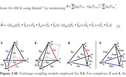
3.6 Cluster Reactivity and Interconversion ... 90
3.7 Electrochemistry and Potential Leveling ... 92
3.9 Ligand Flexibility as Design Element ... 97
3.10 Relation to the Assembly and Turnover of the OEC; Design Implications for Metal-‐ Oxide Clusters ... 98
Conclusions ... 99
Experimental Section ... 100
References ... 123
CHAPTER 4 ... 129
A Synthetic Model of the Mn3Ca Subsite of the Oxygen-‐Evolving Complex in Photosystem II and Progress Toward more Accurate Mn3CaM Models. ... 129
Abstract ... 130
Introduction ... 130
Results & Discussion ... 133
4.1 Initial and Optimized Synthesis of LMnIV3CaO4(OAc)3 (8) ... 133
4.2 Structural Comparison of the Mn3CaO4 Complex 8 and the OEC ... 135
4.3 Electrochemistry of 6 and 8 ... 137
4.4 The Charge Localization Effect of Ca2+ ... 139
4.5 Proposed Formation Intermediates and Relation to Photoactivation of the OEC ... 140
4.6 Design Elements for Functionalizing Mn3CaO4 Toward a Full OEC Model ... 141
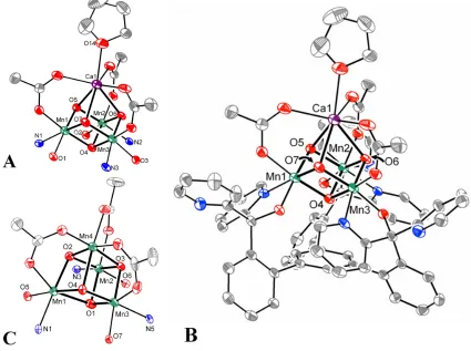
4.7 Synthesis of Asymmetric Mn3CaO4 Complexes ... 143
Conclusions ... 151
Experimental Section ... 152
References ... 169
CHAPTER 5 ... 172
Oxygen Atom Transfer and Oxidative Water Incorporation in Cuboidal Mn3MOn Complexes Based on Synthetic, Isotopic Labeling, and Computational Studies ... 172
Abstract ... 173
Introduction ... 174
Results & Discussion ... 177
5.1 O-‐atom Transfer to Phosphine as Comparative Probe of Mn3MO4 Reactivity. ... 177
5.2 QM studies of O-‐atom Transfer from Mn3MO4 to PMe3. ... 180
5.3 Carboxylate exchange studies. ... 186
5.4 Oxidative incorporation of H2O into 5 ... 187
5.5 Isotopic labeling studies of H2O incorporation. ... 188
Conclusions ... 195
Experimental Section ... 196
References ... 221
APPENDIX A ... 227
Side Products and Other Structures ... 227
Introduction ... 228
A.1 Trinuclear Complexes ... 228
A.2 Mono-‐oxo Complexes ... 230
A.3 Other Mn4O4 Cubane Complexes ... 238
A.4 Other Mn3MO4 Cubane Complexes ... 244
Conclusions ... 247
Experimental Section ... 247
APPENDIX B ... 256
CHAPTER 1
HISTORICAL PERSPECTIVE &GENERAL INTRODUCTION
Published in part as:
1.1 Photosynthesis and Photosystem II
One of the most fascinating and important transformations in nature is the biological generation of O2 by the Oxygen Evolving Complex (OEC) of Photosystem II (PSII) in
cyanobacteria and plants.1 This transformation was responsible for the formation of the
oxygenic atmosphere that has shaped the evolution of life on Earth as we know it. In this process, solar energy is converted to the reducing equivalents and proton gradient necessary to power carbon dioxide fixation and other processes of life, while forming dioxygen as byproduct. The biological catalyst, PSII, has been studied in detail for more than 50 years. Progress in understanding the site of catalysis, the OEC, has depended on advances in several fields, including biochemistry, biophysics, spectroscopy, inorganic chemistry, and computational chemistry. While many properties of the OEC are well documented and generally agreed upon, many aspects of the catalytic site remain controversial, with computational and experimental chemists still pushing the boundaries of our understanding of the OEC. In this chapter, the structural and mechanistic proposals of the OEC as they were reported chronologically and the technologies and methodologies that supported them, with a focus on the insight gained from recent synthetic inorganic work in the field, are reviewed to put this thesis into historical context.
Photosystem II is a 350 kDa homo-dimer in the thylakoid membrane with ca. 20 protein subunits.1f, h PSII absorbs photons that drive the separation of charge, which is
transferred through several redox cofactors. The ultimate electron donor is water, being oxidized to O2 and releasing four electrons and four protons. The chemiosmotic gradient
pheophytin a, quinone A, and quinone B (Figure 1.1). Structurally, the D1 and D2 subunits make up the main membrane-bound core of PSII, with D1 containing much of the electron transfer pathway.2 The other membrane bound subunits mainly function as a light
absorption antenna via a multitude of cofactors to transfer the exciton to P680. There are also a number of extrinsic, water-soluble subunits that bind to the lumenal side of PSII that are proposed to stabilize the binding of the Ca2+ and Cl– cofactors necessary for efficient
oxygen evolution.3
[image:15.612.124.510.276.523.2]a) b)
Figure 1.1 Electron transfer pathway shown in the overall PSII structure given by the 1.9Å
diffraction data.2b (a) distances (b) aliphatic tails of the quinones, PheoD1, and
chlorophylls are not shown for clarity.
1.2 The Oxygen-Evolving Complex: Composition and Kok Cycle
The OEC is located on the lumenal face of PSII with the majority of ligating side chains from the D1 subunit, positioning it approximately 5 Å away from Yz.2b Manganese,
contain Mn since the 1950s,4 although oxygenic photosynthesis has been known to be Mn
dependent for much longer.5 As PSII isolation and purification methodology improved, the
stoichiometry of four for Mn was verified by a number of methods, including quantitative electron paramagnetic resonance (EPR) spectroscopy of released Mn2+, 6 and atomic
absorption spectroscopy.7 The specific importance of Ca2+ over other dications was
proposed in the 1970s based on O2 evolution activities at variable Ca2+ concentrations and
the catalytic ineffectiveness or inhibitory effects of other dications.8 ,3, 9 Given the redox
nature of the catalytic reaction, the role of the redox inactive Ca2+ has been debated.
Notably, the only metal to substitute for Ca2+ and generate a catalytic system, albeit with
lower activity, is Sr2+.10 A single Ca2+ center is required for the restoration of the catalytic
activity.11 The close association of the redox inactive metal with the OEC was supported by
early EPR data on Sr2+ substituted samples.12 Removal of Ca2+ was shown by spectroscopy
to arrest the catalytic cycle at intermediate states and affect electron transfer, further supporting the role of Ca2+ in catalysis.13 More recently, EPR and XAS studies indicate that
Ca2+ is part of the OEC.14
Until recently, Cl– was also thought to be part of the OEC, as it is a native cofactor
for O2 production15 and found to have a 1:1 stoichiometry with the OEC, based on 36Cl–
labeling analysis.16 However, more recent structural work suggests a role as H-bond acceptor
in the secondary coordination sphere of the OEC.2b, 17
The OEC must be reassembled frequently under full solar flux due to photoxidative damage.18 The assembly of the OEC, called photoactivation,4b, 19 requires Mn2+, Ca2+, Cl–,
bicarbonate, water, and photogenerated oxidizing equivalents from P680.20 A mechanism has
site proposed to contain D1-Asp170,21 and is photooxidized in low quantum yield to Mn3+,
giving intermediate one (IM1). The quantum efficiency of this initial oxidation is dependent
on the presence of Ca2+, which can bind either before or after the initial Mn2+.22 Ca2+ is
proposed to bridge to the Mn3+ center through oxide or hydroxide bridges. After binding a
second Mn2+ and photooxidation, a rate limiting protein conformation change affords IM 2
that is quickly transformed into the OEC with additional Mn2+ equivalents in kinetically
unresolved steps that must include deprotonation and incorporation of water as oxide donors.20d, e, 22-23
With respect to the mechanism of catalysis, a dependence of O2 production on the
number of short flashes of light on chloroplasts was discovered as early as the 1960s.24
Dark-adapted chloroplasts gave a spike in O2 production on the third millisecond flash, followed
by shorter spikes every four subsequent flashes until steady state O2 production was
observed. Kok proposed that each flash corresponded to a photooxidative event, with three oxidizing equivalents stored until the fourth flash, upon which four-electron oxidation of water to O2 occurs. In this so-called S-state cycle (Scheme 1.1), S1 is the dark stable state and
S4 is the transiently formed state that releases O2 and relaxes back to S0. The four oxidations
of the OEC have to be negative of Eº´= ca. 0.9V as necessitated by the potential of the P680+; concurrent deprotonation helps keep the overall OEC charge low and thus levels the
potentials of the S-state transition.25 For the S-state cycle two possibilities have been put
forward for the Mn oxidation states: the ‘high’ (Scheme 1.1) and the ‘low’ pathways. The high oxidation state pathway has been supported by electron paramagnetic resonance (EPR),2655Mn electron nuclear double resonance (ENDOR),27 x-ray absorption spectroscopy
(XAS),28 and K
spectroscopic data has also been interpreted to support the lower oxidation state cycle with a MnIIMnIII
2MnIV or MnIII4 S1 state.30
Scheme 1.1. The high oxidation state pathway for the S-state cycle.
Utilizing time-resolved mass spectrometry, Ollinger and Radmer31 and then
Messinger, Wydrzynski and coworkers32 studied the kinetics of substrate water binding to the
OEC throughout the S-state cycle. These studies found that: water is exchangeable through S3, suggesting no intermediate oxidations of water occur;31 in all of the S-states there are
kinetically distinct, fast (40 s–1 for S
3 to ≥ 120 s–1 for S0 & S1) and slow (0.02 s–1 for S1 to 10 s–1
for S0) exchanging substrate waters, consistent with two separate sites of water coordination
to the OEC;32a, c, 33 both substrate waters are bound by the S
2 state;32e and Sr2+ substitution of
Ca2+ gives an increase in rate for the slow exchanging water, suggesting it is bound to Ca2+.32f
There are a number of possible ways to explain the slow and fast exchanging waters, including protonation state, MnIII,IV or Ca2+ coordination, and terminal or bridging ligation
mode. Thus, these studies are an important consideration for many mechanistic proposals for O-O bond formation (see Section 1.4).
1.3 Structural Proposals for the OEC: A Historical Perspective
Structural understanding of the OEC has gradually developed over the last 30 years, with many methods across multiple disciplines being paramount. Although multiple XRD structures are now known and provide the location and amino acid ligands of the OEC,2, 17, 34
S0 S1 S2
S3 S4
–O2, + 2H2O
h! h! h! h!
–e–
, –H+ –e
–
–e–
, –H+ –e–, –2H+
(MnIII
3MnIV) (MnIII2MnIV2) (MnIIIMnIV3)
(MnIV
4) or
(MnIIIMnIV
3O•) (MnIV3MnV)
(MnIV
changes in the structure of the OEC due to reductive X-ray damage have been a concern.35
EPR36 and XAS37—techniques used to study the OEC since the early 1980s38—complement
the XRD data to afford more complete structural information. Crucial for these two methods was the parallel growth in the synthetic inorganic coordination chemistry of manganese, particularly of multinuclear cluster chemistry (Section 1.5).39 The synthetic
systems not only showed what was chemically reasonable to propose for the OEC based on precedent, but just as importantly they acted as spectroscopic benchmarks, providing starting points for hypotheses on how the OEC’s spectra relate to structure. Simple synthetic complexes were also key in benchmarking quantum mechanical (QM) computation,40 which
has emerged as a crucial method in the study of the OEC. QM methods have improved drastically in the last decade to allow for structural hypotheses for every S state and mechanisms for substrate water incorporation and O2 formation.40g, 41
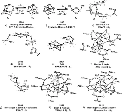
With all of these different methods, and the improvements to each over time, numerous OEC structures have been proposed with a significant amount of disagreement and controversy over the years. The main models discussed over the last 25 years are shown schematically in Figure 1.2. One of the earliest models for the OEC with a specified geometry for the four manganese centers was the Mn4O4 cubane / Mn4O6 adamantane
model proposed by Brudvig and Crabtree in 1986 (Figure 1.2a).42 They proposed that a pseudo-Jahn-Teller distorted Mn4O4 cubane could explain their recent EPR data on the S2
state that suggested two antiferromagnetically coupled dimers ferromagnetically coupled to the other.43 They also posited that a large structural change in the S
2 to S3 transition was
the proposed cubane and adamantane geometries did not prove consistent with extended x-ray absorption fine structure (EXAFS) reported subsequently.45
Figure 1.2. Key structural models of the OEC. The research group, year, main spectroscopic
support, and S-state are included below each structure. Crystal structure resolutions are in parentheses. Dashed lines represent generic coordination sites and could represent amino acids or water. In f through i, Mn numbering combines EXAFS nomenclature (MnA-D)14c
with that of the 2005 and 2011 crystal structures (Mn1-4)2b, 34c in the style of ref. 46.
Mn 1986 Brudvig and Crabtree EPR & Synthetic Models
1987 Christou Synthetic Models & EXAFS
1993 Sauer & Klein
EXAFS ; O OO
Mn Mn
Mn O O Mn O
Mn Mn Mn O O O O Mn – O2
+ 2H2O –4H+, –4e–
Mn Mn Mn O O O O Mn Mn Mn Mn O O Mn OO Mn O O Mn O O O Mn Cl O Mn
O Ca III IV III IV O O
H2O
N Mn O Mn OH2 III IV O O Mn O Mn O IV HN IV 2000 Britt EPR/ENDOR ; 2004 Barber & Iwata
XRD (3.7Å) ;
2011 Shen & Kamiya
XRD (1.9 Å) ; His332 Mn Mn Mn O O O O Ca O Glu189 O O Ala344 O H2O
Mn OH x OHx Asp170 O O Glu333 O O Glu354 O O Asp342 O H2O
O O His332 Mn Mn Mn O O O O Ca O Glu189 O O Ala344 Mn OH2 H2O
Asp170 O O Glu333 O O Glu354 O Asp342 O O O O O O O O O O Mn Mn Mn O O Mn H2O Cl
Ca H2O
2004 Britt EPR/EXAFS ;
2006
Messinger & Zouni & Yachandra EXAFS ; Mn O O O Mn O O Mn Ca
– O2
+ 2H2O –4H+, –4e–
His332 Mn Mn Mn O O O O Ca O O O Ala344 O H2O
Mn OH2 OH Asp170 O O Glu333 O O Glu354 O O Asp342 O O 2011
Messinger & Lubitz & Neese Computation ;
O OH2
Glu189
S4 S0
S4 S0
S1
S2 S2 S1
S1 S1 S2
a) b) c)
d) e) f)
g) h) i)
Another OEC model based on a Mn4O4 cubane was put forward soon after and was
dubbed the “double-pivot” mechanism by Vincent and Christou (Figure 1.2b).47 Here, a
Mn4O2 butterfly structure in the S0 to S2 states was proposed to bind and deprotonate two
water molecules to afford a Mn4O2(OH)2 cubane structure that upon double deprotonation
affords dioxygen and the S0 butterfly structure. Key to the proposal was a synthetically
characterized Mn4O2 structure (Figure 1.4c) that contained Mn-Mn vectors at ca. 2.7 and 3.3
Å, similar to those found in past EXAFS studies.38d, 48 Although further EXAFS studies49
were not consistent with this proposal, synthetic work by the groups of Christou and Dismukes detailed a variety of structural motifs and properties of clusters of these types. (Section 1.5).
A structural model based on oriented-membrane-EXAFS was proposed in 1993 and is generally referred to as the “dimer-of-dimers” model.50 The basic structure is two Mn
2(µ2
-O)2 dimers connected through a mono-µ-O and/or k2-carboxylates (Figure 1.2c). The Cl–
and Ca2+ cofactors were originally proposed to bind Mn and to bridge to the end of one
Mn2O2 dimer unit through a carboxylate, respectively. The dimer-of-dimers served as the
basis for a number of mechanistic proposals, including a metalloradical mechanism51 and a
nucleophilic attack by calcium-ligated hydroxide/water on an electrophilic MnV=O.52
A different structure, the “trimer/monomer,” “3+1”, or “dangling Mn” model (Figure 1.2d,e), was proposed based on EPR experiments. Britt and coworkers posited that the magnetic interaction of the Mn in the dimer-of-dimers model could not explain the high-spin g=4.1 signal and the changes to the g=2 multiline signal upon addition of methanol and ammonia.53 The 3+1 motif, which had been included as a possible structure based on
antiferromagnetically coupled III,IV,IV trimer only weakly coupled to the fourth, “dangling” MnIV. A handful of trimer/monomer arrangements were proposed that fit the EPR and
EXAFS data of the time.26 Soon after this, the first crystal structures of PSII were reported,2a, 34a and although the resolution was only 3.8 Å (2001) or 3.7 Å (2003), the manganese
electron density was consistent with a 3+1 arrangement. In 2004, based on a higher resolution of 3.5 Å, Barber and Iwata proposed a more specific structure: a Mn3CaO4 cubane
with a fourth manganese connected by a cubane oxygen (Figure 1.2f).34b This was consistent
with the EPR proposal and also the Ca K-edge XAS data that suggested a Ca-Mn distance of 3.4 Å.14b In 2005, a higher resolution structure of 3.0 Å was published that reported the OEC
in a 3+1 arrangement as being more distorted and elongated than a cubane motif, without proposing the position for the bridging oxides (the same group published a 2.9 Å PSII structure in 2009 with no change to the OEC geometry).17, 34c
XAS studies in 2005 showed that the x-ray dose used in the XRD analysis of PSII caused reductive damage to the OEC, shedding doubt on the accuracy of the proposed OEC arrangement as based on crystal structures.35 Polarized EXAFS studies at much lower
X-ray dosage on PSII single crystals were used to provide an updated structure of the OEC (Figure 1.2g) with an asymmetric dimer of Mn2O2 diamond cores.14c A different
interpretation of the EXAFS data invoked a cubane with a dangler motif.25d In 2011 a
significantly higher resolution (1.9 Å) crystal structure was published2b with purported X-ray
dosage below the damage level reported in 2005. Here a “chair” geometry of the Mn4CaO5
There has been controversy over the OEC assignment in this recent XRD study because some of the Mn–O bond lengths are not consistent with a supposed S1 oxidation
state of MnIII
2MnIV2. Three explanations have come out in the literature, all based on
computational modeling: the OEC structure is accurate and supports the low-oxidation state Kok cycle, with a MnIII
4 S1 state;30c, 55 X-ray damage has produced a mixture of reduced
oxidation states including S-n states;56 and the observed electron density is a superposition of
two S1 substates in equilibrium by a µ-O migration and proton transfer.57 Another
computational study suggested a similar substate equilibrium for S2, claiming to explain the
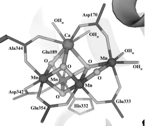
Figure 1.3. The oxygen-evolving complex as described by the 1.9 Å resolution crystal
structure.2b
two S2 EPR signals through changes in the magnetic coupling caused by µ-O migration from
the cubane unit to form a diamond core with the dangling Mn (Figure 1.2i).56a, 58 Although
[image:23.612.180.471.304.555.2]PSII structure, the present data converge toward a cluster with a CaMn3 site (part of a
cubane or distorted cubane) and a dangler Mn center, with bridging oxido moieties.
1.4 Mechanism of O–O bond formation
Paralleling the wide assortment of OEC structures put forward, several mechanisms for O-O bond formation have been proposed.1g, 25d, 36c, 41a, 59 Consistency with XAS, EPR,
XRD, and substrate water exchange studies are required for advancing any mechanism, and developments in these fields have disproved many past proposals, such as the adamantane and double-pivot mechanisms discussed above.42, 45, 47a, 49, 60 As with the structural hypotheses,
some mechanisms have their basis in the chemistry of synthetic transition metal complexes. Copper has been shown to break and form the O-O bond of O2 in an equilibrium between a
CuIII
2(µ2-O)2 diamond core and a CuII2(µ-h2:h2-O2) bridging peroxide.61 Similar proposals for
diamond core O-O bond formation in the OEC exist (Scheme 1.2a); 1d, 47a, 62 however, the
fast and slow water exchange kinetics are difficult to explain by such mechanisms.60
Dinuclear ruthenium water oxidation catalysts have been shown to function through a H-bonding, nucleophilic water attacking a RuV-oxo intermediate,59c, 63 and a Mn
2O2 O2-evolving
catalyst has been proposed to act similarly through a MnV-oxo64 or MnIV-oxyl radical.40d
Water attack on an electrophilic MnIV/V-oxo has likewise been proposed for the OEC, with
the attacking water in a number of different states, both terminal and bridging: as a H-bonding water/hydroxide,32a as Mnn+-bound water/hydroxide,51 or as Ca2+-bound
water/hydroxide (Scheme 1.2b).52, 64a, 65 Brudvig and Batista proposed a Ca2+-OH 2
QM approach used for the OEC and the first coordination shell implicated a different ground state structure.66
Scheme 1.2. Proposed mechanisms for O-O bond formation depicted minimally with
metal-oxo species (top) and as part of the most recent structural models of the OEC (bottom).
The computational work of Siegbahn has supported an oxyl radical (MnIV-O•) in
O-O bond formation at the O-OEC (Scheme 1.2c).40b-d, 41a, 66-67 Others have proposed mechanisms
including radical intermediates as well.68 Both terminal67b (Scheme 1.2c-I) and bridging41a
(Scheme 1.2c-II) oxyl radical intermediates have been discussed, with recent computational work supporting the coupling of a Mn/Mn/Ca-µ3-oxo and a Mn/Ca-µ2-oxyl in the S4 state
(Scheme 1.2c-II).69 Recent 17O-ENDOR studies mapped the substrate water exchange
kinetics onto both the nucleophilic attack or bridging oxyl-coupling mechanisms.46 Overall, a
truly interdisciplinary approach of combining spectroscopy, structural characterization,
Mn O
OHx
M
M=Mn, Ca2+, H+
MnIV O MnIV O
MnIII O MnIII O
MnII O MnII O MnIV MnIV Mn O O O O Ca Mn O
O OH2
H2O OH
IV H
MnIVMn IV Mn O O O O Ca Mn O O
H2O
O
IV V
H
MnIVMn IV Mn O O O O Ca Mn H2O
H2O
O IV IV O Mn Mn Mn O O O O Ca Mn O H2O
HO IV O IV IV IV Mn O Mn O Mn O Mn O M Or
M=Mn, Ca2+, H+
a) b) c)
Diamond Core Nucleophilic
Attack Radical
I
computation, and comparisons to synthetic models has funneled the mechanistic proposals for water oxidation to only a few candidates. Further detailing the mechanism of O-O bond formation is very desirable for both fundamental reasons and application toward the development of practical artificial catalysts. Additional studies from multiple perspectives are necessary to achieve that goal.
1.5 Synthetic OEC Model Coordination Complexes and a General Introduction
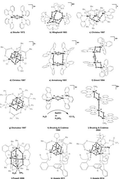
Synthetic manganese coordination clusters have played an important role in our understanding of the OEC, both inspiring the structural and mechanistic hypotheses of their time, and also being targeted due to the OEC structural motifs proposed based on other various analytical techniques. Model complexes, detailed in a number of reviews,39 have been
an instrumental benchmarking tool for XAS, EPR, water exchange rates, and computation. This historically collaborative effort is highlighted by numerous examples over the past 40 years, from the original manganese-bipyridine dimer and tetramanganese dimer-of-dimer models, to the more recent manganese/calcium heterometallic models.
In 1972, Stoufer and coworkers published the X-ray crystal structure of di-µ -oxo-tetrakis(2,2´-bipyridine)dimanganese(III,IV), showing a Mn-Mn distance of 2.716Å and finding strong antiferromagnetic coupling (Figure 1.4a).70 These two observations on a
model complex were utilized to conclude that the Mn2O2 diamond core was a key structural
motif within the OEC: comparison of the S2-state multiline EPR signal to that of the
complex38a, b, 71 supported the idea of an antiferromagnetic MnIIIMnIV pair within the OEC,
and the original OEC EXAFS studies of 198138d found Mn-Mn distances of 2.7 Å,
Mn2O2 dimer using terpyridine rather than bipyridine was reported to oxidize water using
hypochlorite (NaOCl) or oxone (H2SO5) as the stoichiometric oxidant (Figure 1.4h).64
Relevant to mechanistic interpretations for the OEC, the water exchange rates of the MnIII
-O-MnIV units of the bi- and terpyridine manganese dimers were measured by a time-resolved
mass spectrometry technique.60, 72 The exchange rates were much slower (10–3-10-4 s–1) than
those of the OEC (ca. 1 s–1), which is not consistent with mechanisms that invoked bridging
oxo units as substrate water in the OEC.
Wieghardt and coworkers synthesized the first MnIV
4 complex, a Mn4O64+
adamantane stabilized by three chelating 1,4,7-triazacyclononane ligands (Figure 1.4b).73
With this precedent, the adamantane/cubane mechanistic proposal for the OEC invoked access to such a high oxidation state cluster.42 Each MnIV displays a pseudo-octahedral
coordination environment with three µ2-oxido and three terminal N donors. Armstrong and
coworkers further studied the Mn4O6 adamantane core structure, evaluating the effect of
altering the chelating N3 ligand on the basicity of the µ2-oxido moiety and on the pH
dependent reduction potential.74 More recently, the MnIV-O-MnIV water exchange rate of the
adamantane geometry was measured to be ≤10–8 s–1.60
The Mn4O4 cubane geometry was common to both the adamantane/cubane (1986)
and the double-pivot (1987) mechanisms.42, 47bTetramanganese complexes had been isolated
in such a geometry;75 however, these were low oxidation state MnII
4 structures with µ3
-alkoxides rather than oxides. Christou and coworkers synthesized the first cubane complex with µ3-O bridges: [MnIII3MnIVO3Cl6(ImH)(OAc)3]2– (ImH = neutral imidazole; Figure
1.4d).76 Over the following decade, the MnIII
Figure 1.4. Selection of synthetic models relevant to the OEC. N N N O P O P O O O Cl Cl O O O Mn Mn Mn O
O Mn O
N H N N N H N NH HN N HN NH IV IV IV IV 4+ H
b) Wieghardt 1983
Cl Mn Mn Mn Cl Cl Cl O O O Mn O O O O O O Me Me Me N 2– Mn O Mn O Mn Mn N N O O Me O O Me O Me O O Me O O Me O O Me N N Mn Mn Mn O O O O Mn O O O O O O Ph P P P Ph Ph Ph Ph Ph O O P Ph Ph O Ph Ph O Ph Ph Mn O Mn O O Mn O Mn O N N N N N N N N N N N N OH2 OH2 IV IV IV IV 6+
i) Brudvig & Crabtree 2004 N N N N N N N N N N Mn O Mn O IV IV O O Mn O Mn O IV IV
f) Girerd 1994 III IV III III IV III IV III
a) Stoufer 1972
III III
III III
+
c) Christou 1987
d) Christou 1987
g) Dismukes 1997
O O O Mn Mn N N N N N O O O O Ca N O O Me O O Me O O Me O
k) Agapie 2011 IV
IV
IV
l) Agapie 2014
N Mn O O N N N OH2 Mn N N N H2O 3+ Mn Mn Mn Mn O O O O N N N O N N N N N N O N N 4+ N N N Mn N N N N Mn O O 3+
h) Brudvig & Crabtree 1999 e) Armstrong 1991
H2O
NaOCl or H2SO5
1/2 O2 MeO O O N Me Cl
Mn Mn Mn
Mn O N O O O Cl O O N O O Cl Me Me Me O Me Ca Me
H2O OH2
j) Powell 2006
IV IV IV IV IV III IV III II III III III 4+ N N N N N N N N N N N N N H N N N N H H H H N NN N N N N N N N NH Cl Cl Cl Cl N N
N N N
N N N N N N N N N N N N N N N N N N N N N N N N N N N N N N N OH2 OH2 N N N N N N P P P P P P Ph Ph Ph Ph Ph Ph Ph Ph Ph Ph Ph Ph Cl Cl Cl N N
N N N
in great detail, with variation of terminal ligands (Cl–, pyridines, acetylacetonates, etc) and the
anionic µ3-X position (X = Cl
−
,Br–, I−, F−, N 3
−
, O2CR
−
, OMe−, and OH−).76-77 For example,
they were able to synthetically model the S1 to S2 step of the proposed double pivot
mechanism,77a utilizing a MnIII
4O2 butterfly complex (Figure 1.4c) to form a MnIII3MnIVO3Cl
cubane by addition of chloride and disproportionation. In another reactivity study, water was selectively deprotonated and incorporated into the µ3-X position, modeling a key functional
step in OEC photoassembly and turnover.77i
Extensive magnetism77b, d, g, h, k, m and XAS77j studies were performed on the Mn 4O3X
cubanes to test the hypothesis that the OEC was not a high symmetry cubane structure.45b, 48
Although the K-edge XANES and EXAFS spectra looked superficially similar to those of the OEC in the S1 state, detailed analysis indicated that the structural motif contained in
these synthetic clusters did not match the data from the biological system. Also of note, the K-edge energy varied by more than 3 eV for a series of cubanes in the same oxidation state and similar geometry, supporting the notion that in addition to the formal metal oxidation state, the nature of the ligands strongly affects the edge energy. This convolution of effects complicates the interpretation of the edge energies of various clusters and continues to cause disagreement over the oxidation state of the OEC.30b
Other systems that gave some support for the double pivot mechanism were the diarylphosphinate-stabilized MnIII
2MnIV2O46+/MnIIIMnIV3O47+ cubanes synthesized by
Dismukes and coworkers (Figure 1.4g).78 They found that these cubane complexes lose one
phosphinate ligand and a molecule of O2 upon UV photolysis in the gas phase,79 indicating
the ability of a Mn4O4 cubane to form dioxygen as previously proposed for PSII.47a This
further study showed that decomposition to an amorphous manganese oxide provided the active catalyst.81 The Mn
3-µ3-O water exchange rates measured in organic solvent (10–5 s–1)
were one to two orders of magnitude slower than the synthetic complex MnIII-µ
2-O-MnIV
rate (10–3-10-4 s–1) and thus much slower than those found for the OEC.82
As spectroscopic6, 43, 83 and biochemical7 support for a tetramanganese OEC grew,
Mn4 complexes were targeted that contained the 2.7 and 3.3 Å Mn-Mn vectors reported for
the OEC.39a, d For example, alongside the butterfly systems discussed above,84 Armstrong’s
group reported a series of dimer-of-dimer geometries.85 They contained two 2.7 Å Mn-Mn
vectors each, and the EPR of the highest oxidation state dimer—with two MnIII-(µ-O) 2-MnIV
diamond cores (Figure 1.4e)—modeled that of the S1 state. Towards modeling the EXAFS
dimer-of-dimers proposal in 1993,50 complexes such as the MnIV
4 diamond core chain
structure by Girerd and coworkers (Figure 1.4f)86 and the [MnIV-(µ
2-O)2-MnIV]2O
dimer-of-dimers by Brudvig and coworkers (Figure 1.4i)87 were reported.
Based on Ca K-edge XAS data, the calcium ion was proposed to be closely associated with the tetramanganese motif of the OEC, with a Mn-Ca vector of 3.4 Å.14b In
agreement, the 2004 crystal structure proposed an OEC structure displaying a Mn3CaO4
cubane moiety. Calcium is necessary for photoactivation (cluster assembly from Mn2+ in
solution under light) and turnover of the OEC. Synthetic Mn/Ca complexes were targeted to understand the effect of the redox inactive metal on the chemistry of manganese clusters. The first high oxidation state Mn/Ca cluster was isolated in 2005 and contained a Mn4CaO4
motif quite similar to the 2004 crystal structure as part of a high nuclearity Mn13Ca2O10
cluster coordinated by benzoates.88 A Ca K-edge XAS study on this cluster showed a Mn-Ca
synthesized with the correct Mn4Ca metal stoichiometry, although in low oxidation state and
with low oxide content. The first contains a trigonal bipyramidal arrangement of metals with a MnII and Ca2+ at the two vertices and one µ
4-oxide, with a low MnIII3MnII oxidation state
(Figure 1.4j).90 Similar complexes isolated later by the same group showed O
2 evolution in
the presence of O-atom transfer agents and water.91 A more recent cluster displays a MnIII 4
metallocrown moiety with a Ca2+ center coordinated to one side of the crown and chelated
by carboxylates; this cluster contains no bridging oxido ligands.92 Other Mn/Ca structures—
a low oxidation state MnII
4Ca2 cluster93 and a high nuclearity MnIII6Ca2O2 complex94—have
also been reported.
Most of the multinuclear complexes discussed above were synthesized by self-assembly methodology that offers only low control over the geometry and nuclearity of the final complex. Although this manganese cluster chemistry has been invaluable to understanding the OEC, new methods for the controlled synthesis of Mn/Ca complexes are important, especially with the structure of the OEC emerging as a low symmetry Mn4CaO5
cubane/open cubane. In related bioinorganic studies, Holm and coworkers pioneered a synthetic protocol, termed “subsite-specific functionalization,” to study the properties of ubiquitous Fe4S4 biological clusters. A wide array of ligand-differentiated Fe4S4X3X´ and
metal-differentiated Fe3MS4 complexes was accessible using this methodology.95 The basis of
this synthetic strategy is a semi-rigid tridentate ligand design that can accommodate binding three metals of the Fe4S4 core, leaving the fourth metal center open to ligand substitution or
replacement by a heterometal.
based on ligand design was employed by our group to access a series of Mn3MOn OEC
model complexes.96 My contributions to this body of work are the focus of this thesis.
The ligand framework I used was designed to bind three metal centers in close proximity, to accommodate multiple coordination modes, to be oxidatively robust, and to promote site-differentiated functionalization to allow access to 1) models of the OEC and 2) site-differentiated metal-oxido clusters in general. As discussed in Chapter 2, these design criteria led to 1,3,5-tris(2-di(2'-pyridyl)hydroxymethylphenyl)benzene (H3L, or L3–), a
1,3,5-triarylbenzene framework appended with dipyridyl-alcohols in one of the ortho positions of each of the three arenes on the periphery. The variability in the binding mode of dipyridyl ketone and the corresponding hemiacetal or gem-diol is well documented,97 and indeed plays
an important role in the chemistry of this multinucleating ligand L3-, as will be shown
throughout the following chapters. Trimetallic MnII
3, CoII3, and NiII3 species were isolated
upon treatment with M(OAc)2 and base. The three alkoxide moieties bridge between metal
centers, forming a chair-shaped M3O3 ring, and the two pyridines of each aryl arm bind to
two separate metals, resulting in a structure with pseudo-C3 symmetry.96b The magnetism,
redox properties, and ancillary ligand substitution are discussed.
Incorporation of a site-differentiated metal was first studied by addition of a fourth Mn equivalent to the trimanganese(II) complex of ligand L3– in the presence of various oxidants.
A range of complexes could be isolated by varying the Mn salt, oxidant, and solvent used, as delineated in Chapter 3. The various Mn4On (n = 1-4) complexes observed varied in
oxidation state from MnII
4 through MnIIIMnIV3, and were characterized by XAS and EPR
Isolation of a Mn4O4 cubane led us to hypothesize that a heterometallic Mn3CaO4
cubane OEC model could be made using our ligand framework. Indeed, a MnIV 3CaO4
cubane was successfully synthesized (Figure 1.4k), as examined in Chapter 4. These complexes have been instrumental in studying the reactivity and properties of complicated clusters structurally related to the OEC. These studies indicate that a potential role of the redox-inactive metal, Ca2+, is to tune the reduction potential of the cluster. Synthetic
strategies to further functionalize the Mn3CaO4 cluster to better model the low symmetry
Mn4CaO5 OEC are also explored, showing the most accurate OEC model complexes to date
(Figure 1.4l).
Finally, initial reactivity studies of the Mn3MO4 heterometallic cubanes are
communicated in Chapter 5. The site-differentiated metal has a strong affect on not only the reduction potential of the cluster, but also the ability to transfer an O-atom to phosphine, which was studied by computation. In the Mn4O4 case, clean transfer affords a partial cubane
Mn4O3 complex. Interconversion of these species could be accomplished by oxidative water
incorporation into the partial cubane, mimicking a key step in OEC formation and turnover. Additionally, µ-oxido migration was shown to occur through an 18O labeling study within
Mn4O3,4 clusters, supporting recent proposals for equilibria between different structures of
the OEC dependent on oxide migration.
1.6 Conclusion
on PSII provided the motivation for synthetic experiments key to benchmarking and supporting various proposals. These synthetic models in turn inspired new structural and mechanistic proposals and were crucial for testing computational methods as these became powerful enough to study metalloenzymes. More recently, crystal structures have provided atomic coordinates for more powerful computational work. The recent high-resolution crystal structure of PSII has prompted spectroscopic, synthetic, and computational developments. Overall, the interplay of synthetic, structural, spectroscopic, mechanistic, and computational work has led to tremendous insight into the chemistry and properties of manganese clusters relevant to the OEC. Despite these advances, the mechanism of water oxidation remains debated. The development of more accurate models, including of the full OEC, is a direction that will likely provide exciting new insight toward understanding not only the function of the biological system, but also toward delineating the design elements for improved catalysts for artificial photosynthesis.
RE F E R E N C E S
1. (a) Joliot, P.; Kok, B., Oxygen Evolution in Photosynthesis. In Bioenergetics of photosynthesis, Govindjee, Ed. Academic Press: New york, 1975; pp 387-411.(b) Pecoraro (ed.), V. L., Manganese Redox Enzymes. VCH Publishers, Inc.: New York, 1992.(c) Debus, R. J. Biochim. Biophys. Acta 1992,1102, 269-352.(d) Yachandra, V. K.; Sauer, K.; Klein, M. P. Chem. Rev.
1996, 96, 2927-2950.(e) Ort, D. R.; Yocum (eds.), C. F., Oxygenic Photosynthesis: The Light
and Reactivity, Bertini, I.; Stiefel, E.; Valentine, J. S.; Gray, H., Eds. University Science Books: Sausalito, California, 2007; pp 302-318.
2. (a) Zouni, A.; Witt, H.-T.; Kern, J.; Fromme, P.; Krauß, N.; Saenger, W.; Orth, P. Nature
2001, 409, 739-743.(b) Umena, Y.; Kawakami, K.; Shen, J.-R.; Kamiya, N. Nature 2011,
473, 55-U65.
3. Yocum, C. F. Biochim. Biophys. Acta 1991,1059, 1-15.
4. (a) Kessler, E. Arch. Biochem. Biophys. 1955, 59, 527-529.(b) Cheniae, G. M.; Martin, I. F. Biochem. Bioph. Res. Co. 1967,28, 89-95.
5. Emerson, R.; Lewis, C. M. Am. J. Bot. 1939,26, 808-822.
6. Yocum, C. F.; Yerkes, C. T.; Blankenship, R. E.; Sharp, R. R.; Babcock, G. T. Proc. Natl. Acad. Sci. USA 1981,78, 7507-7511.
7. (a) Murata, N.; Miyao, M.; Omata, T.; Matsunami, H.; Kuwabara, T. Biochim. Biophys. Acta 1984,765, 363-369.(b) Ohno, T.; Satoh, K.; Katoh, S. Biochim. Biophys. Acta 1986,852, 1-8. 8. (a) Piccioni, R. G.; Mauzerall, D. C. Biochim. Biophys. Acta 1976,423, 605-609.(b) Piccioni,
R. G.; Mauzerall, D. C. Biochim. Biophys. Acta 1978,504, 384-397.
9. (a) Ghanotakis, D. F.; Topper, J. N.; Babcock, G. T.; Yocum, C. F. Febs Lett. 1984, 170, 169-173.(b) Yocum, C. F. Coordin. Chem. Rev. 2008,252, 296-305.
10. Ghanotakis, D. F.; Babcock, G. T.; Yocum, C. F. Febs Lett. 1984,167, 127-130. 11. Ädelroth, P.; Lindberg, K.; Andreasson, L.-E. Biochemistry 1995,34, 9021-9027. 12. Boussac, A.; Rutherford, A. W. Biochemistry 1988,27, 3476-3483.
13. (a) Boussac, A.; Zimmermann, J.-L.; Rutherford, A. W. Biochemistry 1989, 28, 8984-8989.(b) Sivaraja, M.; Tso, J.; Dismukes, G. C. Biochemistry 1989, 28, 9459-9464.(c) Ono, T.-a.; Inoue, Y. Biochim. Biophys. Acta 1990, 1020, 269-277.(d) Boussac, A.; Sétif, P.; Rutherford, A. W. Biochemistry 1992,31, 1224-1234.
14. (a) Kim, S. H.; Gregor, W.; Peloquin, J. M.; Brynda, M.; Britt, R. D. J. Am. Chem. Soc.
2004, 126, 7228-7237.(b) Cinco, R. M.; Holman, K. L. M.; Robblee, J.; Yano, J.; Pizarro,
15. (a) Arnon, D. I.; Whatley, F. R. Science 1949, 110, 554-556.(b) Izawa, S.; Heath, R. L.; Hind, G. Biochim. Biophys. Acta 1969,180, 388-398.
16. Lindberg, K.; Vanngard, T.; Andreasson, L.-E. Photosynth. Res. 1993,38, 401-408.
17. Guskov, A.; Kern, J.; Gabdulkhakov, A.; Broser, M.; Zouni, A.; Saenger, W. Nat. Struct. Mol. Biol. 2009,16, 334-342.
18. Chow, W. S.; Aro, E.-M., Photoinactivation and Mechanisms of Recovery. In The Light-Driven Water: Plastoquinone Oxidoreductase, Wydrzynski, T. J.; Satoh, K., Eds. Springer: Dordrecht, 2005; Vol. 22, pp 627-648.
19. Cheniae, G. M.; Martin, I. F. Biochim. Biophys. Acta 1971,253, 167-181.
20. (a) Miller, A.-F.; Brudvig, G. W. Biochemistry 1989, 28, 8181-8190.(b) Miller, A.-F.; Brudvig, G. W. Biochemistry 1990, 29, 1385-1392.(c) Burnap, R. L. Phys. Chem. Chem. Phys.
2004, 6, 4803-4809.(d) Bartlett, J. E.; Baranov, S. V.; Ananyev, G. M.; Dismukes, G. C.
Philos. Trans. R. Soc. B-Biol. Sci. 2008, 363, 1253-1261.(e) Dasgupta, J.; Ananyev, G. M.; Dismukes, G. C. Coordin. Chem. Rev. 2008,252, 347-360.
21. Campbell, K. A.; Force, D. A.; Nixon, P. J.; Dole, F.; Diner, B. A.; Britt, R. D. J. Am. Chem. Soc. 2000,122, 3754-3761.
22. Tyryshkin, A. M.; Watt, R. K.; Baranov, S. V.; Dasgupta, J.; Hendrich, M. P.; Dismukes, G. C. Biochemistry 2006,45, 12876-12889.
23. (a) Ananyev, G. M.; Dismukes, G. C. Biochemistry 1997,36, 11342-11350.(b) Zaltsman, L.; Ananyev, G. M.; Bruntrager, E.; Dismukes, G. C. Biochemistry 1997, 36, 8914-8922.(c) Dasgupta, J.; Tyryshkin, A. M.; Baranov, S. V.; Dismukes, G. C. Appl. Magn. Reson. 2010, 37, 137-150.
24. (a) Joliot, P. Biochim. Biophys. Acta 1965, 102, 116-134.(b) Joliot, P.; Joliot, A. Biochim. Biophys. Acta 1968, 153, 625-634.(c) Kok, B.; Forbush, B.; McGloin, M. Photochem. Photobiol. 1970,11, 457-475.
25. (a) Förster, V.; Junge, W. Photochem. Photobiol. 1985, 41, 183-190.(b) Caudle, M. T.; Pecoraro, V. L. J. Am. Chem. Soc. 1997, 119, 3415-3416.(c) Schlodder, E.; Witt, H.-T. J. Biol. Chem. 1999, 274, 30387-30392.(d) Dau, H.; Haumann, M. Coordin. Chem. Rev. 2008, 252, 273-295.
27. Kulik, L.; Epel, B.; Lubitz, W.; Messinger, J. J. Am. Chem. Soc. 2007,129, 13421-13435. 28. Roelofs, T. A.; Liang, W. C.; Latimer, M. J.; Cinco, R. M.; Rompel, A.; Andrews, J. C.;
Sauer, K.; Yachandra, V. K.; Klein, M. P. Proc. Natl. Acad. Sci. USA 1996,93, 3335-3340. 29. (a) Bergmann, U.; Grush, M. M.; Horne, C. R.; DeMarois, P.; Penner-Hahn, J. E.;
Yocum, C. F.; Wright, D. W.; Dubé, C. E.; Armstrong, W. H.; Christou, G.; Eppley, H. J.; Cramer, S. P. J. Phys. Chem. B 1998, 102, 8350-8352.(b) Visser, H.; Anxolabéhère-Mallart, E.; Bergmann, U.; Glatzel, P.; Robblee, J.; Cramer, S. P.; Girerd, J.-J.; Sauer, K.; Klein, M. P.; Yachandra, V. K. J. Am. Chem. Soc. 2001, 123, 7031-7039.(c) Pizarro, S. A.; Glatzel, P.; Visser, H.; Robblee, J. H.; Christou, G.; Bergmann, U.; Yachandra, V. K. Phys. Chem. Chem. Phys. 2004,6, 4864-4870.
30. (a) Kolling, D. R. J.; Cox, N.; Ananyev, G. M.; Pace, R. J.; Dismukes, G. C. Biophys. J. 2012, 103, 313-322.(b) Pace, R. J.; Jin, L.; Stranger, R. Dalton Trans. 2012, 41, 11145-11160.(c) Gatt, P.; Petrie, S.; Stranger, R.; Pace, R. J. Angew. Chem. Int. Ed. 2012, 51, 12025-12028.
31. Radmer, R.; Ollinger, O. Febs Lett. 1986,195, 285-289.
32. (a) Messinger, J.; Badger, M.; Wydrzynski, T. Proc. Natl. Acad. Sci. USA 1995, 92, 3209-3213.(b) Messinger, J.; Hillier, W.; Badger, M.; Wydrzynski, T. Photosynthesis: From Light to Biosphere, Vol Ii 1995, 283-286.(c) Hillier, W.; Messinger, J.; Wydrzynski, T. Biochemistry
1998, 37, 16908-16914.(d) Hillier, W.; Messinger, J.; Wydrzynski, T. Photosynthesis:
Mechanisms and Effects, Vols I-V 1998, 1307-1310.(e) Hendry, G.; Wydrzynski, T. Biochemistry 2002, 41, 13328-13334.(f) Hendry, G.; Wydrzynski, T. Biochemistry 2003, 42, 6209-6217.(g) Hillier, W.; Wydrzynski, T. Coordin. Chem. Rev. 2008, 252, 306-317.(h) Singh, S.; Debus, R. J.; Wydrzynski, T.; Hillier, W. Philos. Trans. R. Soc. B-Biol. Sci. 2008, 363, 1229-1234.
33. Hillier, W.; Wydrzynski, T. Biochemistry 2000,39, 4399-4405.
W. Philos. Trans. R. Soc. B-Biol. Sci. 2008, 363, 1129-1137.(f) Koua, F. H. M.; Umena, Y.; Kawakami, K.; Shen, J.-R. Proc. Natl. Acad. Sci. USA 2013,110, 3889-3894.
35. Yano, J.; Kern, J.; Irrgang, K.-D.; Latimer, M. J.; Bergmann, U.; Glatzel, P.; Pushkar, Y.; Biesiadka, J.; Loll, B.; Sauer, K.; Messinger, J.; Zouni, A.; Yachandra, V. K. Proc. Natl. Acad. Sci. USA 2005,102, 12047-12052.
36. (a) Zheng, M.; Dismukes, G. C. Inorg. Chem. 1996, 35, 3307-3319.(b) Peloquin, J. M.; Britt, R. D. Biochim. Biophys. Acta, Bioenerg. 2001, 1503, 96-111.(c) Britt, R. D.; Campbell, K. A.; Peloquin, J. M.; Gilchrist, M. L.; Aznar, C. P.; Dicus, M. M.; Robblee, J.; Messinger, J. Biochim. Biophys. Acta, Bioenerg. 2004, 1655, 158-171.(d) Haddy, A. Photosynth. Res. 2007, 92, 357-368.
37. (a) Ono, T.-a.; Noguchi, T.; Inoue, Y.; Kusunoki, M.; Matsushita, T.; Oyanagi, H. Science 1992, 258, 1335-1337.(b) Robblee, J.; Cinco, R. M.; Yachandra, V. K. Biochim. Biophys. Acta, Bioenerg. 2001, 1503, 7-23.(c) Sauer, K.; Yano, J.; Yachandra, V. K. Coordin. Chem. Rev. 2008,252, 318-335.(d) Dau, H.; Grundmeier, A.; Loja, P.; Haumann, M. Philos. Trans. R. Soc. B-Biol. Sci. 2008, 363, 1237-1243.(e) Yano, J.; Kern, J.; Pushkar, Y.; Sauer, K.; Glatzel, P.; Bergmann, U.; Messinger, J.; Zouni, A.; Yachandra, V. K. Philos. Trans. R. Soc. B-Biol. Sci. 2008,363, 1139-1147.
38. (a) Dismukes, G. C.; Siderer, Y. Febs Lett. 1980, 121, 78-80.(b) Dismukes, G. C.; Siderer, Y. Proc. Natl. Acad. Sci. USA 1981, 78, 274-278.(c) Kirby, J. A.; Goodin, D. B.; Wydrzynski, T.; Robertson, A. S.; Klein, M. P. J. Am. Chem. Soc. 1981,103, 5537-5542.(d) Kirby, J. A.; Robertson, A. S.; Smith, J. P.; Thompson, A. C.; Cooper, S. R.; Klein, M. P. J. Am. Chem. Soc. 1981,103, 5529-5537.