Continuous Acquisition of MHC:Peptide Complexes
by Recipient Cells Contributes to the Generation of
Anti-Graft CD8
+
T Cell Immunity
L. A. Smyth1,2,3,*,†, R. I. Lechler1,2,†and G. Lombardi1,2,†
1Medical Research Council (MRC) Centre for
Transplantation, King’s College London, London, UK
2
National Institute for Health Research (NIHR)
Comprehensive Biomedical Research Centre, Guy’s and St. Thomas’ NHS Foundation Trust and King’s College London, London, UK
3
School of Health, Sport and Bioscience, University of East London, London, UK
*Corresponding author: Lesley Ann Smyth, [email protected]
†Joint co-authors.
This is an open access article under the terms of the Creative Commons Attribution License, which permits use, distribution and reproduction in any medium, provided the original work is properly cited.
Understanding the evolution of the direct and indirect pathways of allorecognition following tissue trans-plantation is essential in the design of tolerance-promoting protocols. On the basis that donor bone marrow–derived antigen-presenting cells are elimi-nated within days of transplantation, it has been argued that the indirect response represents the major threat to long-term transplant survival, and is consequently the key target for regulation. However, the detection of MHC transfer between cells, and par-ticularly the capture of MHC:peptide complexes by dendritic cells (DCs), led us to propose a third, semidi-rect, pathway of MHC allorecognition. Persistence of this pathway would lead to sustained activation of direct-pathway T cells, arguably persisting for the life of the transplant. In this study, we focused on the contribution of acquired MHC-class I on recipient DCs during the life span of a skin graft. We observed that MHC-class I acquisition by recipient DCs occurs for at least 1 month following transplantation and may be the main source of alloantigen that drives CD8+ cyto-toxic T cell responses. In addition, acquired MHC-class I:peptide complexes stimulate T cell responses in vivo, further emphasizing the need to regulate both pathways to induce indefinite survival of the graft.
Abbreviations: cDC, conventional dendritic cell; DC, dendritic cell; DT, diphtheria toxin; sdLN, skin-draining lymph node
Received 12 November 2015, revised and accepted for publication 26 July 2016
Introduction
The major obstacle to transplantation success is the recog-nition by the recipient immune system of donor major his-tocompatibility complex (MHC molecules) (1,2). Two major pathways have been identified. The first is the “di-rect pathway” in which donor dendritic cells (DCs) present intact donor MHC–peptide complexes to recipient T cells. This pathway is characterized by a very high frequency of T cells specific for alloantigens (3,4). The second, “indirect pathway” of allorecognition involves the presentation of peptides derived from donor MHC molecules by recipient DCs. Based on the fact that donor-derived DCs are elimi-nated within days of transplantation, it has been argued that the indirect response represents the major threat to long-term transplant survival, and is consequently the key target for regulation. However, since we proposed the existence of the third pathway of allorecognition, based on the evidence that intact MHC-class I molecules can be transferred between DC subsets and between DCs and endothelial as well as epithelial cells, this assumption has been challenged (5–8). The discovery of this new pathway of allorecognition suggests that the direct pathway could last for the life of the transplant and pose another chal-lenge for the induction of long-term transplant survival (6).
So far, the contribution of acquired MHC-class I mole-cules has been assessed only at early time points. In both heart and kidney transplant settings, transfer of intact allo-MHC to recipient DCs has been demonstrated at days 1–5 posttransplant for heart (9) and day 8 for kidney (10) in the heart transplant model the transferred MHC led to priming of both humoral and cellular alloimmunity (9).
In addition, the capacity of the transfer of donor-MHC molecules to activate T cells with direct allospecificity has not been tested. Recently, Celli et al (2011) elegantly demonstrated, using intravital imaging, that following skin transplantation, donor-derived dermal DCs migrated out of the transplant and that this occurs before recipient
©Copyright 2016 The Authors. American Journal of Transplantation published by Wiley Periodicals, Inc. on behalf of American Society of Transplant Surgeons
doi: 10.1111/ajt.13996
CD11b+ mononuclear cells, which consisted of neu-trophils, mononuclear cells, and inflammatory DCs, arrived at the transplant site (11). Recipient CD11b+ cells acquired and presented alloantigen (via cross-presentation) to CD8+ T cells in the skin-draining lymph node (sdLN) (11). Given that donor dermal DCs leave the transplant and that host infiltrating CD11b+cells return to the sdLN carrying antigen present in the transplant, in the above model, it is possible that alloantigen in the form of intact allo-MHC class I is acquired in two differ-ent ways. Firstly, recipidiffer-ent residdiffer-ent DCs, presdiffer-ent in the spleen and dLN, could acquire MHC-class I from donor dermal DCs, or secondly recipient graft-infiltrating cells by sampling the parenchymal cells could acquire MHC molecules and then traffic to the sdLN. Given that MHC-class I is present throughout the life span of the graft, it is feasible that acquisition of allo-MHC molecules by recipient DCs drives CD8+T cell responses even at late time points after transplantation.
To test these hypotheses, MHC-class I transfer was evaluated in indirect (also referred to as cross-presentation or cross-priming) pathway deficient recipient mice (12), in the absence and presence of donor-derived DCs.
Materials and Methods
Mice C57BL/6 (H-2b
) mice, 6–10 weeks of age were purchased from Harlan Olac (Bicester, UK). B6.CD11c-GFP-DTR mice (CD11c.DTR.GFP mice), Transgenic B6 mice expressing membrane-bound chicken ovalbumin (Actin-mOVA mice, a kind gift from Dr Randy Noelle), OT-1Rag/and
H-2KBm1
mice were bred and maintained under sterile conditions. B6.Kd were a kind gift from Pat Bucy. B6.Batf3/(Batf3/) mice were a kind gift from Dr Kenneth Murphy (Washington University School of Medicine) (12). Mouse handling and experimental procedures were conducted in accordance with the Home Office Animals Scientific Procedures Act of 1986.
Cell culture medium
Cell cultures were performed in RPMI 1640 (Sigma, Poole, UK) medium supplemented with 100 IU/mL penicillin, 100lg/mL streptomycin, 2 mM
L-glutamine, 0.01M N-2-hydroxyethylpiperazine-N0-2-ethanesulfonic acid, 50lM 2b-mercaptoethanol (Thermo Fisher, Paisley, UK), and 10% heat-inactivated fetal calf serum (FCS) (SERAQ, Sussex, UK). Cells were main-tained at 37°C in a humidified atmosphere with 5% CO2.
Antibodies and flow cytometry
All the mAbs used, unless stated otherwise, were purchased from Affy-metrix eBiosciences, Cheshire, UK. Fluorochrome-conjugated (fluorescein isothiocyanate, phycoerythrin [PE], allophycocyanin [APC]) mAbs against the following mouse cell-surface antigens—CD11c, Va2, and CD8—were used with their relevant isotype controls. For flow cytometry analysis, cells were labeled with fluorochrome-conjugated mAbs for 30 min at 4°C, washed twice in FACS buffer (phosphate-buffered saline/2% FCS/0.1% sodium azide), and analyzed on a FACSCaliburTM
, using the Cell QuestTM
software (Becton Dickinson, Mountain View, CA). Subsequent off-line analysis was performed with FlowJo software (Treestar, Ashland, OR).
Purification and labeling of CD8+T cells
Responder T cells were purified from red blood cell (RBC)–depleted splenocytes isolated from OT-1 Rag/ mice. A single-cell suspension
was obtained by passing spleens and/or pooled lymph nodes through a 70-lm cell strainer (Fisher Scientific, Loughborough, UK). Erythrocytes were lysed using ACK buffer (0.15M NH4Cl/1 mM KHCO3/0.1 mM Na2 -EDTA). The purity of responder T cells was consistently between 90% and 95%. T cells were labeled with 1lM of Vybrant CFDA SE (CFSE (5) and -6)- Carboxyflourescein Diacetate, Succinimidyl Ester) (Thermo Fisher) according to the manufacturer’s instructions before being injected.
Skin grafting
Skin grafting was done according to the technique described by Billing-ham and Medawar (13) with some modifications. Full-thickness donor tail skin (0.5–190.5–1 cm) was grafted on beds prepared on the right lateral flank of recipients. The graft site was protected by a waterproof plaster (Elastoplast, Birmingham, UK), which was removed on day 7. The grafts were observed every day afterwards and considered rejected when no viable skin remained. Graft survival between two groups was compared using the log-rank test.
MHC-class I transfer experiments
Following transplantation, some recipient Batf3/mice were injected i.v.
with 200lg of dsRNA (polyinosinic-polycytidylic acid) to ensure a block in cross-presentation in the presence or absence of 4 ng/g body weight of diphtheria toxin (DT) (Sigma-Aldrich, Dorset, UK) via i.v. and i.p. injection to remove the GFPhi
DCs as described by Prlic et al (14). H-2KBm1 mice that received a transplant were injected with DT only. One day later, 49106 CFSE-labeled T cells isolated from OT-1Rag/mice were adoptively
trans-ferred i.v. T cell proliferation was measured on day 3 following adoptive transfer of the labeled T cells via flow cytometry following staining with an anti-Va2-PE antibody specific to the transgenic T cells and an anti-CD8a -APC.
Additionally, CD11c.DTR.GFP mice received a B6.Kd
skin transplant, and 7 days later mice were treated with picryl chloride (2-chloro-1,3,5-trinitro-benzene, Sigma Aldrich) for 4 days. Spleens were harvested and RBC-depleted splenocytes were stained with antibodies to CD11c and Kd . Expression of Kd
on cells was measured by flow cytometry.
T cell proliferation and cytokine assays
Forin vitrostudies, T cells from OT-1 Rag/mice were isolated using a
CD8+
T cell isolation kit (Miltenyi Biotech, Surrey, UK). The purity of responder T cells was assessed using PE-conjugated anti-CD8 antibodies (clone 53-6.7). The purity of T cells was consistently between 90% and 95%. CD11c selected DCs were isolated using a CD11c isolation kit (Mil-tenyi Biotech) following manufacturers’ instructions. 105purified CD8+T cells and 105CD11c were stimulated in triplicate wells of a 96-well plate. T cell proliferation was measured by [3H] thymidine incorporation after 3 days in culture. Results are shown as mean count per minute of tripli-cate determinationsSD. To measure interferon-c (IFNc) production, culture supernatant, taken from the above cultures, were analyzed using an IFNc-specific enzyme-linked immunosorbent assay (ELISA) kit, follow-ing manufacturer’s instructions (eBioscience). Results are shown as mean pg/mL of triplicate determinationsSD.
Statistical analysis
p<0.01 by **, and p<0.001 by ***, whereas nonsignificant p-values are labeled “ns.” Values of p<0.05 were considered significant.
Results
mOVA-expressing skin allografts are rejected in the absence of CD8a+and CD103+DCs
Rejection of skin expressing OVA, a single minor mis-match antigen, has previously been shown in B6 recipi-ent mice (15). Injection of OVA-specific CD8+ T cells, isolated from OT-1 T cell receptor (TCR)–transgenic mice, into these transplanted B6 mice indicated the presence of OVA antigen in both sdLNs and spleen following skin transplantation (15). Activation of these T cells may be due to recognition of antigen in a variety of ways includ-ing antigen presented by donor DCs, direct recognition,
or cross-presentation by recipient DCs, or by recipient DCs presenting acquired MHC-peptide complexes from the transplanted tissues. To assess the contribution of cross-presentation in this model, we compared the rejec-tion kinetics of Act-mOVA skin in B6 mice and Batf3/ recipient mice (H-2b). Batf3/mice lack CD8a+ conven-tional DCs (cDCs), the DC subset regarded as the main cross-presenters, as well as the nonlymphoid CD103+ migratory cDC population. In comparison to B6 mice, Bat-f3/ mice reject OVA skin transplants at a slower rate (mean survival time was 25 days on B6 recipients com-pared to 32 days on Batf3/recipients, Figure 1A) sug-gesting that either, or both, the CD8a+ and the CD103 DC subset contribute to the rejection of skin transplants in the B6 mice. However, Act-mOVA, but not control B6 skin transplants, were rejected by Batf3/mice even in the absence of these cross-presenting DC subsets
sdLN
Vα2
CFSE
No transplant
Act-mOVA transplant 10
14
21
30
DAY SPLN
0 25 50 75 100 0
10 20
Percent survival 30
40 50 60 70 80 90 100 110
A
B
BATF3–/–+Act–mOVA B6+Act–mOVA BATF3–/-+B6
Days
***
}
}
100 101 102 103 104 0
20 40 60 80 100
100 101 102 103 104 0
20 40 60 80 100
100 101 102 103 104 0
20 40 60 80 100
100 101 102 103 104 0
20 40 60 80 100
100 101 102 103 104 0
20 40 60 80 100
100 101 102 103 104 0
20 40 60 80 100 100 101 102 103 104 0
20 40 60 80 100
100 101 102 103 104 0
20 40 60 80 100
[image:3.595.156.444.258.580.2]***
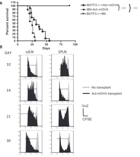
Figure 1: Act-mOVA skin grafts are rejected in the absence of cross-presentation. (A) Batf3/ mice received either an
Act-mOVA or a B6 skin transplant whereas B6 mice received only Act-Act-mOVA skin. Mice were monitored daily, and rejection was deemed as the day when no viable skin remained. ***p<0.001 (Kaplan–Meier). Data are representative of three experiments with five mice per condition. (B) Batf3/ mice were injected with 49106 CFSE-labeled OT-1Rag/T cells on days 10, 14, 21, or 30 post
Act-mOVA skin transplantation. Control mice received CFSE-labeled T cells only (no transplant). After 3 days, proliferation of OVA-specific T cells in the spleen (SPLN) and draining lymph nodes (dLNs) was measured. Cells were stained with antibodies to Va2 and CD8 and CFSE dilution measured on Va2+CD8a+ only cells by flow cytometry. Panels represent the histogram profile of CFSE staining of T
(Figure 1A). Next we measured OVA-specific CD8+T cell response in the Batf3/ recipient mice receiving Act-mOVA skin. Injection of CFSE-labeled CD8+T cells, iso-lated from OT-1Rag/ mice, into transplanted mice on days 10, 14, 21, and 30 after transplantation resulted in T cell proliferation, measured 72 h later by CFSE dilution, at all time points (Figure 1B). The data therefore indicate that OVA antigen was present in the spleen and sdLN for a prolonged period (Figure 1B). Interestingly, even when there was very little skin left, day 30, posttransplant, T cell proliferation was still observed.
MHC-class I molecules are acquired throughout the life span of the transplanted skin
The aforementioned observations suggest that the pre-sentation of OVA by either donor DCs or via the acquisi-tion of intact MHC:peptide complexes by recipient DCs drives T cell proliferation following transplantation in the Batf3/mice. To assess the contribution of MHC-class I:OVA peptide complexes, acquired by recipient DCs, and
to exclude the contribution of donor-derived DCs directly presenting OVA peptide, to CD8+ T cells, Act-mOVA-expressing mice were crossed with CD11c.DTR.GFP mice. In doing so, we created a model in which donor DCs, presenting MHC-class I molecules and OVA pep-tide, could be removed by using DT, following Act-mOVAxDTR/GFP skin transplantation (Figure 2A). Although it has been reported that donor-derived DCs are killed quickly following transplantation in a fully mis-matched setting (16), donor-derived DCs, as measured using CD11c+GFP+ expression, were observed in the spleen of recipient mice up to 14 days after transplanta-tion. However, after 30 days donor DCs were completely absent (Figure 2B). No donor-derived DCs were observed in the dLNs at any time point assessed (data not shown), suggesting that too few of these cells were present in this location in our experimental setup.
To remove donor DCs, recipient Batf3/mice were
trea-ted with DT, as previously published (8), 14 and 30 days CD11c
GFP
Transplant DT
+
-+ +
CD11c
GFP
10
14
30
Day
0
B
100 101 102 103 104 100
101 102 103 104
100 101 102 103 104 100
101 102 103 104
100 101 102 103 104 100
101 102 103 104
100 101 102 103 104 100
101 102 103 104 100 101 102 103 104
100 101 102 103 104
100 101 102 103 104 100
101 102 103 104
A
Batf3
-/-recipient
15 B6OVAxCD11cDTR
Skin transplant
0 14
+/- DT HARVEST SPLN
Batf3
-/-recipient
B6OVAxCD11cDTR Skin transplant
0 Days 10, 14 and 30
HARVEST SPLN
100 101 102 103 104 100
101 102 103 104
F
L2-H
3.08 0.47
6.79 89.7
100 101 102 103 104 100
101 102 103 104
1.91 1.2
5.53 91.4
C
CD11c
CD8
[image:4.595.63.530.51.377.2]Batf3
-/-B6
Figure 2: Donor DCs present after transplantation can be removed following treatment with DT.(A) Batf3/mice received an Act-mOVAxDTR/GFP skin transplant and 14 days later some mice received 4 ng/g body weight of DT. Control mice received no DT. After 24 h the presence of CD11c+ cells expressing GFP was assessed by flow cytometry. (B) Batf3/ mice received Act-mOVAxDTR/GFP a skin transplant and the presence of CD11c+cells expressing GFP was assessed by flow cytometry 10, 14, and 30 days posttransplant. Panels represent the dot-plot profile of CD11c versus GFP expression on live cells for each individual mouse. (C) Spleens isolated from B6 (left) and Batf3/(right) mice 14 days posttransplant were stained with antibodies to CD8 and CD11c.
post skin transplantation. It has been reported that under inflammatory conditions, CD8a+ DCs develop in the Baft3/mice, although very few CD8a+DCs were present in the spleens day 14 posttransplant (Figure 2C). However,
to completely circumvent this possibility, we injected recipi-ent mice with dsRNA intravenously as we, and others, have previously shown that this treatment results in the depletion of CD8a+ DCs (8). Importantly, when
OVA-Batf3
-/-recipient
15
B6OVAxCD11cDTR Skin transplant
0 14
+/- DT CD8+(OT-1)
18 Proliferation Assay dsRNA CFSE Transplant DT dsRNA -+ -+ + + NS NS dLN SPLN Transplant DT dsRNA + + + + + + + -+ -100 101 102 103 104
0 50 100 150
100 101 102 103 104 0
50 100 150
100 101 102 103 104 0
100 200 300 400
100 101 102 103 104 0
100 200 300 400
100 101 102 103 104 0
100 200 300
100 101 102 103 104 0 300 600 900 1200 dLN
SPLN
Batf3-/-recipient
31
B6OVAxCD11cDTR Skin transplant
0 30
+/- DT CD8+(OT-1)
34 Proliferation Assay dsRNA Transplant DT dsRNA -+ -+ + + Transplant DT dsRNA + + + + + + + -+ -NS NS dLN SPLN CFSE SPLN
100 101 102 103 104 0
20 40 60
100 101 102 103 104 0
50 100 150
100 101 102 103 104 0
50 100 150 200
100 101 102 103 104 0 20 40 60 80 100
100 101 102 103 104 0
100 200 300
specific OT-1Rag/ T cells were injected 24 h, or 72 h (data not shown), after depletion of donor DCs using DT and dsRNA treatment, proliferation was observed in both the spleen and the sdLN of the recipient mice (Figures 3A and B). Importantly, no significant differences in the overall T cell responses were observed in the presence or absence of donor DCs. These findings suggest that at days 14 and 30 posttransplantation it was the recipient DCs that were presenting antigen to the T cells in this model. As expected, less T cell proliferation was observed over time due to a diminishing antigen load as the graft was rejected.
Taken together, the data suggest that the acquisition of MHC-class I:peptide complexes from either donor parenchymal cells or dying donor DCs by recipient APCs drives antigen-specific CD8+T cell responses throughout the life span of the transplant.
Recipient-derived DCs acquire MHC-class I molecules from donor cells
To confirm that donor MHC-class I molecules, present on graft tissue, were acquired by recipient DCs and pre-sented to T cells in the dLNs, we repeated the Act-mOVAxDTR/GFP skin transplants using H-2KBm1 recipi-ent mice. We have previously shown that DCs isolated from H-2KBm1 do not cross-present either soluble or cell-associated OVA to OT-1 T cells (1,8). However, these DCs can present acquired MHC:OVA complexes to OT-1Rag/T cells (1,8). Transplanted mice were treated with DT 14 days after transplantation, control mice received no DT, and 1 day later mice received CFSE-labeled OT-1 T cells. As expected from our previous observations in the Baft3/recipients, no significant dif-ferences in the overall T cell responses were observed in the presence or absence of donor DCs (Figure 4A). These findings confirm that in the absence of cross-pre-sentation, recipient DCs acquire donor MHC:peptide and activate CD8+T cells in vivo. This was further confirmed in vitro. DT treatment in transplanted mice (both Batf3/
and H-2KBm1
) was performed at day 12, to remove donor DCs, and 2 days later CD11c+ DCs were isolated from dLNs and co-cultured with T cells isolated from OT-1Rag/ mice. T cell proliferation and cytokine pro-duction (IFNc) were measuredin vitro after 3 days of co-culture (Figure 4B). DCs isolated from the dLNs of both Batf3/ and H-2KBm1
mice treated with DT were cap-able of activating OVA-specific T cells to proliferate and produce IFNc as compared to DCs isolated from
nontransplanted mice (Figure 4B). However, the overall response was less than that seen with transplanted mice not treated with DT, where DCs presenting antigen both directly (donor derived) and via acquisition (recipient DCs) were present. These data confirm that recipient DCs were capable of acquiring MHC:OVA complexes from transplanted tissue and, together with direct pre-sentation of antigen from donor DCs, activates antigen-specific CD8+T cells.
Having established that donor MHC-class I transfer is functional on recipient DCs and drives CD8+T cell activa-tion, we then directly measured the transfer of MHC-class I molecules and addressed whether inflammation influ-ences this phenomenon. To do so, skin transplants from B6.Kd transgenic mice (B6 mice expressing H-2Kd) into CD11c.DTR.GFP recipient mice were performed and the Kdexpression on GFP+ve CD11c+cells was measured by flow cytometry following staining with anti-Kd antibodies. As expected, we found that recipient GFP+CD11c+DCs, present in the spleen, acquired Kd (Figure 4B, middle panel) following transplantation, with an increase in the number of recipient DCs expressing Kd being observed (3.74–16.2%) when the skin irritant picryl chloride was administered to the transplanted Kd skin (Figure 4C, last panel). We conclude that during transplantation recipient DCs can acquire intact allo-MHC-class I molecules from graft parenchyma and that under inflammatory conditions this acquisition is enhanced.
Discussion
In this study we have demonstrated, for the first time, that following transplantation, donor MHC-class I acquisi-tion by recipient DCs occurs throughout the life span of the transplant. We observed, using an OVA antigen skin transplant model, that acquired MHC-class I:OVA com-plexes on recipient DCs were the main source of antigen capable of inducing antigen-specific CD8+ T cells. In addition, we provide evidence for the first time that inflammation of the graft contributes to the transfer of MHC-class I molecules using Kdskin as model antigen. Taken together, the data presented here argue that the direct pathway, as a consequence of acquisition of MHC class I, together with the indirect pathway are major dri-vers of alloimmunity and should be considered when designing tolerance-promoting protocols.
Figure 3: Donor MHC-class I molecules are acquired throughout the life span of the transplant and can induce CD8+T cell
responses. Batf3/ mice received an Act-mOVAxDTR/GFP skin transplant and 14 (A) or 30 days (B) posttransplant; some mice
received 4 ng/g of DT and 200lg of dsRNA (left panels) 24 h prior to injection of CFSE-labeled OT-1Rag/T cells. Control,
trans-planted mice received no DT (middle panels). After 3 days, proliferation of OT-1Rag/T cells, identified using antibodies to Va2 and
CD8, in the spleen (SPLN) and draining lymph node cells (dLNs) was measured by flow cytometry. Control mice received CFSE-labeled OT-1Rag/T cells only (right panels). Histograms shown represent the proliferation of CD8a, Va2, and CFSE-positive cells for
an individual mouse. The percentage proliferation of OT-1Rag/T cells, from five mice per group, is shown. Each dot represents an
In this study, we have shown that removing donor DCs in both the Batf3/and H-2KBm1in vivoskin models did not substantially reduce antigen-specificin vivoT cell
pro-liferation, suggesting that donor DCs may not
monopolize initiation of the direct alloresponse. It should be noted that in our model, DT injection depletes mostly dermal DCs, with minimal effect on epidermal Langer-hans cells. The lack of an essential role for donor DCs in A
transplant rejection has been shown by others using sev-eral murine transplantation models including heart, skin, and kidney. One example was highlighted using a model similar to ours. Garrod et al made use of the CD11c.DTR mice to selectively deplete donor DCs in heart allografts using DT (16). These authors did not report a delay in rejection by removing donor DCs, suggesting that donor DCs, in contrast to what has previously been reported, are not essential for initiating alloimmune responses.
As discussed in the Introduction, intact allo-MHC-class I may be acquired by several different routes, including delivery of these molecules by skin donor dermal DCs to recipient resident DCs. It has been reported that donor DCs that migrate out of transplanted organs, in a fully mismatched setting, are quickly killed by natural killer cells in the secondary lymphoid tissues of the recipient (16). In the skin model described here, donor DCs are present up to 14 days posttransplant, suggesting that acquisition of MHC class I by resident recipient DCs from these donor cells may occur and contribute to the rejection of OVA skin transplants. It is also possible that recipient B cells also acquire donor MHC class, and this warrants further investigation.
Depleting recipient DCs has been shown to significantly prolong graft survival (16). Our transplant data suggest that this may be due to the removal of recipient DCs that have acquired donor MHC-class I molecules. Our data seem to be in contrast to the work presented by Kurts et al (17). These authors showed that when TCR-transgenic OT-1 T cells, specific for an OVA peptide pre-sented by H-2Kb, were injected into mice transgenic for OVA expressed in the pancreas, the transferred T cells divided vigorously in the draining lymph node. This was presumed to result from the capture and processing of OVA by trafficking DCs. However, if the OVA-transgenic mice were made chimeric with H-2KBm1 bone marrow
(KBm1 cannot present the OVA peptide to OT-1 T cells), no OT-1 T cell division was seen. This suggested that the trafficking of KBm1 expressing DCs did not acquire intact complexes of Kbwith OVA peptides from the pan-creatic b cells in sufficient quantities to induce OT-1 T cell proliferation (17). This observation led to the idea that MHC-class I transferin vivomay be more efficient under inflammatory conditions as compared with the steady state. Indeed, we demonstrate the importance of inflam-mation by using the skin irritant picryl chloride. This treat-ment led to a fivefold increase in acquired MHC-class I molecules in a skin transplant setting, further extending our previous findings (5). As immunosuppressive drugs are used extensively following organ transplantation, testing whether MHC transfer occurs under these condi-tions will help elucidate the role of the semidirect path-way in solid organ rejection.
An important question that has not yet been addressed is how the transfer of MHC-class I:peptide complexes occurs in vivo. Indeed, whether the transfer occurs directly via cell-to-cell interaction including membrane “nibbling” (18,19) or through production of either apop-totic bodies or exosomes warrants further investigation (20,21). Whether specific receptors are involved in the uptake of MHC:peptide complexes and whether inflam-mation influences any of the aforementioned mecha-nisms are questions that need further investigation. Although bone marrow–DCs have been shown to pro-duce exosomes containing MHC-class I molecules (22,23), Wakim and Bevan observed using an in vitro transwell system that direct contact, rather than exo-somes release and acquisition, was important for the transfer of MHC-class I molecules (24). However, given that this experimental system may limit exosome trans-fer, the question of whether exosomes or apoptotic bod-ies released from donor DCs or parenchymal cellsin situ contribute to the semidirect pathway is something that
Figure 4: Acquisition of donor MHC-class I molecules by recipient DCs activates antigen-specific T cells. (A) H-2KBm1mice
received an Act-mOVAxDTR/GFP skin transplant and 14 posttransplant mice received 4 ng/g of DT (left panels) 24 h prior to injection of CFSE-labeled OT-1Rag/T cells. Control, transplanted mice received no DT (middle panels). After 3 days, proliferation of OT-1Rag/T
cells, identified using antibodies to Va2 and CD8, in the spleen (SPLN) and draining lymph node cells (LNs) was measured by flow cytometry. Control mice received CFSE-labeled OT-1Rag/T cells only (right panels). Histograms shown represent the proliferation of
CD8a, Va2, and CFSE-positive cells for an individual mouse. The percentage proliferation of OT-1Rag/T cells, from three mice per
group, is shown. Each dot represents an individual mouse. Statistical analysis using an unpaired t-test with no significant difference is denoted as NS. (B) H-2KBm1(Bm1, black column) and Batf3/(white column) mice received an Act-mOVAxDTR/GFP skin transplant
and 12 days later some mice received 4 ng/g of DT. Forty-eight hours following DT injection, dLNs were harvested and CD11c+DCs
were isolated. CD11c+DCs were used to stimulate OT-1 T cells at a 1:1 ratioin vitroand proliferation was measured on day 3 using3H incorporation. Control CD11c+DCs were isolated from unmanipulated mice. Data shown represent a pool of three individual experiments and mean (CPM)1 SEM are shown. IFNcpresent in culture supernatant was measured using a specific ELISA and is expressed in pg/mL. Data shown represent a pool of three individual experiments and mean (CPM)1 SEM are shown. Statistical analysis using an unpaired t-test with significance denoted as p<0.001 by ***. No significant difference is denoted as NS. (C) CD11c.DTR.GFP mice received a B6.Kdskin transplant and 7 days later some mice received a picryl chloride application to the grafted tissue. Four days follow-ing treatment, RBC-depleted splenocytes were stained with antibodies to CD11c and Kd. Splenocytes from nontransplanted and non-treated mice were used as controls. The data represent the profile of Kdexpression on viable CD11c+GFP+cells. Statistical analysis
warrants further investigation. This has recently been addressed with respect to donor DCs (25).
In summary, we have demonstrated for the first time that MHC transfer lasts at least for the life of the trans-plant, activating CD8+ T cells with specificity against MHC-class I:peptide complexes transferred from the graft to recipient antigen presenting cells for the duration of the graft. Furthermore, we have proven that inflamma-tion of the graft further increased the amount of MHC transfer. Altogether, the data presented here extend our previous observations, further emphasizing that both the indirect and the direct pathway of allorecognition are likely to persist for the life of a transplant. Consequently, both pathways need to be regulated if transplantation tol-erance is to be achieved.
Acknowledgments
This project was funded by a Programme Grant from the British Heart Foundation. The research was also supported by the National Institute for Health Research (NIHR) Biomedical Research Centre based at Guy’s and St Thomas’ NHS Foundation Trust and King’s College London.
Disclaimer
The views expressed are those of the author(s) and not necessarily those of the NHS, the NIHR, or the Depart-ment of Health.
Disclosure
The authors of this manuscript have no conflicts of inter-est to disclose as described by the American Journal of Transplantation.
References
1. Smyth L, Harker N, Turnbull W, et al. The relative efficiency of acquisition of MHC:peptide complexes and cross-presentation depends on dendritic cell type. J Immunol 2008; 181: 3212–3220. 2. Safinia N, Afzali B, Atalar K, Lombardi G, Lechler RI. T-cell
alloimmunity and chronic allograft dysfunction. Kidney Int Suppl 2010; 119: S2–S12.
3. Whitelegg AM, Oosten LE, Jordan S, et al. Investigation of pep-tide involvement in T cell allorecognition using recombinant HLA class I multimers. J Immunol 2005; 175: 1706–1714.
4. Lindahl KF, Wilson DB. Histocompatibility antigen-activated cyto-toxic T lymphocytes. I. Estimates of the absolute frequency of killer cells generatedin vitro. J Exp Med 1977; 145: 500–507. 5. Herrera OB, Golshayan D, Tibbott R, et al. A novel pathway of
alloantigen presentation by dendritic cells. J Immunol 2004; 173: 4828–4837.
6. Smyth LA, Herrera OB, Golshayan D, Lombardi G, Lechler RI. A novel pathway of antigen presentation by dendritic and
endothelial cells: Implications for allorecognition and infectious diseases. Transplantation 2006; 82(1 Suppl): S15–S18.
7. Smyth LA, Afzali B, Tsang J, Lombardi G, Lechler RI. Intercellu-lar transfer of MHC and immunological molecules: MolecuIntercellu-lar mechanisms and biological significance. Am J Transplant 2007; 7: 1–8.
8. Smyth LA, Hervouet C, Hayday T, et al. Acquisition of MHC:pep-tide complexes by dendritic cells contributes to the generation of antiviral CD8+ T cell immunityin vivo. J Immunol 2012; 189: 2274–2282.
9. Sivaganesh S, Harper SJ, Conlon TM, et al. Copresentation of intact and processed MHC alloantigen by recipient dendritic cells enables delivery of linked help to alloreactive CD8 T cells by indi-rect-pathway CD4 T cells. J Immunol 2013; 190: 5829–5838. 10. Brown K, Sacks SH, Wong W. Coexpression of donor peptide/
recipient MHC complex and intact donor MHC: Evidence for a link between the direct and indirect pathways. Am J Transplant 2011; 11: 826–831.
11. Celli S, Albert ML, Bousso P. Visualizing the innate and adaptive immune responses underlying allograft rejection by two-photon microscopy. Nat Med 2011; 17: 744–749.
12. Hildner K, Edelson BT, Purtha WE, et al. Batf3 deficiency reveals a critical role for CD8a+dendritic cells in cytotoxic T cell immunity. Science 2008; 322: 1097–1100.
13. Billingham RE, Brent L, Medawar PB. Actively acquired toler-ance of foreign cells. Nature 1953; 172: 603–606.
14. Prlic M, Hernandez-Hoyos G, Bevan MJ. Duration of the initial TCR stimulus controls the magnitude but not functionality of the CD8+ T cell response. J Exp Med 2006; 203: 2135–2143. 15. Ehst BD, Ingulli E, Jenkins MK. Development of a novel
trans-genic mouse for the study of interactions between CD4 and CD8 T cells during graft rejection. Am J Transplant 2003; 3: 1355–1362.
16. Garrod KR, Liu FC, Forrest LE, Parker I, Kang SM, Cahalan MD. NK cell patrolling and elimination of donor-derived dendritic cells favor indirect alloreactivity. J Immunol 2010; 184: 2329–2336. 17. Kurts C, Heath W, Carbone F, Allison J, Miller J, Kosaks H.
Con-stitutive class I-restricted exogenous presentation of self
anti-gensin vivo. J Exp Med 1996; 184: 923–930.
18. Harshyne LA, Zimmer MI, Watkins SC, Barratt-Boyes SM. A role for class A scavenger receptor in dendritic cell nibbling from live cells. J Immunol 2003; 170: 2302–2309.
19. Harshyne LA, Watkins SC, Gambotto A, Barratt-Boyes SM. Den-dritic cells acquire antigens from live cells for cross-presentation to CTL. J Immunol 2001; 166: 3717–3723.
20. Morelli AE. The immune regulatory effect of apoptotic cells and exosomes on dendritic cells: Its impact on transplantation. Am J Transplant 2006; 6: 254–261.
21. Robbins PD, Morelli AE. Regulation of immune responses by extracellular vesicles. Nat Rev Immunol 2014; 14: 195–208. 22. Zitvogel L, Regnault A, Lozier A, et al. Eradication of established
murine tumors using a novel free vaccine: Dendritic cell-derived exosomes. Nat Med 1998; 4: 594–600.
23. Andre F, Chaput N, Schartz NE, et al. Exosomes as potent cell-free peptide-based vaccine. I. Dendritic cell-derived exosomes transfer functional MHC class I/peptide complexes to dendritic cells. J Immunol 2004; 172: 2126–2136.
24. Wakim L, Bevan M. Cross-dressed dendritic cells drive memory CD8+ T-cell activation after viral infection. Nature 2011; 471: 629–632.