Management of Gingival Melanin Pigmentation
Using Four Different Techniques: A Split Mouth Study
Surg Lt Cdr (D) Oliver Jacob*, Lt Col Manab Kosala*, Sqn Ldr Navneet*, Capt Jacqueline Jacinta Dias**
* Division of Periodontology, Department of Dental Surgery and Oral Health Sciences, Armed Forces Medical College, Pune, Maharashtra ** 9 CDU, YOL Camp, Kangra, Himachal Pradesh
DOI: 10.29322/IJSRP.9.12.2019.p9607
http://dx.doi.org/10.29322/IJSRP.9.12.2019.p9607
Abstract- Context: Dark or black coloured gum is one of the aesthetic concern of patients reporting to dental clinics. The normal colour of the gingiva is coral pink. Dark pigmentation of the gums occurs due to excessive melanin deposition in gingival epithelium which can be treated using several techniques. With this as the back ground our study was formulated with the objective to compare the treatment outcome of gingival depigmentation with four different techniques i.e. Er:YAG laser, Diode laser, scalpel abrasion technique and split thickness slicing using scalpel. 10 patients, with no systemic comorbidities visiting the OPD with concerns regarding unpleasant smile due to dark coloured gingiva were selected for the study. After thorough Phase-I therapy, split mouth depigmentation by four different techniques was carried out. Patients underwent depigmentation using Diode LASER at 910nm on the right lower quadrant, Er: YAG LASER on the right upper quadrant, Split thickness slicing procedure was employed on the left upper quadrant and surgical blade abrasion on the lower left quadrant using No 15 BP blade. Patients were evaluated for pain and discomfort for 7 days following surgery using VAS. Patients were evaluated for 28 days for healing and recurrence after 3 and 6 months. Results: Patient discomfort was lesser in scalpel side compared to LASER side. Healing was comparable with LASERs and Scalpel technique. Isolated recurrence of pigmentation was seen in the side with surgical scraping technique. Conclusion: LASERs are more effective as compared to conventional scalpel.
Index Terms- Depigmentation, Lasers, Scalpel
I. INTRODUCTION
he normal colour of the gingiva is coral pink [1]. However it is seen that part of the population is affected by dark coloured gingiva due to excessive melanin pigmentation produced by melanocytes present in the basal layers of the epithelial cells. This pigmentation is seen more prominent in certain ethnic groups [2] [3]. The excessive display of gums in patients with gummy smile and pigmented gingiva only add on to unesthetic appearance and social concerns for individuals.
The demand for esthetic surgery has grown by leaps and bounds in the recent years. Thus necessitating the development of techniques to alter the colour of the gingiva such as free mucosal grafts, scalpel abrasion, Split thickness slicing by scalpel, bur abrasion, cryosurgery, electro-surgery. Recently with the development of several laser systems: Erbium family of lasers,
Diode lasers, Nd:YAG lasers and CO2 lasers has also proven
efficacy in depigmentation [4].
One of the first techniques developed was the scalpel technique [4]. A portion of the sub epithelial connective tissue with complete epithelium was abraded and allowed to heal by secondary intention. The second technique being a split thickness slicing technique where the entire epithelium and a part of the sub epithelial connective tissue is sliced from the gingiva to expose the connective tissue and healing is achieved by secondary intention. This technique has been long referred to as “Gold Standard” in treatment of gingival pigmentation [5]. Recently, with the addition of lasers in the treatment modality for gingival depigmentation a lot of research has been directed towards this modality to compare with the existing modalities in terms of patient comfort and recurrence of pigmentation. Diode lasers and Er: YAG lasers are the choice amongst lasers for depigmentation procedures. All the available modalities have their own advantages and disadvantages. Recurrence being one of the major concerns [6]. Our study was aimed at evaluating the clinical outcomes by using two scalpel techniques and two lasers used for depigmentation procedures.
II. AIM&OBJECTIVES
To compare the treatment outcome of gingival depigmentation with two types of LASERs against two different types of scalpel techniques with the objectives to evaluate the level of post-operative discomfort, post-operative wound healing and the intensity and extent of recurrence of pigmentation.
III. MATERIAL&METHODS
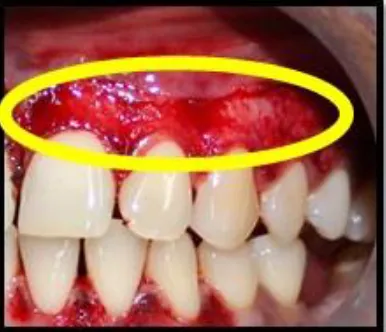
The study was carried out in a Tertiary care government hospital. The study was carried out as a split mouth study on adult patients concerned with black coloured gums causing unesthetic smile. The duration of study was 6 months. Pre-clinical parameters include Dummett Gupta Oral Pigmentation Index score [2] and Hedin Pigmentation Extent Score [7]. The post treatment parameters include pain and discomfort to the patient using Visual Analog Scale [8], epithelial wound healing based on peroxide test [9], pigmentation score using Dummett Gupta Oral Pigmentation Index score [2] and Hedin Pigmentation Extent Score [7]. Surgical scalpel BP blade no 15 (Fig 1), Syneron Lite touch Dental Hard and soft tissue Er:YAG laser (Fig 2) and Dental diode laser at 980 nm wavelength (Fig 3).
International Journal of Scientific and Research Publications, Volume 9, Issue 12, December 2019 69 ISSN 2250-3153
http://dx.doi.org/10.29322/IJSRP.9.12.2019.p9607 www.ijsrp.org
Patient between the age group of 18 to 35 years with gender nonspecific, concerned about unpleasing smile due black gums, with good oral hygiene and willing for minor surgical procedure were included in the study population. Patients with thin gingival biotype chronic smokers and tobacco chewers, pigmentation associated with syndromes, uncontrolled diabetics, patients with auto-immune disorders, untreated periodontal conditions, patients on prolonged use of anti-malarial, tricyclic anti-depressants, oral contraceptive pills and minocycline therapy, patients with melanogenic tumors, pregnant or lactating women and non- compliant patients were excluded from the study Ten Patients who came to the General OPD, who meet the inclusion and exclusion criteria, were selected and examined for participation in this clinical study. At baseline gingival pigmentation based on the Dummett-Gupta Oral pigmentation Index [2] and Hedin Oral Pigmentation extent index score was evaluated for the patients [7]. The descriptive data of the study population is given in Table 1 and the mean indices in Table 2. Following Phase-I therapy, all selected patients underwent depigmentation procedure using the four different techniques in one sitting under adequate local anesthesia. The upper right quadrant was assigned for Er: YAG laser technique using brush stroke in contact mode, lower right quadrant was assigned for diode laser at 980 nm in contact mode and using brush stroke (Fig 4 & 5). The upper left quadrant was assigned to Split thickness slicing technique (Figs 6, 7 & 8) and lower left quadrant was assigned to scalpel abrasion technique. Bard Parker Blade no 15 was used for the scalpel techniques on the left side (Fig 9). The depigmentation procedure was carried out from central incisor to premolar region in all four quadrants. Gingival epithelia from the mucogingival junction to the marginal gingival was denuded and the interdental papilla also was included. Periodontal pack was placed at the site after procedure for 07 days. Oral hygiene instructions was given to all the patients. Restriction on consumption of hot and spicy foods for 01 week was advised and analgesics prescribed. Chlorhexidine mouth was (0.2%) was used for chemical plaque control for 01 week post-operative.
Post-operative evaluation of pain and discomfort was assessed using the Visual Analog Scale (8) after 4 hours, 24 hours, 7 days, 12 days and 21 days. Wound healing was assessed using the Hydrogen peroxide test as given by Merucha et al in 1998 [9]. On the 7th day after surgery the periodontal pack was removed and
3 % hydrogen peroxide was placed on the surgical wound. The presence of effervescence showed evidence of activity of the sub epithelial catalase enzyme that degrades H2O2 to release O2 and
thus the effervescence. This indicated the complete epithelial coverage of the surgical wound and hence completion of the wound healing process [9]. The patients were assessed for wound healing on all four quadrants using the peroxide test until the test result proved to be negative.
Post op the patients were then assessed after 6 months for recurrence (Fig 10 & 11). The Dummet Oral Pigmentation Index for intensity of pigmentation [2] and Hedin index for extent of pigmentation [7] was used to assess the recurrence in all four quadrants.
The data was collected and stored in MS Excel data sheet, the statistical analysis was carried out using IBM SPSS version 21.0 software.
IV. RESULTS
Post op evaluation of the patient discomfort was assessed using the visual analog scale after 4 hours when the effect of Local anesthesia wore out then after 24 hours, 7 days, 12 days and 21 days. The assessment was carried out using non-parametric equivalence of repeated measurements-ANOVA namely Friedman’s Test. The results were plotted on a graph (Fig 12). It showed significant difference in values intra group in the different time periods (p=0.001). However inter group interaction was not significant. (Table 3)
Wound healing test was carried out using the peroxide test as described by Phillipe Merucha et al in 1998 [9]. The assessment as described in methodology aforementioned was carried out and a value of 1 was allotted to positive test i.e. presence of effervescence and 0 to negative result i.e. absence of effervescence. The values were statistically analysed using a chi-square test for 7 days, 12 days, 21 days and 28 days (Table 4). The results showed that at the end of 7 days when the epithelialisation is supposed to be completed, the side where scalpel was used showed 60% of patients with complete epithelialization whereas 30% of the laser side showed completion of epithelial formation (Table 4). After 12 days the scalpel side showed complete epithelialization in all patients and 70% of the patients showed complete epithelialization (Table 4). By day 28 the epithelialization was completed in all groups (Table 4). This showed a statistically significant variation in wound healing in the laser and scalpel groups. The scalpel groups showed faster wound healing as compared to the laser groups. In the inter scalpel group of scalpel stripping and scalpel abrasion there was no significant difference between the groups and between the laser groups i.e. diode laser and Er: YAG groups there was no significant difference between the groups.
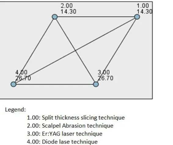
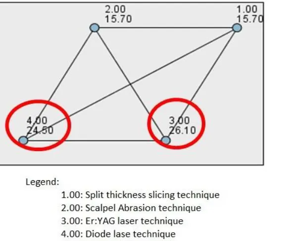
The patients were evaluated after 6 months for recurrence of pigmentation and the assessment was carried out using the Dummett and Hedin indices for intensity and extent of repigmentation. The statistical analysis was carried out using Kruskal-Wallis test for independent samples. The analysis showed significant difference in recurrence amongst the groups. The scalpel groups showed faster incidence of recurrence as compared to laser groups. The laser group showed less intensity of the repigmentation as compared to scalpel group, pair wise comparison chart (Fig 13). There was significant difference between extent of intensity amongst the groups. The scalpel groups showed more extensive recurrence as compared to laser groups. However among the laser groups the Er: YAG group showed a statistically significant difference and lesser intensity of recurrence as compared to diode laser group, pairwise comparison chart (Fig 14), showing clinical evidence that Er: YAG laser is more effective than diode laser or scalpel techniques for reducing recurrence of pigmentation.
V. DISCUSSION
melanophores and melanophages phagocytose these pigments and are present in the epithelium.
Several modalities exist for the treatment of pigmentation such as free gingival autografts, cryosurgery, electro-surgery, scalpel abrasion and split thickness slicing, Lasers and chemical such as 90% phenol. However, these methods have their own disadvantages. Free mucosal graft has its inherent disadvantages of having a variation in the colour of the graft and adjacent tissues and a second surgical site morbidity [10]. The depth of depigmentation achieved is not predictable using cryo-surgery and chemical methods. The disadvantages of the electro-surgery being the heat dissipation and thermal damage to adjacent tissues and bone, if the depth is not controlled [10].
Scalpel abrasion is the simplest method, with control over the depth of abrasion and being less technique sensitive. The split thickness slicing has an advantage of predictable depth of the de-epithelialisation. However, the technique needs to be performed on thick gingival biotype as there is risk of exposure of the roots and the underlying alveolar bone. This is a highly technique sensitive method and is time consuming. Both the scalpel techniques have the disadvantages of excessive bleeding and requires the placement of periodontal pack.
Er: YAG and diode lasers are new additions to the laser family. Er:YAG functions on the principle of ablation of the cells by absorption of laser photons by water molecules. Thus, causing explosion of these water molecules in the cells and thus cellular ablation [11]. Diode laser functions on the principle of absorption of laser photons by pigments and subsequent ablation of the cells. Pain and discomfort experienced by the patients analysed on Visual Analog Scale is a subjective assessment. This scale is charted on a scale of 100mm [8]. Several Studies have shown that pain and discomfort was comparatively lesser with use of lasers as compared to scalpel [12] [13][14][15]. Our study showed a slightly higher discomfort on the laser side as compared to the scalpel groups. However, the over-all reduction in pain was comparatively similar in all groups.
Depigmentation procedure is undertaken by de-epithelization of the gingiva by split thickness slicing or abrasion. During this process the epithelium is completely scrapped off with a part of the sub epithelial connective tissue. Epithelium generally regenerates by the seventh day and forms the normal barrier [16]. Complete epithelialization blocks the activity of sub epithelial connective tissue enzyme catalase that degrades hydrogen peroxide to water and oxygen showing effervescence when applied over open wounds. This test proves the completion of epithelial barrier as described by Phillipe Marucha et al in his study in 1998 [9]. Our study used 3% H2O2 to check the integrity
of the epithelial barrier. Lasers due to their ablative nature on the cells have shown slower wound healing as compared to scalpel technique [17] and this has been confirmed by our study.
Recurrence of pigmentation has been a cause of concern amongst patients, clinicians and researchers. The exact cause or mechanism of recurrence is not known, however it is hypothesized that the melanocytes which are present in the adjacent sites can cause recurrence of pigmentation. However, the intensity and extent of repigmentation is questionable and variable in different patients [6]. Studies have shown consistent recurrence in pigmentation after several months [18]. Our study showed comparative results as shown in the aforementioned results.
Comparison of diode laser and Er: YAG showed statistically that Er: YAG group showed lesser extent of pigmentation as compared to the diode laser group (Fig 8).
VI. CONCLUSION
Depigmentation of gingival pigmentation has been one of the clinical necessities of the modern day practice of Perio-Esthetic procedures. With the advent of lasers as a new tool at the hands of the clinician, depigmentation procedures has become less technique sensitive procedure with predictable and long term success rates. Recurrence is a cause of concern to many clinicians however the extent and degree of repigmentation seen in the scalpel and laser groups show significant difference with the balance tipped towards the laser group especially Er: YAG as shown in our study. Though split thickness slicing has been considered for long as a “Gold Standard” [5] the newer modalities such as lasers are now considered superior in clinical outcomes [19].
Within the limitations of our study, the effectiveness of Er: YAG laser has shown clinically and statistically significant results favouring the modality. However, a large scale randomized controlled trial and systematic review of the several modalities in split mouth study design is warranted to establish the clear cut advantage of Er: YAG lasers over other lasers and modalities of depigmentation of gingiva.
APPENDICES
Appendix 1: Tables 1 through 4 Appendix 2: Figures 1 through 14
REFERENCES
[1] Gupta G, Kumar A, Khatri M, Puri K, Jain D, Bansal M. Comparison of two different depigmentation techniques for treatment of hyperpigmented gingiva. J Indian Soc Periodontology 2014;18(6):705-9.
[2] Dummett C. Estimating the Epidemiology of Oral Pigmentation. Journal of the National Medical Association 1964; 56(5):419–20.
[3] Dummett C, Barens G. Oromucosal Pigmentation : an Updated Literary J Periodontol 1971; 42 (11) :726–36.
[4] Roshna T, Nandakumar K. Anterior Esthetic Gingival Depigmentation and Crown Lengthening: Report of a Case. J Contemp Dent Pract 2005; (6)3:139-147.
[5] Hegde R, Padhye A, Sumanth S, Jain AS, Thukral N. Comparison of Surgical Stripping; Erbium-Doped:Yttrium, Aluminum, and Garnet Laser; and Carbon Dioxide Laser Techniques for Gingival Depigmentation: A Clinical and Histologic Study. J Periodontol. 2013;
[6] Perlmutter S, Tal H. Repigmentation of the Gingiva Following Surgical Injury. J. Periodontol 1986;57(1):48–50.
[7] Hedin CA. Smokers’ Melanosis Occurrence and Localization in the Attached Melanosis. Arch Dermatol 1977; 113:1533-1538.
[8] Beilin Y. The Visual Analog Scale for Pain Clinical Significance in Postoperative Patients. Anesthesiology 2001; 95:1356–61.
[9] Avagehi MEF. Mucosal Wound Healing Is Impaired by Examination Stress Subject Characteristics and Timing of Wounds Whole Blood Assay for IL-1 β. Psychosomatic Medicine 1998; 60:362-365.
[10] Pooja S, Bhongade ML. Comparative evaluation of effectiveness of surgical blade , electrosurgery , free gingival graft and diode laser for the management of gingival hyperpigmentation. Sch J. Dent. Sci., 2017;4(2):68–73. [11] Pavlic V, Brkic Z, Marin S, Cicmil S, Gojkov-vukelic M, Aoki A, et al.
International Journal of Scientific and Research Publications, Volume 9, Issue 12, December 2019 71 ISSN 2250-3153
http://dx.doi.org/10.29322/IJSRP.9.12.2019.p9607 www.ijsrp.org
[12] Doshi Y, Khandge N, Byakod G, Patil P. Management of Gingival Pigmentation with Diode Laser : Is It a Predictive Tool ? International Journal of Laser Dentistry 2012; 2(1):29–32.
[13] Suragimath G, Lohana MH, Varma S. A Split Mouth Randomized Clinical Comparative Study to Evaluate the Efficacy of Gingival Depigmentation Procedure Using Conventional Scalpel Technique or Diode Laser. J lasers Med Sci.; 2016;7(4):227–32.
[14] Bhardwaj A, Uppoor AS, Naik DG. A comparative evaluation of management of melanin pigmented gingiva using a scalpel and laser. J Interdiscip Dentistry 2014;4:135-9.
[15] Bhardwaj A, Grover HS, Lal S. Gingival Depigmentation with Scalpel and Diode Laser. World Journal of Dentistry 2012; 3(4):359–62.
[16] Newman MG, Takei HH. Newman and Carranza’s Clinical Periodontology 13th Ed: page 616
[17] Akira A, Koji M, Frank S, Anton S, Raymond A. Yukna, Aristeo A. Takasaki, Georgios ER, Yoichi T, Katia M, Sasaki, Jorge lZ, Geena K, Donald J. Coluzzi, Joel MW, Yoshimitsu A, Isao I & Yuichi I. Periodontal and peri-implant wound healing following laser therapy. Perio 2000. 2015; 68 (Table 1):217–69.
[18] Mahajan G, Kaur H, Jain S, Kaur N, Sehgal NK, Gautam A. To compare the gingival melanin repigmentation after diode laser application and surgical removal J Indian Soc Periodontol. 2017; 21(2): 112–118.
[19] Yi HL, Yu KT, Chun TL, Wen CC, Chiung FH, Mao SH, Hsein KL. Systematic Review of Treatment Modalities for Gingival Depigmentation :
A Random-Effects Poisson Regression Analysis. Journal of Esthetic and Restorative Dentistry 2014;26(3):162–78.
AUTHORS
First Author – Surg Lt Cdr (D) Oliver Jacob, BDS, Armed
Forces Medical College, Pune, [email protected].
Second Author – Lt Col Manab Kosala, MDS, Armed Forces
Medical College, Pune, [email protected].
Third Author – Sqn Ldr Navneet, BDS, Armed Forces Medical
College, Pune, [email protected]
Fourth Author – Capt Jacqueline Jacinta Dias, MDS, 9 CDU,
YOL Camp, Kangra, HP [email protected]
Correspondence Author – Surg Lt Cdr (D) Oliver Jacob, BDS,
Armed Forces Medical College, Pune, [email protected].
Appendix 1: TABLES
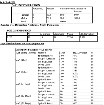
PATIENT POPULATION
Frequency Percent Valid Percent Cumulative Percent
Females 8 80.0 80.0 80.0
Males 2 20.0 20.0 100.0
Total 10 100.0 100.0
Table 1: Gender wise Descriptive Analysis of Study Population
AGE DISTRIBUTION
N Minimum Maximum Mean Std. Deviation
[image:4.612.69.485.311.734.2]AGE 10 18 39 27.90 7.578
Table 2: Age distribution of the study population
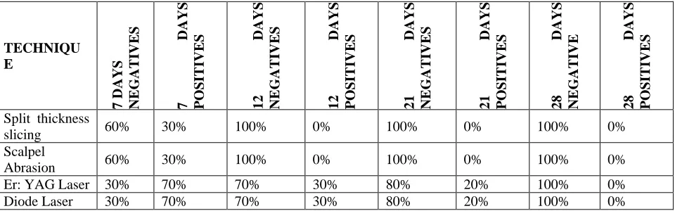
Descriptive Statistics: VAS Scores
VAS (Time PostOp) Method Mean Std. Deviation N
VAS (4hrs)
Split thickness 3.60 .699 10 Scalpel Abrasion 3.60 .699 10 Er: Yag Laser 4.10 .876 10
Diode Laser 4.10 .876 10
VAS (24hrs)
Split thickness 1.80 .789 10 Scalpel Abrasion 1.80 .789 10 Er: Yag Laser 2.60 1.075 10
Diode Laser 2.60 1.075 10
VAS (7 Days)
Split thickness .40 .516 10 Scalpel Abrasion .40 .516 10 Er: Yag Laser 1.20 .919 10
Diode Laser 1.20 .919 10
VAS (12 Days)
Split thickness .10 .316 10 Scalpel Abrasion .10 .316 10
Er: Yag Laser .10 .316 10
Diode Laser .10 .316 10
Table 3: Statistical Data of VAS scores at different intervals
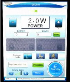
TECHNIQU E
7 DAYS NEGA
TI
VES
7
D
AYS
POSITI
VES
12
D
AYS
NEGA
TI
VES
12
D
AYS
POSITI
VES
21
D
AYS
NEGA
TI
VES
21
D
AYS
POSITI
VES
28
D
AYS
NEGA
TI
VE
28
D
AYS
POSITI
VES
Split thickness
slicing 60% 30% 100% 0% 100% 0% 100% 0%
Scalpel
Abrasion 60% 30% 100% 0% 100% 0% 100% 0%
Er: YAG Laser 30% 70% 70% 30% 80% 20% 100% 0%
Diode Laser 30% 70% 70% 30% 80% 20% 100% 0%
Table 4: Wound Healing Test results on Chi-square test Appendix 2: FIGURES
Scalpel Abrasion .00 .000 10
Er: Yag Laser .00 .000 10
International Journal of Scientific and Research Publications, Volume 7, Issue 8, August 2017 73 ISSN 2250-3153
[image:6.612.36.295.55.255.2]www.ijsrp.org
FIG 1: Bard Parker Blade no 15 FIG 2: Syneron Er:YAG Laser system
[image:6.612.37.176.290.452.2]FIG 3: Diode Laser system
[image:6.612.39.210.515.661.2]FIG 5: Depigmentation using Diode laser on the right side lower quadrant
FIG 6: Incision for split thickness slicing
FIG 7: Excised portion of split thickness slice from the mucosa
[image:7.612.39.269.387.513.2] [image:7.612.40.233.549.715.2]International Journal of Scientific and Research Publications, Volume 7, Issue 8, August 2017 75 ISSN 2250-3153
[image:8.612.39.284.55.225.2]www.ijsrp.org
[image:8.612.38.445.264.449.2]FIG 9: Shows depigmentation by scalpel abrasion technique by Bard Parker blade no. 15
FIG 10: Shows comparison at baseline and post-operative recall 6 months later on the laser side
[image:8.612.38.466.493.674.2]FIG 12: Shows the graphical representation of the reduction of Pain in the various groups plotted along the X Axis and time periods on the Y axis by estimated marginal means of measures
FIG 13: Shows the graphical representation of the intensity of recurrence of pigmentation amongst the groups after 6 months: 1-Split
[image:9.612.55.338.472.710.2]International Journal of Scientific and Research Publications, Volume 7, Issue 8, August 2017 77 ISSN 2250-3153
[image:10.612.50.345.60.304.2]www.ijsrp.org
FIG 14: Shows the graphical representation of the extent of recurrence of pigmentation amongst the groups after six months: 1-Split

![1 (2 Methylpropyl) 1H imidazo[4,5 c]quinolin 4 amine](data:image/gif;base64,R0lGODlhAQABAIAAAP///wAAACH5BAEAAAAALAAAAAABAAEAAAICRAEAOw==)