The Association of Fibroblast Growth Factor-2 (FGF-2)
Expression with Benign, Borderline, and Malignant
Phyllodes Tumours of the Breast
Rizmeyni Azima*, Delyuzar, Betty
Department of Anatomical Pathology, Faculty of Medicine, Universitas Sumatera Utara, Medan, Indonesia. *Corresponding author Faculty of Medicine, Universitas Sumatera Utara, Medan, Indonesia
DOI: 10.29322/IJSRP.9.12.2019.p9657
http://dx.doi.org/10.29322/IJSRP.9.12.2019.p9657
Abstract- Phyllodes tumours is a rare biphasic tumours, this is
composed of neoplastic stromal cells and evolution containing epithelium. The stromal tumour is more cellular and increased. Phyllodes tumours are far less common than fibroadenoma and de novo arising, and not from previous fibroadenoma tumours. The bad changes are the feared malignant are increased stromal cellularity, anaplasia, high mitotic activity, rapidly increasing tumour size, and infiltative edges. The incidence of phyllodes is <1% of all breast neoplasm in 0,1-0,5%, with the most incidence occurring at the age of 30- 40 years old.
Objective: This study was aimed to analyze the association of Fibroblast Growth Factor-2(FGF-2) with benign, borderline, and malignant phyllodes tumours of the breast.
Methods: This is an observational analytic study with cross sectional approach, involving 35 paraffin block samples from phyllodes tumours. In this study we found FGF-2 expression in 35 samples, consist of 15 samples of benign phyllodes tumours, 8 sample of borderline tumours, and 12 samples of malignant phyllodes tumours. The specimen of this study were embedded in paraffin and each section was cut from each block. Then FGF-2 staining were done. The positivity for FGF-2 were scored by using the following parameters. FGF-2 counts among 3 groups of cases were analyzed using statistic software.
Result: There were association between FGF-2 expression with benign, borderline, and malignant phyllodes tumours.
Conclusion: The FGF-2 expression in malignant phyllodes tumours has the highest mean value, followed by borderline phyllodes tumours, and benign phyllodes tumours have the lowest FGF-2 expression values.
Index Term: FGF-2, Phyllodes Tumours.
I. INTRODUCTION
hyllodes tumours is a rare biphasic tumours of the breast. The incidence of phyllodes tumours account for 0,3-0,9% of all primary tumours of the breast.1 Phyllodes tumours is biphasic
neoplasm originally named cystosarcoma phyllodes by Johannes Muler in 1838, a term that is to be avoided because of its malignant conotations. Phyllodes tumours commonly occurs in middle-aged and older woman. Very few patients are younger than 25 years age, which is in striking contrast with the age
distribution of fibroadenoma tumours. However, phyllodes tumours can certainly occur in young adults and even in adolescents, and therefore, the diagnosis can’t be excluded on the basis of age.1-3 Phyllodes tumours increased in Asian countries,
in Singapore this tumours occurred 6,92% of all malignancies in the breast and occur at a younger ages, 25-30 years old. Although rarely found, there have been reports of this tumours in men. The
frequency of these tumours, based on changes in
histopathological features (gradations) is 75% benign, 16% borderline, and 9% malignant. Although it has been reported, there is rarely a synchronous or metachronous presence in this tumours.4-7 It also difficult to diagnose phyllodes tumours
because of subclassification resembling fibroadenomas. The prognostic value of immunohistochemical markers of tumour growth, angiogenesis, and malignant transformation has been confirmed in many tumours, including breast carcinoma. Studies have shown significant differences in FGF-2 expression for benign and malignant phyllodes tumours of the breast.9 FGF-2 is
a very potent stimulator for proliferation, migration of endothelial cells, and tube formation in vitro and also high angiogenic in a number of tissues in vivo.10 The existence of
FGF can be obtained by two methods, namely: first by conducting immunohistochemical examination of tumour tissue and by examining the Enzyme-Linked Immunosorbent Assay (ELISA) of blood serum, then FGF levels will be obtained. From this examination will be obtained a response from the FGF receptor. Increases in both FGF and FGF receptors have important significance in tumour growth.11 In a previous study,
Dacic et al., only examined the expression of FGF-2 in benign and malignant phyllodes breast tumours, and did not evaluate it in phyllodes borderline breast tumours.9 In addition, there is still
very little research that addresses the problems mentioned above, causing the existing data is also limited. In Indonesia, especially in North Sumatera, there are no studies that analyze the appearance of expression and its relationship with phyllodes tumours.
II. MATERIAL AND METHODS
Medan General Hospital, and Department of Anatomical Pathology in Pirngadi Medan Hospital. The research was held from July 2019 until November 2019, after approved by the Universitas Sumatera Utara and H. Adam Malik General Hospital Health Research Ethics Committee. All samples were obtained through surgical procedure. Inclusion criteria were phyllodes cases with adequate clinical data, available and undamaged formalin-fixed paraffin embedded tissue block with sufficient tumor tissue. Detailed clinical data were obtained from medical records or pathology archives consisting of age, sex, and size of the tumor. Histological type were determined independently by researchers through hematoxylin and eosin stained slides examination.
Histochemistry protocol and interpretation
The tissue sections were deparaffinized and rehydrated before pretreatment. Endogenous peroxidase was blocked with hydrogen peroxide followed by antigen retrieval. FGF-2 (GTX84502, GeneTex, California, America) mouse monoclonal antibodies was used as primary antibody. Diagnostic BioSystems (Diagnostic BioSystems, Pleasanton, CA, USA) polymer kit was used for detection. The reaction was visualized with diaminobenzidine and counterstained with Mayer's hematoxylin followed by dehydration, clearing, and mounting. Positive control was colon. Expressions of FGF-2 were determined independently by researchers. The expression in cytoplasm was analyzed. FGF-2 expressions were determined independently by researchers. Nuclear and cytoplasmic positivity for FGF-2 by using the following parameters: 0 (no staining) 1+ (1-35% positive cells), 2+ (35-70% positive cells), and 3+ (70-100% positive cells).9
Statistical analysis
Statistical analysis was performed using SPSS software package version 22.0 (SPSS Inc., Chicago) with 95% confidence interval and Microsoft Excel 2010. Categorical variables were presented in frequency and percentage. Saphiro-Wilk test was applied to find out the normality continue data. The association between FGF-2 expresion with benign, borderline, and malignant phyllodes tumours is assessed by Kruskal-Wallis, and continue with post hoc Dunn test to assessed the differences between the groups. The p-values < 0.05 were considered significant.
III. RESULT
Patients' characteristics
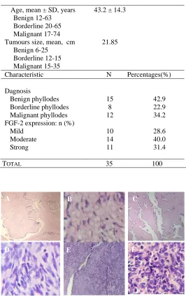
[image:2.612.312.578.73.498.2]The mean age for phyllodes tumour patients was 43.2 (±14.3) years. The most common in 12-63 years age group, 34 patients (97.1%) were females, only 1 patients (2.9%) were males. The mean size of tumours was 21.85 cm which is 6-25 cm for benign phyllodes, 12-15 cm for borderline phyllodes, and 15-35 cm for malignant phyllodes tumours. The number of the patients benign phyllodes was 15 (42.9%), borderline phyllodes was 8 (22.9%), and malignant phyllodes was 12 (34.2%). The histological subtypes of benign phyllodes was the majority of this case. Clinical basic characteristic of phyllodes patients were summarized in table 1. Representative H&E sections are shown in figure 1.
Table 1. Characteristic of phyllodes patients
Age, mean ± SD, years Benign 12-63
Borderline 20-65 Malignant 17-74
Tumours size, mean, cm Benign 6-25
Borderline 12-15 Malignant 15-35
43.2 ± 14.3
21.85
Characteristic N Percentages(%)
Dagnosis
Benign phyllodes Borderline phyllodes Malignant phyllodes
15 8 12
42.9 22.9 34.2 FGF-2 expression: n (%)
Mild Moderate Strong
10 14 11
28.6 40.0 31.4
TOTAL 35 100
Figure 1. Histological type. A, Benign phyllodes (HE, 40x). B, Benign phyllodes (HE, 400x). C, Borderline phyllodes (HE, 40x). D, Borderline phyllodes (HE, 400x). E, Malignant phyllodes (HE, 40x). F, Malignant phyllodes (HE, 400x).
FGF-2 expression
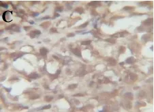
The intensity of FGF-2 expression in nuclear and cytoplasm are shown in figure 2.
B
A C
E
D F
H I
[image:2.612.326.564.623.706.2]Figure 2. Immunoistochemistry FGF-2 expression. A, Mild intensity B, Moderate intensity. C. Strong intensity.
Association between FGF-2 expression in benign, borderline, and malignant phyllodes tumours
[image:3.612.112.225.57.139.2]The number of cases for positive FGF-2 expression was found more in malignant phyllodes tumour. In this study, a statistical test was performed to determined the relationship between FGF-2 expression and benign, borderline, and malignant phyllodes breast tumours, the a statistical test was performed to determined it.
Tabel 2. Association between FGF-2 expression in benign, borderline, and malignan phyllodes tumours
Diagnosis FGF-2 expression p-value* Mild Moderate Strong
n % n % n %
Benign 9 60.0 5 33.3 1 6.7 0.0008
Borderline 0 0 6 75.0 2 25.0
Malignant 1 8.3 3 25.0 8 66.7
*Kruskal-Wallis test
In this study, we found that the test p-value 0.0008 was obtained, it can be concluded thet there is a relationship between FGF-2 expression and phyllodes tumours.
Table 3. The differences FGF-2 expressions between group lesions of benign, borderline, and malignant phyllodes
tumours
The differences of FGF-2 expression p-value*
Benign vs Borderline 0.012
Benign vs Malignant 0.0001
Borderline vs Malignant 0.169
There were differences in the FGF-2 values between groups of benign phyllodes tumour lesions with malignant phyllodes tumour lesions with p<0.001, while between groups of benign tumour lesions with borderline lesion with malignant there are no differences in values with p-values of 0.012 and 0.169, respectively.
IV. DISCUSSION
The incidence of breast phyllodes tumours is very rare, with the most incidence occurring at the age of 30 to 40 years, even in one study reported phyllodes tumours occurred at an older age, ie 45-54 years. In a research, Tan et al. reported that phyllodes tumours were 6.92% of all malignancies in the breast and
occurred at a younger age of 25-30 years.1 In some previous
study found that phyllodes tumours occurs at a younger age of 19-66 years, with a mean ± sd 40.11 ± 12.06 and a median of 38.9, according to the young age in the study conducted by Ben et al., with a mean of 39,5 (14-71) and Nurhayati’s study with a mean of 42 (16-78).19 In this study the mean age of the sufferers
was 43.2 ± 14.3 years with the youngest age is 12 years, and the oldest age is 74 years. And most occur at the age of 12-63 years.
Karim et al., reported that although it was rarely found, phyllodes tumours had been reported report in men.12 This was in
line with this study where of the 35 samples studied, there was only one male samples (2.9%), while 34 other samples were female (97.1%). The frequency of these tumour events based on changes in histopathological features is 75% benign, 16% borderline, and 9% malignant.12 Tan et al., reported that the
relative proportions of benign phyllodes tumours were 60-70%, borderline 15-20%, and malignant at 10-20%.1 Xiaofang et al.,
in their study reported from 52 patients there were 64% benign phyllodes tumours 25% borderline phyllodes tumours, and 6% malignant phyllodes tumours.13 From this study we found 15
benign phyllodes tumours (42.9%), 8 borderline phyllodes tumours (22.9%), and 12 malignant phyllodes tuomors (34.2%). The FGF-2 expression in this study was found to be mild in 10 samples (28.6%), moderate expression in 14 samples (40%), and in strong expression there were 11 samples (31.4%).
Phyllodes tumours can be clearly seen if it rapidly enlarges. Rapid enlargement does not always indicates malignancy. Looks shiny with stretched skin surface accompanied by widening of the skin surface veins. In some cases that are nor handled properly, skin ulcers can occur due to tissue ischemia. Although proper skin changes in breast tumours always show signs of malignancy, but not phyllodes tumours, ulcer on the skin can occur in benign, borderline, or malignant lesions. Nipple retraction is not common. Ulceration indicates tissue necrosis due to large tumour suppression. Large size of tumours can also cause necrosis with bleeding.14,15,16 In one study it was reported
that changes in clonality in stromal cells lead to benign phyllodes tumours and progression to monoclonal progression in epithelial and stromal cells in borderline and malignat. 7 Approximately
10-40% of tumours of this type have a risk of local recurrence and spread systemically.5 This corroborates the results of the study
why delay in handling from patients and tumour recurrence can cause the percentage of malignancy to increase. Research on 8.567 breast tumour patients in 1969-1993 only found 31 cases of phyllodes tumours (0.37%). Overall, 2.1 cases per million women, very rare in men. Most phyllodes tumour cases occur in the 4th decade, rarely in adolescents, can occur at any age.
Tumours are usually benign but local recurrence can occur and sometimes can spread systematically; rarely bilateral (either synchronous or metachronous).20-22 Several previous studies
found that tumours to be less than 5 cm in size, therefore, the diagnosis cannot be made based solely on tumour size. Prolonged gaps (leaf-like appearance) in the across section are typical signs of phyllodes tumours.17 In this study, the average tumour size
was 21.85 cm, with a range of 6-35 cm. In benign phyllodes tumours the tumour size ranges from 6-25 cm, borderline 12-15 cm, and malignant phyllodes tumours 15-35 cm. Calhoun et al., in their study found that the phyllodes tumour size ranged from 1-40 cm. This research indicate that most phyllodes tumours are
[image:3.612.28.305.325.400.2]large, however, there are also small-sized phyllodes tumours. In a previous study conducted by Flynn et al., and Calhoun et al., it was reported that phyllodes tumours were generally large (>2-3 cm).15,16,18 This is suitable with the results of this study where it
was found that phyllodes tumours size ranged from 6-35 cm. This study used the WHO classification breast tumours involving 35 samples of phyllodes tumour consisting of 15 samples of benign phyllodes tumours, 8 samples of borderline phyllodes, and 12 samples for malignant phyllodes tumours.
In a research conducted by Parker, found that phyllodes tumour are more often in the upper lateral quadrant with the same tendency between the right and left breast.23 In this study
also found that of the 35 samples examined, there werw 28 (80%) patients with tumour location in the upper lateral quadrant and 7 (20%) patients with tumour location in the lower lateral quadrant. It is not yet known exactly why the location of this phyllodes tumour is predominantly in the upper lateral quadrant, suggesting that it may be caused by more glands in the upper lateral quadrant of the breast.
In this study the authors performed FGF-2 staining on benign, borderline, and malignant phyllodes breast tumour sample with the aim of improving the accuracy of diagnosis so that management can be carried out appropriately. Dacic et al., in their study stated thar of the 23 samples studied, there were 14 samples of benign phyllodes tumours and 9 samples of malignant phyllodes tumours. In that study it was stated that there were differences in the expression of FGF-2 between benign phyllodes breast tumour and malignant phyllodes tumours. FGF-2 expressions are more expressed in the stroma than in the epithelium in both benign and malignant phyllodes tumours.9
FGF-2 is expressed in benign breast tumours and malignant breast tumours. FGF-2 enhances tumour growth through the proliferation effect of tumour cells undergoing the degeneration of the surrounding matrix, or the formation of new blood vessels.9,24
In their study, dacic et al., showed that FGF-2 is expressed in the cytoplasm in the stroma and epithelial components in most tumours. FGF-2 has also been detected in normal breast tissue (myoepithelial cells and extracellular perivascular matrix) and in malignant breast tumours. This shows that mitogenic polypeptide which stimulates plasminogen activator and has been detected in tumours that have invaded. It was further said that there was a significant correlation between FGF-2 expression and benign and malignant phyllodes breast tumours. In addition, FGF-2 is also involved in the regulation of angiogenesis and synthesis of various proteases.9
Granato et al., in their study stated that the percentage of FGF-2 immunopositive breast tumours varies greatly in different tumours, from negative to positive in all tumour areas. In the majority of cases (84&) were positively expressed in the cytoplasm, and only a small proportion (16%) were expressed in the cell nucleus and cytoplasm of tumour cells.24 In line with the
research conducted by Granato et al., in this study was found that the expression of FGF-2 in benign, borderline, and malignant phyllodes tumours varied in value, and performed in the cytoplasm and nucleus of tumour and stromal cells.
In this study, for FGF-2 expression, from the 15 benign phyllodes tumour samples, 9 (60%) samples were mild expressed, 5 (33.3%) samples were moderately expressed, and 1
(6,7%) sample was strongly expressed. For borderline phyllodes tumours, from 8 samples, there were no sample that were mildly expressed, 6 (75%) samples were moderately expressed, and 2 (25%) samples were strongly expressed. Whereas in malignant phyllodes tumours, from 12 samples, there were 1 (8.3%) samples that were mildly expressed, 3 (25%) samples were moderately expressed, and 8 (66.7%) samples were strongly expressed. From the results of this study it was also found that there were differences in values between the groups of benign phyllodes tumour lesions with malignant phyllodes with p<0.001, while between groups of benign tumour lesions with borderline and groups of borderline lesions with malignant there were no differences in values of 0.012 and respectively 0.169.
V. CONCLUSION
For FGF-2 expression, from the 15 benign phyllodes tumour samples, 9 (60%) samples were mild expressed, 5 (33.3%) samples were moderately expressed, and 1 (6,7%) sample was strongly expressed. For borderline phyllodes tumours, from 8 samples, there were no sample that were mildly expressed, 6 (75%) samples were moderately expressed, and 2 (25%) samples were strongly expressed. Whereas in malignant phyllodes tumours, from 12 samples, there were 1 (8.3%) samples that were mildly expressed, 3 (25%) samples were moderately expressed, and 8 (66.7%) samples were strongly expressed.
COMPETING INTERESTS
The authors have no relevant financial interest in the products or companies described in this article.
ACKNOWLEDGMENT
We thank to all staff members in Department of Anatomical Pathology, Universitas Sumatera Utara/H. Adam Malik General Hospital, and RSU Pirngadi Medan, Indonesia for their help and cooperation.
ETHICAL APPROVAL
Health Research Ethical Committee, University of Sumatera Utara, Medan, Indonesia approved this study.
REFERENCES
[1] Tan PH, Tse G, Lec A, Simpson JF, Hanby AM, Fibroepithelial tumours: Phyllodes tumours, In: Lakhani SR, Ellis IO, Schnitt SJ, Tan PH, Vijver MJ, editors. WHO Classification of Tumour of the Breast, 4th Edition, France; IARC Press, Lyon, 2012, p 143-7.
[2] Rosai J. Rosai and Ackerman’s Surgical Pathology, 10th Edition. Volume 1. Missouri: Mosby Elsevier; 2011. Chapter 36, Breast; p 1449-52.
[3] Yanhong Z and Celina GK, Phyllodes Tumor of the Breast, Histopathologic Features, Differential Diagnosis, and Molekular/Genetic Updates, Department of Pathology and Laboratory Medicine, University of California, and the Department of Pathology, University of Michigan, 2016. [4] Lester SC. The Breast. In: Kumar V, Abbas AK, Aster JC, Editors Robbins and Cotran Pathologic Basic of Disease, 9th Edition. Philadelphia: Saunders; 2015 p 1051-67.
[5] Azamris, Tumor Phyllodes, 2014, CDK 212, 41(1), p 40-2.
Mechanisms, Department of Pathology, University of Massachusetts Medical School and UMass Memorial Medical Center, Worcester, 2014. [7] Fairuz Q, Tumorigenesis Tumor Filodes Payudara serta Peranan Estrogen
dan Progesteron sebagai Faktor Hormonal, 2015, JMJ, 3(2) p 140-51. [8] Matthew S, Thaer K, Christhoper M, Kazuaki T, Update on the Diagnosis
and Management of Malignant Phyllodes Tumors of the Breast, 2017, Elsevier.
[9] Dacic S, Kounelis S, Kouri E, Jones MW.: Immunohistochemical Profile of Cystosarcoma Phyllodes of the Breast: A Study of 23 Cases. The Breast Journal, Volume 8 (6), 2002, p 376-81.
[10] Dunn IF, Heese O, Black PM. Growth Factors in Glioma Angiogenesis: FGFs, PDGF, EGF, and TGFs. Journal of Neuro-Oncology. 2000; 50:121-37.
[11] Risfandi M.: Hubungan Kadar Fibroblast Growth Factor 2 (FGF-2) Serum dengan Derajat Meningioma Pada Penderita Meningioma di RSUP H. Adam Malik Medan, 2015.
[12] Karim RZ, Garega SK, Yang YH, Spillane A, Camalt H, Scolver RA, et al., Phyllodes Tumours of the Breast: A Clinicopathological Analysis of 65 Cases from a Single Institution Breast. Breast (Edinburgh, Scotland), 2009, 18(3): p 165-70.
[13] Guillot E, Couturaud B, Reyal F. Management of Phyllodes Breast Tumours. Breast J. 2011; 17(2) p 129-37.
[14] Azamris, Tumor Phyllodes, 2014, CDK 212, 41(1), p 40-2.
[15] Flynn LW and Borgen PI. Phyllodes Tumor: About this Rare Cancer. Community Oncology. 2006:3 p 46-8.
[16] Calhoun KE. Phyllodes Tumours, In: Harris JR, Lipmann ME, Morrow M, Osborne CK, editors. Disease of the Breast, 4th Edition. Lipincott William & Wilkins; 2009. P 781-92.
[17] Juanita and Sungowati NK. Malignant Phyllodes Tumour of the Breast. Indon J Med Sci. 2008:1 p 101-4.
[18] Tan EY TH, Hoon TP, Yong WS, Wong HB, Go WH, et al., Recurrent Phyllodes Tumours of the Breast: Pathological Features and Clinical Implications. ANZ J Surg, 2006: 76(6), p 476-80.
[19] Quzwain F and Suryawati B, Correlation between Immunoexpression of ER-α, ERβ, and PR and Grading in Phyllodes Tumour of the Breast, Anatomical Pathologhy Department, Medical Faculty Universitas
Padjajaran Bandung, Jurnal Kedokteran Brawijaya, Vol.29 (3), 2017, p 238-43.
[20] Flynn LW, Borgen PI. Phyllodes tumor: About this rare cancer. Community Oncology. 2006;3: p 46-8.
[21] Calhoun KE, et al. Phyllodes tumors. In: Harris JR, Lippman ME, Morrow M, Osborne CK, editors. Diseases of the breast. 4th ed. Lipincott Williams & Wilkins; 2009. p 781-92.
[22] Juanita, Sungowati NK. Malignant Phyllodes Tumour of the Breast. Indon J Med Sci. 2008;1: p 101-4.
[23] Parkers SJ, Harries SA. Phyllodes tumor. Postgrad Med J. 2001; 77: p 428-35.
[24] Granato AM, Nanni O, Falcinni F, Folli S, Mosconi G, Paola F, et al., Basic fibroblast growth factor and vascular endothelial growth factor serum levels in breast cancer patients and healthy women: useful as diagnostic tools?: Department of Medical Oncology, Division of Oncology and Diagnostics, Pierantoni Hospital, Forlì, Italy, Breast Cancer Research Vol 6 (1), 2003. p R38-45.
AUTHORS
First Author – dr. Rizmeyni Azima, Resident of Department of
Anatomical Pathology, Faculty of Medicine, Universitas
Sumatera Utara, Medan, Indonesia, email ID:
Second Author – DR. dr. Delyuzar, M.Ked.(PA), Sp.PA(K),
Department of Anatomical Pathology, Faculty of Medicine, Universitas Sumatera Utara, Medan, Indonesia.
Third Author – DR. dr. Betty, Sp.PA(K), Department of
Anatomical Pathology, Faculty of Medicine, Universitas Sumatera Utara, Medan, Indonesia
Correspondence Author – dr. Rizmeyni Azima, resident of
Department of Anatomical Pathology, Faculty of Medicine, Universitas Sumatera Utara, Medan, Indonesia, email ID: