N A N O E X P R E S S
Open Access
Self-assembly of gold nanoparticles to silver
microspheres as highly efficient 3D SERS
substrates
Shouhui Chen
1,2†, Peng Huang
2†, Zhihua Wang
2, Zhe Wang
2, Magdalena Swierczewska
2, Gang Niu
2,
Daxiang Cui
1*and Xiaoyuan Chen
2*Abstract
Herein we report a simple, one-pot, surfactant-free synthesis of 3D Ag microspheres (AgMSs) in aqueous phase at room temperature. The 3D AgMSs act as supports to fix the gold nanoparticles (GNPs) in 3D space via the
interaction between the carboxyl groups of GNPs and the Ag atoms of AgMSs. The ensemble of AgMSs@GNPs with high surface-enhanced Raman scattering (SERS) activity and sensitivity can be an ideal 3D substrate choice for practical SERS detection applications. The simple self-assembly strategy may be extended to other metallic materials with great potentials in SERS, catalysis, and photoelectronic devices.
Keywords:Self-assembly, Gold nanoparticles, Silver microspheres, SERS
Background
Ensembles of inorganic nanoparticles, which display unique collective properties that are different from those of both the individual nanoparticles and bulk materials, are of much scientific and technological interest [1-5]. The ensembles have the following potential advantages: (1) to display new electronic, magnetic, and optical properties as a result of interactions between the exci-tons, magnetic moments, or surface plasmons of indi-vidual nanoparticles; (2) to improve the mechanical properties of composite materials; and (3) to allow mul-tiple tasks to be performed simultaneously or in se-quence. Typically, the self-assembly of noble-metal nanoparticles has attracted much attention because of their unique plasmon resonance and their tremendous applications in the area of optical waveguides [6], superlensing [7], photon detection [8], and surface-enhanced Raman scattering (SERS) [9-12]. Recently, the
SERS effect based on noble-metal ensembles is of par-ticular interest because of its extraordinary ability to detect a wide variety of chemical/biological species at extremely low concentrations even down to the single-molecule level [9].
Gold nanoparticles (GNPs) have been widely used as Raman active substrates because of their good biocom-patibility and strong SERS enhancement [13-18]. How-ever, it should be mentioned that the particles tend to aggregate during aging, which results in an unwanted re-duction of the active surface area [19,20]. To address this issue, the fixation of GNPs in one-dimensional (1D), 2D, or 3D spaces can avoid the aggregation of the parti-cles as SERS substrates. Tsukruk et al. assembled GNPs onto 1D silver nanowires and 2D silver nanoplates to create bimetallic nanostructures as efficient single-nanoparticle Raman markers [21]. Li et al. developed a 2D GNP monolayer film as SERS substrate by the self-assembly of nanoparticles at a liquid/liquid interface [22]. Zhang et al. reported that GNPs dispersed on the grapheme oxide (GO, 2D) and reduced graphene oxide (RGO, 2D) supports exhibit excellent SERS and catalytic performance compared with the metal nanoparticles alone [23]. Qian et al. prepared the self-assembled 3D-ordered GNP precursor composite (SiO2/GNPs) arrays as SERS nanoprobes [24]. Choi et al. reported a highly * Correspondence:[email protected];[email protected]
†Equal contributors
1
Department of Bio-Nano-Science and Engineering, National Key Laboratory of Nano/Micro Fabrication Technology, Key Laboratory for Thin Film and Microfabrication of Ministry of Education, Institute of Micro-Nano Science and Technology, Shanghai JiaoTong University, Shanghai 200240, China
2
Laboratory of Molecular Imaging and Nanomedicine (LOMIN), National Institute of Biomedical Imaging and Bioengineering (NIBIB), National Institutes of Health (NIH), Bethesda, MD 20892, USA
ordered SERS-active surface that is provided by a 3D GNP array based on thermal evaporation of gold onto an indium tin oxide (ITO) surface through a nanoporous alumina mask [25]. This SERS-active surface was applied to analyze the intracellular state. Therefore, the develop-ment of appropriate support materials to fix GNPs is very important in practical SERS detection applications.
Recently, 3D Ag microspheres (AgMSs), which contain special fine structure, large specific surface area, and micron-sized particles, have been applied as SERS sub-strates [19,26]. For example, Zhao et al. prepared 3D AgMSs with nanotextured surface morphology by a sim-ple, sonochemical, surfactant-free method. Due to their special structural features with nanoscale corrugations, the obtained 3D silver microstructures showed a struc-turally enhanced SERS performance [19]. Zhang et al.
developed hierarchical assemblies of silver nanostruc-tures as highly sensitive SERS platforms by an acid-directed assembly method [26]. Our group also used proteins [27] and microorganisms [28] as templates to synthesize AgMSs and hollow porous AgMSs, respect-ively. However, the controlled synthesis of AgMSs with clean rough surface is still a significant challenge.
[image:2.595.60.539.89.195.2]Herein, we report a simple,‘one-pot’, surfactant-free syn-thesis of 3D Ag microspheres by using silver nitrate (AgNO3) along with L-ascorbic acid (L-AA) as a reducer in aqueous phase at room temperature. The present method provides a facile and rapid route to the large-scale synthesis of 3D AgMSs with nanotextured surface morphology. The GNPs were successfully assembled on the clean rough sur-face of AgMSs via the interaction between the carboxyl groups of GNPs and the silver atoms of AgMSs (Figure 1).
Figure 1Schematic representation of the self-assembly between gold nanoparticles (GNPs) and Ag microspheres (AgMSs) via the coupling between the carboxyl groups of GNPs and the silver atoms of AgMSs.
[image:2.595.57.540.469.705.2]Methods
Experimental section
Preparation of gold nanoparticles
Briefly, 50 mL (0.2 mg/mL) of chloroauric acid (Sigma-Aldrich) was heated to boiling point, and then 1.2 mL (10 mg/mL) of sodium citrate (Sigma-Aldrich) was added. Boiling lasted for 5 min until the solution became dark red in color. After cooling down to room temperature, 20μL of GNPs was used for the analysis using transmis-sion electron microscopy (TEM).
Zeta potential of the assemblies prepared at different molar ratios of Ag microspheres to gold nanoparticles
Typically, 2.5 mL of 5 mM AgNO3aqueous solution was added to 95 mL of deionized (DI) water in a 150-mL beaker. Then, 2.5 mL of 5 mM L-AA (Sigma-Aldrich) was added into the above-mentioned solution under vig-orous stirring at room temperature. The system was stirred vigorously under ambient conditions for 4 h. The color of the solution rapidly changed from colorless to
gray. The resulting product was collected by centrifuga-tion, washed three times with DI water and ethanol, and then dispersed in ethanol for further use.
Preparation of the assemblies of GNPs to AgMSs
AgMSs (10.8 mg) was dispersed in 0.9 mL of ethanol so-lution, then 100μL of different concentrations of GNPs (0.4, 0.2, 0.1, 0.02, and 0.01 mg) were mixed with AgMSs solution under ultrasonic interaction, respectively. After 10 min, the resulting product was collected by centrifu-gation at 1,000 rpm for 5 min and washed twice with DI water and then dispersed in 1 mL DI H2O for further use.
Preparation of Raman samples
A total of 200 μL of GNPs to AgMSs (AgMSs@GNPs) was immersed in ethanol solutions containing 200μL of 2-mercaptopyridine (2-Mpy) (10 to 7 M) under ultra-sound for 10 min. After 2-Mpy molecules (Sigma-Aldrich) were adsorbed on the AgMSs@GNPs, the samples were washed twice with DI water and ethanol by centrifugation and finally dispersed in 10μL ethanol. Then, an aliquot of 10μL of 2-Mpy-loaded AgMSs@GNPs in ethanol solution was dropped onto a Si wafer. The dropped solution was spread evenly into a circle. After evaporation of ethanol under the dry N2, the sample was measured by a simple Raman instrument for six times. All of the experiments were carried out at room temperature.
Characterization
[image:3.595.57.291.89.257.2]The UV-visible spectra were recorded in a Shimadzu UV-2450 UV-visible spectrophotometer (Shimadzu Co. Ltd., Beijing, China) from 300 to 600 nm. DI water was used as the blank. SEM images were taken on a ZEISS-ULTRA 55 scanning electron microscope (Carl Zeiss AG, Oberkochen, Germany). For TEM, a drop of aque-ous solution containing the samples was placed on the carbon-coated copper grids and dried under an infrared lamp for 30 min. The micrographs were obtained using
Figure 3Histogram showing the size distribution of Ag microspheres.Gaussian curve is represented by a red line.
[image:3.595.58.540.562.705.2]a JEOL JEM-2010 transmission electron microscope (JEOL Ltd., Tokyo, Japan) operating at an accelerating voltage of 200 kV. Electron diffraction patterns were also recorded for the selected area. The surface charge of the samples was performed on NICOMP 380ZLS (Zeta potential/particle sizer; Agilent Technologies Inc., Santa Clara, CA, USA) system. SERS spectra of 2-Mpy-loaded AgMSs@GNPs were recorded by a simple Raman instrument (BWS415 B&W Tek Inc., Newark, DE, USA).
Results and discussion
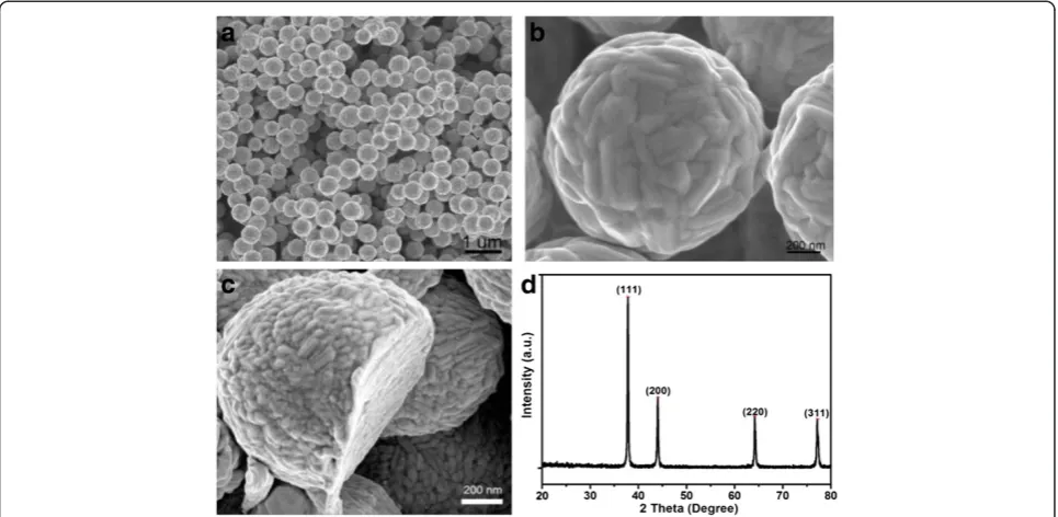
[image:4.595.57.292.88.298.2]In a typical synthesis of AgMSs, 2.5 mL of 5 mM aque-ous solution of AgNO3was added to 95 mL of deionized water in a 150 mL beaker. Then, 2.5 mL of 5 mM L-AA was added into the above-mentioned solution under vig-orous stirring at room temperature. The system was stirred vigorously under ambient conditions for 4 h. During the whole process, there was no addition of any surfactants and/or organic solvents, and L-AA plays dual roles as both reducing and capping agent. Figure 2a shows the scanning electron microscopy (SEM) images of the AgMSs obtained from a typical experiment. The as-synthesized AgMSs are quasi-spherical with large quantity and good uniformity. The average overall diam-eter of Ag microspheres was 1.26 ± 0.11 μm, estimated by measuring 200 randomly selected spheres in the en-larged SEM images. The corresponding histogram of AgMSs shows the particle size distribution fitted by a Gaussian curve (Figure 3). The magnified SEM image (Figure 2b) indicates that these microspheres possess walnut-like rough morphologies with many trenches on their surfaces. To investigate the structure of AgMSs, the AgMSs were cut using a vibratome (UltraPro 5000; Leica Biosystems Inc., Weltzar, Germany) and observed by SEM, as shown in Figure 2c. It can be seen that the AgMSs are solid inside. Figure 2d is the X-ray diffraction (XRD) pattern of AgMSs. The peaks are assigned to diffractions from the (111), (200), (220), and (311) planes of face-centered cubic (fcc) Ag phase, respectively, which were in good agreement with the reference (JCPDS
Figure 5TEM image of gold nanoparticles dispersed in water.
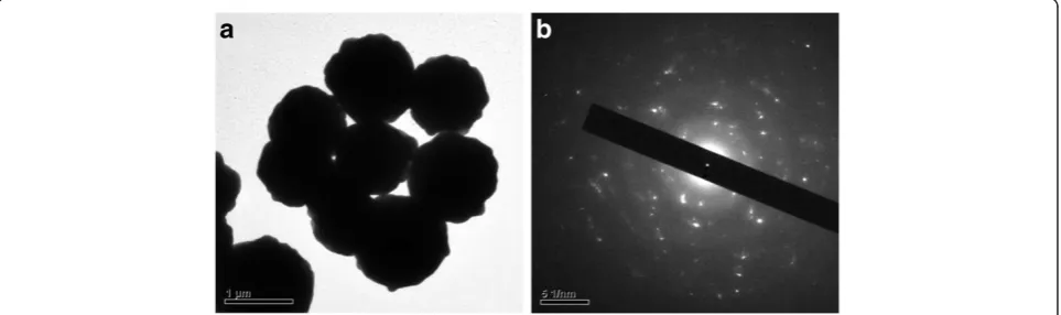
[image:4.595.60.539.485.715.2]04-0783). These planes with sharp peaks indicate that the AgMSs are all well crystallized. The peaks can be easily indexed to a pure cubic phase of silver. Meanwhile, no other impurity peaks were detected, suggesting the high purity of AgMSs. TEM is also performed to observe the morphologies of the as-prepared AgMSs (Figure 4a). The morphology of AgMSs is quasi-spherical, and the size is approximately 1.26μm. There are some convex struc-tures on the edges of microspheres, indicating that their surfaces are very rough. The results are consistent with the observation of SEM. The selected area electron diffrac-tion (SAED) pattern (Figure 4b) further reveals that the AgMSs are crystalline with fcc structure and independent orientations.
Gold nanoparticles were synthesized according to our previous report [29]. TEM image of GNPs is shown in Figure 5, indicating that the GNPs are spherical and monodisperse with an average diameter of 15 nm. Based on the interaction between the carboxyl groups and sil-ver atoms, the GNPs were successfully assembled on the surface of AgMSs [30]. Figure 6a,b,c,d clearly reveals that GNPs are homogeneously distributed on the surface of AgMSs. As can be seen, there are no changes in the shape and size of GNPs and AgMSs after self-assembly. With the increase of GNP concentration, the number of GNPs on the surface of AgMSs is also increased. When the molar ratio of AgMSs/GNPs is 100:20, the surface of AgMSs is completely coated by GNPs (Figure 6b).
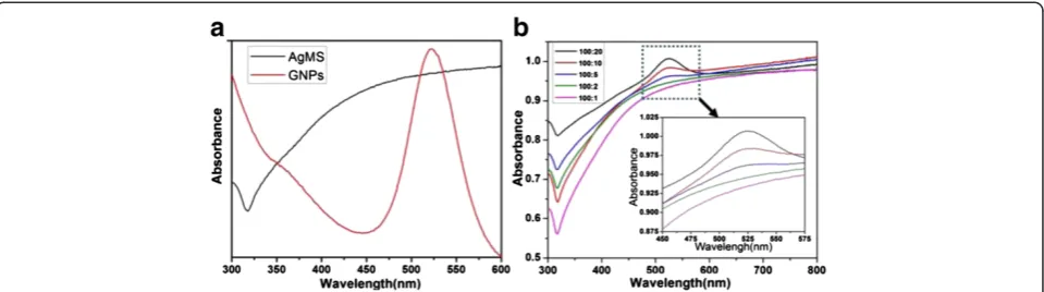
To further testify the self-assembly between GNPs and AgMSs, the assemblies were also detected by a UV–vis spectrophotometer. As shown in Figure 7a, there is a strong absorption band in 350 to 600 nm for AgMSs. The broad half-peak width indicates that the size of AgMSs is bigger than nanoscale, which agrees with SEM and TEM observations. The absorption spectrum of GNPs displays a characteristic surface plasmon reson-ance band at approximately 520 nm. Figure 7b shows the UV–vis spectra of the assemblies prepared at differ-ent AgMSs/GNPs molar ratios. With the increase of
[image:5.595.59.539.89.223.2]GNP concentration, the intensity of the characteristic band at approximately 520 nm in the assemblies is also gradually increased. This is attributed to the increase of GNPs on the surface of AgMSs. The assemblies are negatively charged and display a GNP concentration-dependent increase of negative charges on the surface (Figure 8). The above facts suggest that the GNPs were successfully assembled on the surface of AgMSs.
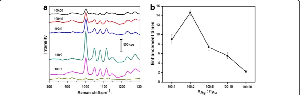
[image:5.595.306.538.533.704.2]To evaluate the SERS activities of the assemblies of GNPs to AgMSs (AgMSs@GNPs), we chose 2-Mpy as targeted probing molecules [28]. Figure 9a shows the representative SERS spectra of 2-Mpy molecules on the assembled substrates of AgMSs to GNPs. All spectra ex-hibit peaks at 1,001, 1,049, 1,080, and 1,114 cm−1, which are assigned to the characteristic peaks of 2-Mpy mole-cules. Figure 9b shows the corresponding enhancement of the assembled substrates at different molar ratios of AgMSs to GNPs relative to 2-Mpy on pure AgMSs. Compared with the SERS activity of pure AgMSs, all AgMSs@GNPs exhibit obvious enhancement of SERS sig-nal in varying degrees. The most significant enhancement
Figure 8Zeta potential of the assemblies prepared at different molar ratios of Ag microspheres to gold nanoparticles.
of SERS signal is found atnAg/nAuratio of 100:2, which is about 14-fold higher than that of pure AgMSs. Further increase ofnAg/nAuratio leads to decrease of SERS sig-nal, which is likely due to the decreased nanogaps with increased gold particle deposition onto the surface of AgMSs. Several reasons can account for the enhanced Raman scattering signal: (1) The 3D assemblies of AgMSs@GNPs with huge, rough, and clean surface can absorb more molecules; (2) There are abundant‘hotspots’ at the nanoparticles junctions to amplify the local E-fields as well as the Raman signal; and (3) AgMSs support the GNPs in 3D space to avoid the aggregation of the particles during application as SERS substrates.
Conclusions
In summary, we report a simple, one-pot, surfactant-free synthesis of 3D AgMSs in aqueous phase at room temper-ature. The 3D AgMSs act as supports to fix the GNPs in 3D space via the interaction between the carboxyl groups of GNPs and the Ag atoms of AgMSs. The ensemble of AgMSs@GNPs with high SERS activity and sensitivity can be an ideal 3D substrate choice for practical SERS detec-tion applicadetec-tions. The simple self-assembly strategy may be extended to other metallic materials with great poten-tials in SERS, catalysis, photoelectronic devices, etc.
Competing interests
The authors declare that they have no competing interests.
Authors’contributions
SHC carried out the preparation of AuNPs, AgMSs, AgMSs@GNPs assembly, Raman and XRD, characterization, drafted the manuscript, PH modified the draft of manuscript, ZHW carried out the UV and SEM Characterization. ZW checked the manuscript grammar. MS participated in the analysis of Raman results. GN gave many advices for this manuscript. DXC and XYC designed of the study and guided this work. All authors read and approved the final manuscript.
Acknowledgments
This work was supported in part by the Intramural Research Program (IRP), National Institute of Biomedical Imaging and Bioengineering (NIBIB), National
Institutes of Health (NIH), the National Key Basic Research Program (973 Project) (2010CB933902 and 2011CB933100), National 863 Hi-tech Project (2007AA022004), Important National Science & Technology Specific Projects (2009ZX10004-311), National Natural Scientific Fund (nos. 81225010, 20771075, 20803040, and 81028009), New Century Excellent Talent of Ministry of Education of China (NCET-08-0350), and Shanghai Science and Technology Fund (10XD1406100).
Received: 6 March 2013 Accepted: 17 March 2013 Published: 12 April 2013
References
1. Nie ZH, Fava D, Kumacheva E, Zou S, Walker G, Rubinstein M:Self-assembly of metal–polymer analogues of amphiphilic triblock copolymers.
Nat Mater2007,6:609–614.
2. Nie ZH, Petukhova A, Kumacheva E:Properties and emerging applications of self-assembled structures made from inorganic nanoparticles.
Nat Nanotechnol2010,5:15–25.
3. Nie ZH, Li W, Seo M, Xu SQ, Kumacheva E:Janus and ternary particles generated by microfluidic synthesis: design, synthesis, and self-assembly.
J Am Chem Soc2006,128:9408–9412.
4. Xia YS, Nguyen TD, Yang M, Lee B, Santos A, Podsiadlo P, Tang ZY, Glotzer S, Kotov N:Self-assembly of self-limiting monodisperse supraparticles from polydisperse nanoparticles.Nat Nanotechnol2011,6:580–587.
5. He D, Hu B, Yao QF, Wang K, Yu SH:Large-scale synthesis of flexible free-standing SERS substrates with high sensitivity: electrospun PVA nanofibers embedded with controlled alignment of silver nanoparticles.
ACS Nano2009,3:3993–4002.
6. Maier SA, Kik PG, Atwater HA, Meltzer S, Harel E, Koel BE, Requicha AA: Local detection of electromagnetic energy transport below the diffraction limit in metal nanoparticle plasmon waveguides.Nat Mater
2003,2:229–232.
7. Fang N, Lee H, Sun C, Zhang X:Sub-diffraction-limited optical imaging with a silver superlens.Science2005,308:534–537.
8. Konstantatos G, Clifford J, Levina L, Sargent EH:Sensitive solution-processed visible-wavelength photodetectors.Nat Photonics2007, 1:531–534.
9. Zhu ZN, Meng HF, Liu WJ, Liu XF, Gong JX, Qiu XH, Jiang L, Wang D, Tang ZY: Superstructures and SERS properties of gold nanocrystals with different shapes.Angew Chem Int Ed Engl2011,50:1593–1596.
10. Lu G, Li H, Liusman C, Yin ZY, Wu SX, Zhang H:Surface enhanced Raman scattering of Ag or Au nanoparticle-decorated reduced graphene oxide for detection of aromatic molecules.Chem Sci2011,2:1817–1821. 11. Lu G, Li H, Wu SX, Chen P, Zhang H:High-density metallic nanogaps
fabricated on solid substrates used for surface enhanced Raman scattering.Nanoscale2011,4:860–863.
[image:6.595.58.539.90.243.2]12. Braun G, Pavel I, Morrill AR, Seferos DS, Bazan GC, Reich NO, Moskovits M: Chemically Patterned microspheres for controlled nanoparticle assembly in the construction of SERS hot spots.J Am Chem Soc2007,129:7760–7761.
13. Emory SR, Haskins WE, Nie SM:Direct observation of size-dependent optical enhancement in single metal nanoparticles.J Am Chem Soc1998, 120:8009–8010.
14. Krug JT, Wang GD, Emory SR, Nie SM:Efficient Raman enhancement and intermittent light emission observed in single gold nanocrystals.
J Am Chem Soc1999,121:9208–9214.
15. Qian XM, Peng XH, Ansari D, Yin QG, Chen GZ, Shin DM, Yang L, Young A, Wang DM, Nie SM:In vivotumor targeting and spectroscopic detection with surface-enhanced Raman nanoparticle tags.Nat Biotechnol2008, 26:83–90.
16. Liu HN, Li S, Liu LS, Tian L, He NY:An integrated and sensitive detection platform for biosensing application based on Fe@Au magnetic nanoparticles as bead array carries.Biosens Bioelectron2010,26:1442–1448. 17. Huang P, Lin J, Li ZM, Hu HY, Wang K, Gao G, He R, Cui DX:A general
strategy for metallic nanocrystals synthesis in organic medium.
Chem Commun2010,46:4800–4802.
18. Li S, Liu H, Jia YY, Deng Y, Zhang LM, Lu ZX, He NY:A novel SNPs detection method based on gold magnetic nanoparticles array and single base extension.Theranostics2012,2:967–975.
19. Zhang MF, Zhao AW, Sun HH, Guo HY, Wang DP, Li D, Gan ZB, Tao WY: Rapid, large-scale, sonochemical synthesis of 3D nanotextured silver microflowers as highly efficient SERS substrates.J Mater Chem2011, 21:18817–18824.
20. Zhang MF, Zhao AW, Guo HY, Wang DP, Gan ZB, Sun HH, Li D, Li M:Green synthesis of rosettelike silver nanocrystals with textured surface topography and highly efficient SERS performances.Cryst Eng Comm
2011,13:5709–5717.
21. Gunawidjaja R, Kharlampieva E, Choi I, Tsukruk V:Bimetallic nanostructures as active Raman markers: gold-nanoparticle assembly on 1D and 2D silver nanostructure surfaces.Small2009,5:2460–2466.
22. Wang MH, Hu JW, Li YJ, Yeung ES:Au nanoparticle monolayers: preparation, structural conversion and their surface-enhanced Raman scattering effects.Nanotechnology2010,21:145608.
23. Huang J, Zhang LM, Chen B, Ji N, Chen FH, Zhang Y, Zhang ZJ: Nanocomposites of size-controlled gold nanoparticles and graphene oxide: formation and applications in SERS and catalysis.Nanoscale2010, 2:2733–2738.
24. Rao YY, Chen QF, Dong J, Qian WP:Growth-sensitive 3D ordered gold nanoshells precursor composite arrays as SERS nanoprobes for assessing hydrogen peroxide scavenging activity.Analyst2010,136:769–774. 25. El-Said WA, Kim TH, Kim H, Choi JW:Analysis of intracellular state based
on controlled 3D nanostructures mediated surface enhanced Raman scattering.PLoS One2011,6:e15836.
26. Zhang B, Xu P, Xie XM, Wei H, Li ZP, Mack NH, Han XJ, Xu HX, Wang HL: Acid-directed synthesis of SERS-active hierarchical assemblies of silver nanostructures.J Mater Chem2010,21:2495–2501.
27. Huang P, Yang D, Zhang C, Lin J, He M, Bao L, Cui DX:Protein-directed one-pot synthesis of Ag microspheres with good biocompatibility and enhancement of radiation effects on gastric cancer cells.Nanoscale2011, 3:3623–3626.
28. Yang DP, Chen SH, Huang P, Wang XS, Jiang WQ, Pandoli O, Cui DX: Bacteria-template synthesized silver microspheres with hollow and porous structures as excellent SERS substrate.Green Chem2010,12:2038–2042. 29. Yang H, Li D, He R, Guo Q, Wang K, Zhang XQ, Huang P, Cui DX:A novel
quantum dots-based point of care test for syphilis.Nanoscale Res Lett
2010,5:875–881.
30. Weddemann A, Ennen I, Regtmeier A, Albon C, Wolff A, Eckstädt K, Mill N, Peter MKH, Mattay J, Plattner C:Review and outlook: from single nanoparticles to self-assembled monolayers and granular GMR sensors.
Beilstein J Nanotechnol2010,1:75–93.
doi:10.1186/1556-276X-8-168
Cite this article as:Chenet al.:Self-assembly of gold nanoparticles to
silver microspheres as highly efficient 3D SERS substrates.Nanoscale
Research Letters20138:168.
Submit your manuscript to a
journal and benefi t from:
7 Convenient online submission 7 Rigorous peer review
7 Immediate publication on acceptance 7 Open access: articles freely available online 7 High visibility within the fi eld
7 Retaining the copyright to your article