BIROn - Birkbeck Institutional Research Online
Ruiz-Rizzo, A.L. and Sorg, C. and Napiórkowski, N. and Neitzel, J. and
Menegaux, A. and Muller, Hermann and Vangkilde, S. and Finke, K. (2018)
Decreased cingulo-opercular network functional connectivity mediates the
impact of aging on visual processing speed. Neurobiology of Aging 73 , pp.
50-60. ISSN 0197-4580.
Downloaded from:
Usage Guidelines:
Please refer to usage guidelines at or alternatively
Accepted Manuscript
Decreased cingulo-opercular network functional connectivity mediates the impact of aging on visual processing speed
Adriana L. Ruiz-Rizzo, Christian Sorg, Natan Napiorkowski, Julia Neitzel, Aurore Menegaux, Hermann J. Müller, Signe Vangkilde, Kathrin Finke
PII: S0197-4580(18)30338-5
DOI: 10.1016/j.neurobiolaging.2018.09.014
Reference: NBA 10376
To appear in: Neurobiology of Aging
Received Date: 6 March 2018 Revised Date: 10 August 2018 Accepted Date: 11 September 2018
Please cite this article as: Ruiz-Rizzo, A.L., Sorg, C., Napiorkowski, N., Neitzel, J., Menegaux, A., Müller, H.J., Vangkilde, S., Finke, K., Decreased cingulo-opercular network functional connectivity mediates the impact of aging on visual processing speed, Neurobiology of Aging (2018), doi: https://doi.org/10.1016/ j.neurobiolaging.2018.09.014.
This is a PDF file of an unedited manuscript that has been accepted for publication. As a service to our customers we are providing this early version of the manuscript. The manuscript will undergo copyediting, typesetting, and review of the resulting proof before it is published in its final form. Please note that during the production process errors may be discovered which could affect the content, and all legal disclaimers that apply to the journal pertain.
M
AN
US
CR
IP
T
AC
CE
PT
ED
ACCEPTED MANUSCRIPT
Title:
Decreased cingulo-opercular network functional connectivity mediates the impact of aging on visual
processing speed
Authors and affiliations:
Adriana L. Ruiz-Rizzo1,2
, Christian Sorg1,3
, Natan Napiorkowski1,2
, Julia Neitzel1,2
, Aurore
Menegaux1,2
, Hermann J. Müller1,2
, Signe Vangkilde4
, Kathrin Finke1,5
1
Department of General and Experimental Psychology, Ludwig-Maximilans-Universität München,
Munich (Germany)
2
Graduate School of Systemic Neurosciences, GSN LMU Munich, Munich
3
TUM-Neuroimaging Center,TUM-NIC, Technische Universität München, Munich
4
Department of Psychology, Center for Visual Cognition, University of Copenhagen, Copenhagen
(Denmark)
5
Hans Berger Department of Neurology, Jena University Hospital, Jena (Germany)
Corresponding author:
Adriana L. Ruiz Rizzo. Department of General and Experimental Psychology,
Ludwig-Maximilans-Universität München, Leopoldstraße 13, 80802 Munich, Germany. Phone: +49 89 2180 72569,
email: [email protected].
M
AN
US
CR
IP
T
AC
CE
PT
ED
ACCEPTED MANUSCRIPT
Abstract
The neural factors that account for the visual processing-speed reduction in aging are incompletely
understood. Based on previous reports of age-related decreases in the intrinsic functional
connectivity (iFC) within the cingulo-opercular network and its relevance for processing speed, we
hypothesized that these decreases are associated with age-related reductions in visual processing
speed. We used a whole-report task and modeling based on Bundesen’s ‘theory of visual attention’ to
parameterize visual processing speed in 91 healthy participants from 20 to 77 years old. iFC was
estimated using independent-component analysis of resting-state fMRI data. From the clusters within
the cingulo-opercular network exhibiting age-related decreased iFC, we found a cluster in the left
insula to be particularly associated with visual processing speed and to mediate the age effect on
visual speed. This mediation was not observed for age-related decreased iFC in other networks or for
other attentional parameters. Our results point to the iFC in the cingulo-opercular network,
represented by the left insula, as being a relevant marker for visual processing-speed changes in
aging.
Keywords: Cingulo-opercular network; functional connectivity; healthy aging; processing speed;
M
AN
US
CR
IP
T
AC
CE
PT
ED
ACCEPTED MANUSCRIPT
1. Introduction
A decline in processing speed represents a fundamental aspect of cognitive aging (Salthouse, 1996).
In particular, a reduction of visual processing speed, or the rate of information encoding into visual
short-term memory (VSTM), has previously been established in both healthy (Espeseth et al., 2014;
Habekost et al., 2013; McAvinue et al., 2012; Wiegand et al., 2014); and, more severely,
pathological aging (Bublak et al., 2011; Ruiz-Rizzo et al., 2017) using a computational approach
based on Bundesen’s (1990) ‘theory of visual attention’ (TVA). TVA permits the contribution of
visual processing speed to the efficiency of visual selection to be quantitatively estimated –
independently of motor speed as well as other visual-attention functions such as VSTM capacity or
the visual threshold, which also degrade during aging. The TVA-based visual processing-speed
parameter, C, is estimated from a participant’s performance in a psychophysical (letter) whole-report
task (Bundesen, 1990; Habekost et al., 2014), where C provides a measure of the individual’s rate of
visual information uptake in elements (i.e., letters) per second. While the reduction of visual
processing speed is generally thought to be brought about by neural aging, the underlying brain
mechanisms are only incompletely understood. In this study, we aimed to shed light on this issue by
investigating the role of the cingulo-opercular network in the reduction of visual processing speed as
a result of aging.
The cingulo-opercular network, also referred to as a ‘salience’ (e.g., Seeley et al., 2007) or
‘ventral attention’ (e.g., Yeo et al., 2011) network, is centered on the anterior insula and the anterior
cingulate cortex (Dosenbach et al., 2008; Dosenbach et al., 2007; Menon and Uddin, 2010; Seeley et
al., 2007). It is thought to play a central role in ‘tonic alertness’: the endogenous ability to maintain
an appropriate level of arousal (Posner and Petersen, 1990; Sturm and Willmes, 2001). Thus, for
instance, the spontaneous activity fluctuations of the cingulo-opercular network, measured using
functional magnetic resonance imaging (fMRI), have been shown to correlate with markers of tonic
M
AN
US
CR
IP
T
AC
CE
PT
ED
ACCEPTED MANUSCRIPT
(Schneider et al., 2016). Further, the cingulo-opercular network displays sustained activity during
perceptual processing (Sestieri et al., 2014), with its activity level relating to the speed of stimulus
detection (Coste and Kleinschmidt, 2016). Importantly in the present context, intrinsic functional
connectivity (iFC) in the cingulo-opercular network increases with rising demands on tonic alertness
(Sadaghiani and D'Esposito, 2015) – where iFC refers to the coherence of the infra-slow (i.e., 0.01 –
0.1 Hz) spontaneous neural activity, typically measured with BOLD-
(blood-oxygenation-level-dependent-) fMRI during resting state (De Luca et al., 2006; Fox and Raichle, 2007; Raichle, 2015).
In fact, tonic alertness has been proposed to relate closely to visual processing speed C (e.g.,
Bundesen et al., 2015). This is evidenced, for example, by tonic alertness influencing visual
processing speed in a TVA-based whole-report task (Matthias et al., 2010). Also, raising tonic
alertness through psychostimulant medication increases visual processing speed (Finke et al., 2010).
And of note, iFC in the cingulo-opercular network has been found to be related to visual processing
speed in healthy young adults (Ruiz-Rizzo et al., 2018).
Age-related decreases of the iFC in the cingulo-opercular network have been reported
previously and were found to be related to a reduced global cognitive state (He et al., 2014) and
lowered visuospatial and executive abilities (Onoda et al., 2012). Given that a significant amount of
variance in diverse cognitive tasks (employed in the previous reports) can be accounted for by visual
processing speed especially in old age (e.g., Deary and Stough, 1996), we hypothesized that reduced
iFC in the cingulo-opercular network would be particularly associated with a decrease in visual
processing speed. Based on the documented associations between age and both reduced iFC in the
cingulo-opercular network (He et al., 2014; Onoda et al., 2012) and visual processing speed
(Espeseth et al., 2014; Habekost et al., 2013; McAvinue et al., 2012; Wiegand et al., 2014), we
further explored whether the reduced iFC would mediate the effect of age on visual processing
M
AN
US
CR
IP
T
AC
CE
PT
ED
ACCEPTED MANUSCRIPT
To investigate the association between iFC in the cingulo-opercular network and the
age-related decrease of visual processing speed, we examined whether individuals’ iFC would correlate
with their visual processing speed C (as estimated from performance in a in a TVA-based task
requiring participants to report as many letters as possible from briefly presented letter arrays
(so-called whole-report task). To assess the concurrent criterion validity of this association, we further
examined whether the results would generalize to a clinically well-established measure of visual
processing speed: the Trail Making Test, form A (TMT-A; Tombaugh, 2004). To take account of
individual differences in brain size that could influence the relationship between iFC and behavior,
the correlations were computed controlling for brain volume (Smith et al., 2004; Smith et al., 2002).
Next, we performed two lines of control analysis. First, we checked whether the association between
the cingulo-opercular network and visual processing speed is a specific one, as hypothesized, rather
than being part of a more global relationship between changes in iFC and visual attention functions
in general. That is, given the well-documented age-related decreases in iFC in the default-mode and
dorsal-attention networks (see, e.g., Andrews-Hanna et al., 2007; Damoiseaux et al., 2008; Ferreira
and Busatto, 2013), age-related reductions in visual processing speed might conceivably be
associated with a general age-related decrease of iFC, rather than with that of the cingulo-opercular
network exclusively. To check for this, (a) we examined whether visual processing speed C also
relates to the iFC in other networks; and (b) we assessed whether the iFC in the cingulo-opercular
network relates to other visual attention functions previously reported to change with aging, such as
the visual perceptual threshold and VSTM storage capacity (see, e.g., McAvinue et al., 2012).
Second, we controlled for potentially relevant confounds in the association between iFC in the
opercular network and visual processing speed. In particular, given that iFC in the
cingulo-opercular network has previously been found to be related to anxiety (see, e.g., Seeley et al., 2007)
and anxiety level might in turn affect processing speed, we controlled for anxiety using the state-trait
M
AN
US
CR
IP
T
AC
CE
PT
ED
ACCEPTED MANUSCRIPT
we controlled for were vascular risk and gray-matter density, both of which are influenced by aging
and might also affect processing speed (e.g., D'Esposito et al., 2003; Lu et al., 2013). Following
these control analyses, we went on to explore whether the iFC in the cingulo-opercular network
mediates the well-established relation between age and visual processing speed, as hypothesized.
2. Material and methods
2.1. Participants
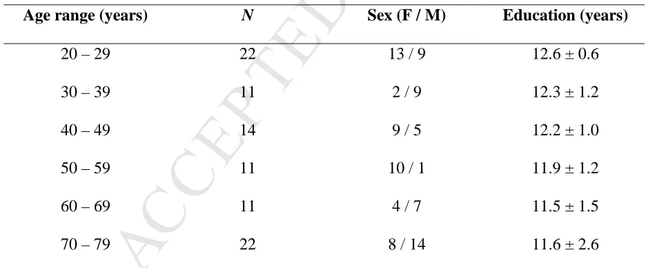
The current study included 91 healthy adults in the age range from 20 to 77 years (mean age: 48.8 ±
19.2 years; 46 females; mean education: 12.0 ± 1.6 years; 4 left-handed; Table 1). The study was
approved by the LMU Munich ethics committee, and written informed consent was obtained from all
participants. Initially, 108 adults (19 to 78 years old) of the Munich INDIREA aging cohort1 took
part in the study. The Mini Mental State Examination (MMSE; Folstein et al., 1975) was used for
screening for cognitive impairments in participants from 60 years onwards (i.e., a MMSE score
below 27), and the Beck Depression Inventory (BDI; Beck et al., 1996) for screening for symptoms
of depression in all participants (i.e., a BDI score above 19). Seventeen participants were excluded
because they asked to be withdrawn from the study (n = 9) or because of incomplete or unreliable
data (n = 3; 1 exhibited significant head motion in the resting-state fMRI session, for 1 TVA-based
parameters fitting was inadequate, and 1 had a MMSE score below 27), uncorrected visual acuity
decreases (n = 2), or symptoms of depression (n = 3).From the remaining 91 participants, those in
the older adults group had no indication of cognitive impairment (mean MMSE score: 29 ± 0.9), and
all were free of previous or current psychiatric or neurological disorders, psychiatric or neurological
medication, diabetes, color blindness, or current symptoms of depression (mean BDI score: 5.2 ±
4.8). In one session, participants underwent resting-state fMRI at the Department of Neuroradiology,
1
M
AN
US
CR
IP
T
AC
CE
PT
ED
ACCEPTED MANUSCRIPT
Klinikum rechts der Isar, Munich (Germany). In a separate, psychophysical-testing session, visual
attention functioning was assessed using the whole-report task. In the psychophysical-testing session,
participants also completed the TMT and MMSE, and filled out demographic, anxiety, and BDI
questionnaires. Session order depended on individual participants’ convenience. The average time
between sessions was 2.6 months.
2.2. Assessment and estimation of visual processing speed C
A whole-report task, based on TVA (Bundesen, 1990), was used to estimate visual processing speed
C. On each trial, four red letters were briefly presented to participants, who were instructed to
verbally report, in any order, all letters they were fairly certain they had seen. Stimuli were randomly
chosen from the set of letters (A, B, D, E, F, G, H, J, K, L, M, N, O, P, R, S, T, V, X, Z). Letters
were arranged in the form of a semicircle, with a radius of 5.3° of visual angle, on either the right or
the left of a central fixation point, as shown in Figure 1. To ensure balanced visual stimulation in
both hemifields, targets were accompanied by four blue symbols (composed of random letter parts;
see Figure 1 for an example) of the same luminance displayed on the semicircle on the other side of
fixation. Visual stimuli were 1.3° of visual angle in diameter, and both letters and symbols appeared
only once in a particular trial.
The task included 10 blocks of 40 trials each (400 trials in total), with targets presented in the
left and right hemifields, respectively, in half of the blocks. Five individually adjusted stimulus
exposure durations were determined in a short pre-test and introduced in the subsequent whole-report
task. To start with, the participant was presented with one adjustment trial displayed for 80 ms, with
stimulus exposure terminated by post-display masks (see below). If s/he reported at least one letter
correctly, the exposure duration was decreased by 10 ms, and this procedure continued for the next
15 adjustment trials. These (16) adjustment trials were divided into 4 blocks, with each 4 adjustment
post-M
AN
US
CR
IP
T
AC
CE
PT
ED
ACCEPTED MANUSCRIPT
display masks (see below for details) presented for 250 ms (yielding 12 trials per block). The
exposure duration was decreased until the (shortest) duration was established at which the participant
could no longer report one letter. If this point was reached before the last of the 16 adjustment trials,
the exposure duration was kept constant for the few remaining adjustment trials. Setting the exposure
duration so short was meant to ensure that we would obtain a valid estimate of the visual perceptual
threshold parameter. Then, based on the shortest exposure duration, four longer values were
additionally chosen to allow for variability in letter report performance across the whole range from
near-floor to near-ceiling, and thus render the TVA-based parameter estimation more precise. On
masked trials, the displays were shown for one of the five durations and immediately followed by
masks (a scattered patch of red and blue squares, 1.3° in size) presented for 900 ms at each stimulus
location, so as to avoid visual persistence effects. In addition to trials with masked display exposure,
we introduced unmasked trials (without post-display masks), to increase the variability of effective
exposure times (by allowing for an additional component of iconic memory buffering; Sperling,
1960) and thus ensure reliable and valid TVA parameter fitting. Specifically, on unmasked trials,
displays were presented at one of two exposure durations: one was the same as the second shortest
masked duration and the other one was 200 ms2. Thus, overall, trial displays were presented for
seven effective exposure durations, five masked and two unmasked. A block of 40 trials (with
hemifield blocked) thus consisted of 15 masked trials, with 3 trials for each of the 5 set exposure
durations; 3 unmasked trials with the second shortest duration; and 22 unmasked trials with 200-ms
duration. Trials were presented in random order within each block.
Participants were tested in a sound-attenuated chamber (Industrial Acoustics Company) with
a dim light placed behind them. Stimuli were presented on a 24'' LED screen with an 800px x 600px
resolution and a 100-Hz refresh rate. The viewing distance was kept constant at about 65 cm. At the
2
M
AN
US
CR
IP
T
AC
CE
PT
ED
ACCEPTED MANUSCRIPT
beginning of each block, a black screen with a white arrow appeared pointing towards the side on
which the stimuli would appear for that specific block. After stimulus presentation, a white question
mark appeared in the center of the screen prompting the start of the verbal report. In each trial, the
experimenter entered the reported letters in the reported order and manually started the next trial. The
measure of interest was report accuracy (at a given effective exposure duration). At the end of each
block, a feedback bar was presented that signaled whether the report accuracy of the actually
reported letters ranged between 70% and 90%. If the report accuracy was outside this range, oral
instructions were given by the experimenter. If it was above 90%, the participant was told that he/she
should try to report more letters, even if not absolutely sure that they are correct. If it dropped below
70%, the participant was told to try to refrain from guessing and report only those letters he/she was
relatively sure to have seen. The whole-report task lasted about 45 minutes.
Visual processing speed C was estimated by modeling the participant’s report accuracy as a
function of the effective exposure duration, using a maximum likelihood-fitting algorithm
(Bundesen, 1990; Dyrholm et al., 2011; Kyllingsbaek, 2006). The TVA-based fitting procedure
models the probability of correct letter report in terms of an exponential growth function with
increasing (effective) exposure duration. The slope of the function at its origin represents the visual
processing speed or parameter C, that is, the rate of visual information uptake (in elements per
second). Additionally, two other parameters were estimated, that in our analyses served for testing
whether the relationship between the iFC in the cingulo-opercular network and visual processing
speed is a specific one, namely: parameter t0, indicating the visual perceptual threshold, that is, the
longest ineffective exposure duration (in ms) below which information uptake is effectively zero;
and parameter K, indicating the maximum number of elements that can be simultaneously
represented in visual short-term memory (VSTM)3. With aging, t0 exhibits an increase whereas K
3 An additional parameter, parameter
M
AN
US
CR
IP
T
AC
CE
PT
ED
ACCEPTED MANUSCRIPT
shows a decrease (Espeseth et al., 2014). Critically, the TVA-based estimation of parameter C (visual
processing speed) is mathematically independent of that of t0 and K. Accordingly, the measure of C
allows controlling for potential influences of changes in threshold and VSTM storage capacity and
has, thus, a high cognitive specificity. In summary, the computational model used had 6 degrees of
freedom (df): C, 1 df; t0, 1 df; K, 3 df (the reported K value is the expected K given a particular
distribution of the probability that, on a given trial, K = 1, 2, 3, or 4); and µ, 1 df. For those
participants whose t0 was estimated to be below 0 (i.e., 11 in total), we re-fitted the data fixing t0 at
04.
2.3. Paper-and-pencil assessment of visual processing speed: Trail Making Test A (TMT-A)
Participants’ visual processing speed was also assessed using a standard neuropsychological
measure, the TMT-A, to test whether the association between iFC in the cingulo-opercular network
and the age-induced reduction in visual processing speed can also be shown when an established
paper-and-pencil task is used. The TMT-A5 measures visual scanning speed in terms of the time
required to connect circles with numbers in ascending order (Reitan and Wolfson, 1985; Tombaugh,
2004) and is widely used in Alzheimer’s disease screening batteries (e.g., Weintraub et al., 2009).
We examined the association of the time to complete the TMT-A with the iFC of the
cingulo-opercular network.
2.4. State-Trait Anxiety Inventory (STAI)
Given previously reported associations between the iFC in the cingulo-opercular network and
anxiety, we also assessed anxiety in our sample and used it as an additional control variable. The
4
Crucially, when the data of these 11 participants are refitted without the limitation of t0 being at minimum 0, the mean change in C values was 2.12 (±2.62). Post-hoc analyses showed that constraining the model to t0 at a minimum of 0 does not impact the conclusions of our study or its validity.
5
M
AN
US
CR
IP
T
AC
CE
PT
ED
ACCEPTED MANUSCRIPT
STAI Form X (Laux et al., 1981; Spielberger et al., 1970) consists of two (Barnes et al., 2002)
self-report scales of 20 items each that measure state and trait anxiety.
2.5. MRI data acquisition
MRI data were acquired on a Philips Ingenia 3T system (Netherlands), using a 32-channel SENSE
head coil. Functional MRI T2*-weighted data were collected for 12.5 min while participants’ rested
with their eyes closed, after having been told not to fall asleep. We checked that participants had not
fallen asleep by directly asking them immediately after finishing the sequence. Foam padding was
used to constrain participants’ head motion while scanning, and earplugs and headphones were
provided to reduce adverse effects of scanner noise. Six hundred volumes of BOLD-fMRI signal
were acquired from each individual, using a multiband (Feinberg and Setsompop, 2013) echo-planar
imaging (EPI) sequence, with a 2-fold in-plane SENSE acceleration (SENSE factor, S = 2) and an
M-factor of 2 (Preibisch et al., 2015). Other fMRI acquisition parameters were: repetition time, TR =
1,250 ms; time to echo, TE = 30 ms; phase encoding, PE direction: anterior-posterior; flip angle =
70º; field of view, FOV = 192 mm2; matrix size = 64 x 64, 40 slices; slice thickness = 3.0 mm;
interslice gap 0.3 mm; reconstructed voxel size = 3 x 3 x 3.29 mm3. A high-resolution T1-weighted
anatomical volume was acquired using a 3D magnetization prepared rapid acquisition gradient echo
(MPRAGE) sequence with the following parameters: TR = 9 ms; TE = 4 ms; inversion time, TI = 0
ms; flip angle = 8º; 170 sagittal slices; FOV = 240 mm2; matrix size = 240 x 240; reconstructed
voxel size = 1 mm isotropic.
2.6. MRI Data Analysis
2.6.1. Resting-state fMRI data preprocessing
Six hundred resting-state fMRI volumes per individual were preprocessed using the Data Processing
M
AN
US
CR
IP
T
AC
CE
PT
ED
ACCEPTED MANUSCRIPT
analysis of resting-state fMRI based on MATLAB (R2016b; MathWorks Inc.; Natick, MA, USA).
To start with, the first five volumes were discarded to compensate for T1 saturation effects. Next, the
data were slice-timing-corrected, realigned, reoriented to the AC-PC axis, and co-registered to the
individual structural images. Nuisance variables (i.e., six head motion parameters, white matter,
CSF, and global signals) were regressed out from the functional data. Data were normalized to MNI
(Montreal Neurological Institute) space with a 2-mmisotropic voxel size and smoothed using a
4-mm full-width-at-half-maximum (FWHM) Gaussian kernel (Chao-Gan and Yu-Feng, 2010). Given
the relevance of controlling the signal-to-noise ratio (SNR) in studies of aging (D'Esposito et al.,
2003), we examined the temporal SNR of the fMRI time series (Murphy et al., 2007) in relation to
age, but found no significant association (r(89) = -0.07, p = 0.528). The mean frame-wise
displacement values (Power et al., 2012) were used as a control variable in the statistical analyses.
2.6.2. Independent component analysis and dual regression
The preprocessed resting-state fMRI data were analyzed by employing probabilistic
independent-component analysis (ICA) with 20 dimensions in FSL MELODIC (Beckmann and Smith, 2004;
Smith et al., 2004), following previous ICA-based studies (e.g., Onoda et al., 2012; Ruiz-Rizzo et al.,
2018; Smith et al., 2009). Group ICA permits the identification of spatiotemporal signals
representing intrinsic brain networks, besides signals reflecting physiological noise or
scanner-related artifacts (Nickerson et al., 2017). The preprocessed data were normalized for voxel-wise
mean and variance and then reduced to a 20-dimensional subspace by probabilistic principal
component analysis. Subsequently, data were decomposed into time courses and spatial maps by
optimizing for non-Gaussian spatial distributions using a fixed-point iteration technique (Hyvarinen,
1999). Next, we performed dual regression (Beckmann et al., 2009; Filippini et al., 2009) using these
M
AN
US
CR
IP
T
AC
CE
PT
ED
ACCEPTED MANUSCRIPT
Dual regression is a multivariate approach that yields individual spatial maps with associated
time courses and works in two stages, namely, a spatial and a temporal regression. First, in the
spatial regression, the group independent component maps obtained from ICA are regressed onto
each participant’s preprocessed dataset, resulting in one time course per component that is further
normalized by its standard deviation (Nickerson et al., 2017). Second, in the temporal regression, the
time courses obtained are regressed again onto each participant’s dataset, thus resulting in one spatial
map per component. Because these spatial maps can accurately localize differences in the shape (i.e.,
spatial pattern) and amplitude (i.e., normalized time course) of a particular network (see, e.g.,
Nickerson et al., 2017), they can be used for the group-level statistical analyses. Next, we selected
the components that represented the networks on which we would conduct the group-level statistical
analyses.
2.6.3. Network selection
To select the components of interest, we performed a spatial cross-correlation between all the 20
independent components and the templates of the 7-network parcellation reported by Yeo et al.
(2011), using the fslcc command in FSL. We identified as the cingulo-opercular network the
component with the highest spatial correlation coefficient with the ‘ventral attention’ network of Yeo
et al. (2011) (component 7, r = 0.23; see Introduction for different naming). We similarly identified
the default mode and dorsal attention networks (component 2, r = 0.25, and component 8, r = 0.30,
respectively). Other templates obtained from parcellations similar to ours (i.e., ICA; Allen et al.,
2011) further confirmed our selection (cingulo-opercular with IC55_salience of Allen et al.: r = 0.43;
default mode with IC53_posterior_default_mode: r = 0.39; and dorsal attention with
IC34_attention_left, r = 0.41).
M
AN
US
CR
IP
T
AC
CE
PT
ED
ACCEPTED MANUSCRIPT
2.7.1. Multiple regression analysis of age on iFC in the cingulo-opercular network
To replicate the age-related decrease in iFC in the cingulo-opercular network (e.g., He et al., 2014;
Onoda et al., 2012), we carried out a voxelwise linear regression analysis. We did so because, based
on our data-driven approach (i.e., ICA and dual regression), not all voxels within the
cingulo-opercular network of each participant equally represent the ICA-derived group cingulo-cingulo-opercular
network (see, e.g., Smith et al., 2014). Each voxel’s iFC value (i.e., Z-value) represents how much its
time course resembles the network’s time course relative to all the other voxels within an
individual’s brain. Therefore, the inferences that we derive from our results will remain at the
network level.
The voxelwise regression of age on the iFC in the cingulo-opercular network, conducted
using SPM12 (http://www.fil.ion.ucl.ac.uk/spm/), included participants’ education, mean
volume-to-volume head motion (i.e., frame-wise displacement), and sex as regressors of no interest. We
repeated this analysis, once on the iFC in the default mode network and once on the iFC in the dorsal
attention network to later assess the specificity of the role of the cingulo-opercular network for visual
processing speed C. Clusters were considered significant at p < 0.05 family-wise error (FWE)
corrected for multiple comparisons at the cluster level (height voxelwise threshold p < 0.001).
2.7.2. Partial correlation of iFC clusters affected by age with visual processing speed and control
measures
Next, we extracted the Eigenvariate (i.e., average iFC values) of the significant age-related clusters
from a sphere 5 mm in radius around the peak, to examine iFC in relation to the visual processing
speed C estimates. This analysis was supplemented by the respective analysis with the
paper-and-pencil TMT-A visual processing-speed task. In order to control the specificity, the Eigenvariate was
additionally examined in relation to the other visual attention parameters (i.e., VSTM storage
M
AN
US
CR
IP
T
AC
CE
PT
ED
ACCEPTED MANUSCRIPT
partial correlations were conducted using participants’ brain volume to account for individual
differences in brain size that could influence the relationship between iFC and behavior (see
Supplementary Material for details on the estimation of brain volume; Smith et al., 2004; Smith et
al., 2002). All reported p values are based on two-tailed tests, unless otherwise specified.
2.7.3. Analysis of a mediating effect of the iFC in the cingulo-opercular network in the relationship
between age and visual processing speed decline
Based on previous reports of associations between age and visual processing speed (e.g., McAvinue
et al., 2012), between iFC in the cingulo-opercular network in young adults (Ruiz-Rizzo et al., 2018),
and between age, iFC in the cingulo-opercular network, and cognitive functioning (e.g., He et al.,
2014; Onoda et al., 2012), we hypothesized that the iFC in the cingulo-opercular network might
mediate the association between age and visual processing speed. Accordingly, we examined for
such a role of the cingulo-opercular network iFC by conducting a mediation analysis. First, the total
effect of age on visual processing speed was estimated with a simple linear regression. Second, this
total effect was deconstructed into an indirect effect – through the iFC in the cingulo-opercular
network – and a direct effect. Third, the indirect effect, reflecting the significance of the mediation,
was evaluated using bootstrap confidence intervals (Hayes, 2012; Hayes and Rockwood, 2017;
Mackinnon and Fairchild, 2009; Preacher and Hayes, 2004) at 95% level of confidence and based on
10,000 replication samples. The indirect effect represents an estimate of the amount of change in
visual processing speed per year of age.
3. Results
3.1. Visual processing speed in aging
Means and standard deviations of TVA parameter visual processing speed C obtained from the
M
AN
US
CR
IP
T
AC
CE
PT
ED
ACCEPTED MANUSCRIPT
did not differ in these measures (both p-values > 0.445). As expected (e.g., McAvinue et al., 2012),
both measures were significantly correlated with age. Finally, and also as expected, both measures
were significantly correlated (r(88) = -0.28, p = 0.008; one young participant lacked TMT-A data),
with higher visual processing speed C being associated with less time needed to complete the
TMT-A.
3.2. Functional connectivity in the cingulo-opercular network in aging
The cingulo-opercular network encompassed frontal regions including the inferior frontal and middle
frontal gyri bilaterally, anterior and middle cingulate cortex; insular regions; the superior temporal
gyrus; parietal regions including supramarginal gyrus, inferior parietal lobule, and precuneus; and
subcortical regions including the basal ganglia, thalamus, amygdala, brain stem, and cerebellum
(one-sample t-test, p < 0.05 FWE-corrected at the cluster level; Figure 2).
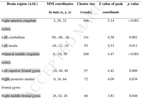
In a voxelwise multiple regression analysis, we found significantly reduced iFC in the right
anterior and bilateral middle cingulate cortex, left insula, right middle frontal gyrus, left cerebellum,
and the right posterior medial and left superior frontal gyri with increasing age (controlled for the
individual mean volume-to-volume head motion, gender, and education, p < 0.05 FWE-corrected at
the cluster level; Figure 3 and Table 3).
3.3. The iFC in the cingulo-opercular network is associated with visual processing speed C
Within the cingulo-opercular network, the iFC values of the left insula cluster correlated significantly
with the visual processing speed C estimates while controlling for normalized brain volume (r(88) =
0.33, p = 0.002; Figure 4). None of the other clusters correlated significantly with visual processing
speed C in the entire sample (all p-values > 0.244).
The iFC values of the left insular cluster were also significantly related to the time needed to
cingulo-M
AN
US
CR
IP
T
AC
CE
PT
ED
ACCEPTED MANUSCRIPT
opercular network’s clusters correlated significantly with performance in the TMT-A (bilateral
middle cingulate cortex: r = -0.37; and right posterior medial frontal gyrus: r = -0.34; both p-values <
0.007, Bonferroni-corrected).
3.4. Control analyses
3.4.1. Specificity of the role of the iFC in the cingulo-opercular network for visual processing speed
C. For the default-mode network, age-related decreased iFC was found in the right middle temporal
gyrus and the right cuneus. However, none of these clusters’ iFC values correlated significantly with
visual processing speed C while controlling for brain volume (both p-values > 0.118). For the
dorsal-attention network, age-related decreased iFC was found in the left superior, middle, and inferior
(pars triangularis) frontal gyri, left inferior parietal lobule, left precuneus, and right middle temporal
gyrus. As with the default-mode network clusters, none of the dorsal-attention network clusters’ iFC
values correlated significantly with visual processing speed C while controlling for brain volume (all
p-values > 0.157).
To test whether the iFC of the left insula cluster was associated specifically with visual
processing speed (and not generally with visual attention), we examined its correlation with VSTM
capacity K and perceptual threshold t0, controlling for brain volume. Similar to visual processing
speed C, VSTM storage capacity K was significantly correlated with age, and a non-significant trend
was also seen for the visual perceptual threshold t0 (Table 2). However, none of the correlations
between iFC in the cingulo-opercular network and these measures was significant (both p-values >
0.125), thus supporting the specificity of the association between the iFC in the cingulo-opercular
network with visual processing speed. Likewise, none of the other iFC clusters of the
cingulo-opercular network correlated significantly with K (all p-values > 0.355) or t0 (all p-values > 0.202).
3.4.2. Control of potential confounds in the association between iFC in the cingulo-opercular
p-M
AN
US
CR
IP
T
AC
CE
PT
ED
ACCEPTED MANUSCRIPT
values > 0.073). Furthermore, the iFC of the left insula cluster did not significantly correlate with the
anxiety scores derived from both STAI scales (both p-values > 0.230). Controlling for each of the
anxiety scores did also not affect the correlation between visual processing speed C and the iFC of
the left insula cluster (both p-values < 0.003). Similarly, controlling for possible vascular risk or gray
matter density within the left insula cluster did not compromise the association between the iFC of
the left insula cluster and visual processing speed C (see Supplementary Material).
3.5. The cingulo-opercular network mediates the age-related reduction in visual processing speed
Finally, we explored whether the iFC in the cingulo-opercular network mediated the association
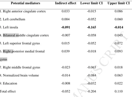
between age and visual processing speed. We included all age-decreased cingulo-opercular network
clusters (Table 3), along with normalized brain volume and education, as potential mediators (i.e.,
multiple mediation; Preacher and Hayes, 2008) of the relationship between age and visual processing
speed. Table 4 lists the indirect effects (through the iFC in the cingulo-opercular network) of age on
visual processing speed for each of those potential mediators as well as the corresponding bootstrap
intervals at 95% level of confidence. In line with the partial correlation results presented in the
previous section, only the left insula cluster was found to significantly mediate the association
between age and visual processing speed [direct effect of age on visual processing speed: b = -0.060,
standard error = 0.060, 95% confidence interval (-0.178, 0.059); see also Table 4]. Figure 5 depicts
the path model of the mediation for the left insula cluster only. This model suggests that every year
that age increases results in a decrease of visual processing speed C of 0.09 through the sequential
effects of age on iFC in the left insula cluster (blue arrow) and of iFC in the left insula cluster on
visual processing speed (red arrow).
In contrast to the TVA-based parameter of visual processing speed, as measured with whole
-M
AN
US
CR
IP
T
AC
CE
PT
ED
ACCEPTED MANUSCRIPT
0.063 – 0.079) nor any other cluster of the cingulo-opercular network mediated the association
between age and visual processing speed as measured by the TMT-A.
Control analyses were carried out to test the specificity of the relation between iFC in the
cingulo-opercular network on the one hand and visual processing speed C on the other. First, testing
analogous mediation models for the default-mode and, respectively, the dorsal-attention network’s
age-relevant clusters yielded no significant results (see Supplementary Material). Second, the left
insula cluster also did not mediate the association between age and VSTM capacity K (indirect
effect: -0.002, CI: -0.006 – 0.000) or between age and visual perceptual threshold t0 (indirect effect:
0.029, CI: -0.056 – 0.136).
4. Discussion
In this study, we investigated whether age-related decreases of iFC in the cingulo-opercular network
are related to those in visual processing speed. First, we identified regions with age-related decrease
of the iFC in the cingulo-opercular network in a group of participants of widely varying age. We
found that, among these clusters, iFC in the left insula was significantly associated with a parametric,
TVA-based measure of visual processing speed. This was confirmed by the results from a
paper-and-pencil test of visual processing speed. Furthermore, the iFC in the respective cluster significantly
mediated the effect of age on visual processing speed as measured using the TVA-based paradigm,
but not as measured with the paper-and-pencil task. Taken together, our results suggest that the iFC
in the cingulo-opercular network is of particular relevance for understanding visual processing speed
reductions in healthy aging.
4.1. Mediator role of the iFC in the cingulo-opercular network in the visual processing speed
M
AN
US
CR
IP
T
AC
CE
PT
ED
ACCEPTED MANUSCRIPT
Our results (Table 2) replicate previous findings concerning decreases in both visual processing
speed (Espeseth et al., 2014; Habekost et al., 2013; McAvinue et al., 2012; Salthouse, 1996;
Tombaugh, 2004) and the iFC in the cingulo-opercular network (Figure 3) (He et al., 2014; Meier et
al., 2012; Onoda et al., 2012) over the course of normal aging. More importantly, they demonstrate
that a decreased iFC in the cingulo-opercular network is associated with the age-related reduction in
visual processing speed (Figure 4). Specifically, the left insular iFC within the cingulo-opercular
network was significantly related to visual processing speed and further found to mediate the
association between age and speed (Table 4, Figure 5). The anterior insula is a hub of the human
brain (Power et al., 2013) and, in particular, an anchor of the cingulo-opercular network (e.g., Seeley
et al., 2007). It plays a crucial role in allowing switching between the activity of the central executive
network and of the default mode network (Sridharan et al., 2008). Accordingly,the anterior insula
has also been proposed to play an integrative role between brain networks involved in external and
internal processing (e.g., Menon and Uddin, 2010), permitting the individual to prepare for external
or internal processing in the presence of visual stimuli (e.g., Riedl et al., 2016). A comprehensive
review on the role of the anterior insula in perceptual tasks proposed that it mediates heightened
alertness in sensory cortices, thus rendering the individual more sensitive to sensory stimulation
(Sterzer & Kleinschmidt, 2010). In the visual domain, according to TVA (Bundesen, 1990), this
would directly relate to visual processing speed. Although in line with a prominent role of the
anterior insula for visual processing speed, our results should not be interpreted as indicating a
specific role of a circumscribed region within the cingulo-opercular network. Given that the insula
iFC values are relative values (i.e., with respect to the other voxels of an individual’s brain) within a
network, we can only state that the left insula cluster best represented the cingulo-opercular network
association with age and visual processing speed. Accordingly, we keep the interpretation of our
M
AN
US
CR
IP
T
AC
CE
PT
ED
ACCEPTED MANUSCRIPT
The results of the current study are in line with previous documentations of (i) significant
associations between task-evoked (Coste and Kleinschmidt, 2016; Sadaghiani and D'Esposito, 2015)
and spontaneous (Sadaghiani et al., 2010; Schneider et al., 2016) fMRI BOLD activity of the
cingulo-opercular network and the level of tonic alertness; of (ii) a close association between tonic
alertness and TVA parameter visual processing speed C (Bundesen et al., 2015; Finke et al., 2010;
Matthias et al., 2010; Vangkilde et al., 2013); and of (iii) significant differences between young
participants with relatively high and relatively low visual processing speed C especially in the iFC in
the cingulo-opercular network (Ruiz-Rizzo et al., 2018). In addition to these prior studies, we now
provide evidence for the crucial role of the cingulo-opercular network for the decrease of visual
processing speed across adult age. The mediating role of iFC in the cingulo-opercular network
between age and visual processing speed (Table 4) suggests that it is not aging per se that, in a
deterministic manner, gives rise to the well-established decrements in visual processing speed.
Instead, it implies that, even at an advanced age, individuals might exhibit relatively ‘normal’ visual
processing speed (comparable to that of younger individuals), given ‘normal’ (i.e., youth-like) iFC in
the cingulo-opercular network.
We found the relationship of the iFC in the cingulo-opercular network to visual processing
speed to be additionally supported by a measure that is widely used in the clinical settings for
assessing processing speed: performance in the TMT-A also correlated significantly with the iFC of
the left insula. Additionally, other clusters, such as the middle cingulate cortex, and the posterior
medial frontal gyrus correlated with performance in the TMT-A. But unlike with our TVA-derived
measure of visual processing speed C, the association between age and performance on the TMT-A
was not mediated by the iFC in any cluster within the cingulo-opercular network that showed
age-related decrease in iFC. This is likely attributable to the fact that, as in most standard processing
speed tasks, performance level in the TMT-A heavily relies on additional functions, such as other
M
AN
US
CR
IP
T
AC
CE
PT
ED
ACCEPTED MANUSCRIPT
prone to age-related decline, age-related decrease in TMT-A performance is likely related to a
relatively broad range, or more general, brain changes on the one hand. On the other, the TVA-based
measure, with its high cognitive specificity is optimally suited to identifying the particular brain
changes contributing to the reduction of visual processing speed.
Visual processing speed has long been known to explain a substantial part of the variability in
different, speed-dependent cognitive tasks and fluid intelligence, especially in older adults (e.g.,
Deary et al., 2001; Deary et al., 2010; Deary and Stough, 1996). Moreover, the TVA visual
processing-speed parameter C has been suggested to represent a quantitative measure of an
individual, latent parameter (Finke et al., 2005) with substantial influence on cognitive capabilities.
Our results thus indicate that the previously established links between the iFC of the left insula in the
cingulo-opercular network and general cognitive measures in older adults (e.g., He et al., 2014;
Onoda et al., 2012) are mediated by a reduction in this more specific, basic function.
4.2. Specificity of the association between the iFC in the cingulo-opercular network and visual
processing speed
As not only the iFC in the cingulo-opercular network is known to decrease with aging, a number of
control analyses were run in order to confirm the specificity of the relation between iFC in the
cingulo-opercular network and visual processing speed. In particular, we looked at iFC in the default
mode and dorsal attention networks (Andrews-Hanna et al., 2007; Damoiseaux et al., 2008; Ferreira
and Busatto, 2013). However, we found no evidence for the iFC in these two networks to contribute
to the variance in visual processing speed. Thus, rather than supporting a general relationship
between age-related decreases in iFC across diverse networks and visual processing speed
decrements, our results indicate that such a relationship is particular for the iFC in the
M
AN
US
CR
IP
T
AC
CE
PT
ED
ACCEPTED MANUSCRIPT
Similarly, as not only visual processing speed, but also other visual attention functions, such
as the visual perceptual threshold and VSTM storage capacity, change with aging (Espeseth et al.,
2014; McAvinue et al., 2012), we also looked at the relationship of the iFC in the cingulo-opercular
network and these functions. The cognitive purity of the different measures obtained with
TVA-based testing permitted us to identify a specific role for reductions in visual processing speed. Our
results showed that the iFC of the cingulo-opercular network was only associated with visual
processing speed C. Of theoretical importance, this is in line with a central assumption of the neural
interpretation of TVA (NTVA, Bundesen et al., 2005), namely, that the different visual attention
parameters reflect distinct neural processes that contribute independently to the individual attentional
performance. Thus, it is likely that the brain mechanisms underlying changes in VSTM capacity and
the perceptual threshold are distinct from those in visual processing speed. Of note, as the
TVA-based testing does not rely on motor speed, our results can also not be explained by age-related
changes in motor performance.
Furthermore, iFC in the cingulo-opercular network is known to relate not only to cognitive
measures, but also to anxiety (Seeley et al., 2007). As anxiety might influence visual processing
speed, as indicated, for example, by reduced processing speed C in patients with major depression
(Gögler et al., 2017), we analyzed whether anxiety might be a critical confound in our findings.
However, the STAI (trait and state) anxiety scores failed to correlate with iFC in the left insula
cluster of the cingulo-opercular network.
Additional potential confounds of our results are both vascular (e.g., reactivity or pathology
of the blood vessels; D'Esposito et al., 2003) and structural changes (e.g., changes in gray matter; Lu
et al., 2013) that occur with aging. However, the association between iFC and visual processing
speed still held while controlling for those factors. Thus, our results point to variations in the intrinsic
functional organization of the cingulo-opercular network as critical for reductions in visual
M
AN
US
CR
IP
T
AC
CE
PT
ED
ACCEPTED MANUSCRIPT
4.3. Limitations
Our study has a number of limitations. First, the results are based on cross-sectional and correlational
data, which does not allow firm inferences to be drawn regarding the directional relationship(s)
among aging, iFC, and visual processing speed. Second, the mediation analysis would need to be
replicated in an independent, longitudinally assessed sample. And third, such analyses should
additionally include other potential mediators derived from further brain or environmental measures.
For example, it remains to be determined whether structural connectivity changes (e.g., as measured
by tractography) within the cingulo-opercular network or with other networks underlie the changes
in iFC.
4.4. Conclusion
Our results indicate a mediator role of the iFC in the cingulo-opercular network in the impact of
aging on visual processing speed. They thus indicate that it is not simply aging per se that, in a
deterministic manner, gives rise to the well-established decrements in visual processing speed.
Rather, they imply that, even at an advanced age, individuals might exhibit relatively ‘normal’ visual
processing speed (comparable to that of younger individuals), given ‘normal’ (i.e., youth-like) iFC in
the cingulo-opercular network. Future longitudinal studies could attempt to identify whether, among
older individuals, changes in iFC and visual processing speed occur at similar or different time
points. To conclude, the evidence presented here, for the first time, points to a significant role of the
iFC in the cingulo-opercular network in the attentional processing capacity of healthy aging
individuals.
M
AN
US
CR
IP
T
AC
CE
PT
ED
ACCEPTED MANUSCRIPT
This work was supported by the European Union’s Seventh Framework Programme for research,
technological development and demonstration (INDIREA, grant no. ITN-2013-606901 to H.J.M and
K.F.), by the Alzheimer Research Initiative e.V. (AFI) (Grant number 12819 to K.F. and C.S.); and
the German Research Foundation (grant no. FI 1424 to K.F. and grant no. SO 1336 to C.S.).
6. Acknowledgments
We thank Dr. Anders Petersen for providing the TVA modeling scripts and Dr. Petra Redel for
organizational support.
7. References
Andrews-Hanna, J.R., Snyder, A.Z., Vincent, J.L., Lustig, C., Head, D., Raichle, M.E., Buckner, R.L. 2007. Disruption of large-scale brain systems in advanced aging. Neuron 56(5), 924-35. doi:10.1016/j.neuron.2007.10.038. Barnes, L.L., Harp, D., Jung, W.S. 2002. Reliability generalization of scores on the Spielberger state-trait anxiety
inventory. Educational and Psychological Measurement 62(4), 603-18.
Beck, A.T., Steer, R.A., Brown, G.K. 1996. Manual for the Beck Depression Inventory-II. Second Edition ed. The Psychological Corporation, San Antonio, TX.
Beckmann, C.F., Mackay, C.E., Filippini, N., Smith, S. 2009. Group comparison of resting-state FMRI data using multi-subject ICA and dual regression. Neuroimage 47(Suppl 1), S148.
Beckmann, C.F., Smith, S.M. 2004. Probabilistic independent component analysis for functional magnetic resonance imaging. IEEE Trans Med Imaging 23(2), 137-52. doi:10.1109/TMI.2003.822821.
Bublak, P., Redel, P., Sorg, C., Kurz, A., Forstl, H., Muller, H.J., Schneider, W.X., Finke, K. 2011. Staged decline of visual processing capacity in mild cognitive impairment and Alzheimer's disease. Neurobiol Aging 32(7), 1219-30. doi:10.1016/j.neurobiolaging.2009.07.012.
Bundesen, C. 1990. A theory of visual attention. Psychol Rev 97(4), 523-47.
Bundesen, C., Habekost, T., Kyllingsbaek, S. 2005. A neural theory of visual attention: bridging cognition and neurophysiology. Psychol Rev 112(2), 291-328. doi:10.1037/0033-295X.112.2.291.
Bundesen, C., Vangkilde, S., Petersen, A. 2015. Recent developments in a computational theory of visual attention (TVA). Vision Res 116(Pt B), 210-8. doi:10.1016/j.visres.2014.11.005.
Chao-Gan, Y., Yu-Feng, Z. 2010. DPARSF: A MATLAB Toolbox for "Pipeline" Data Analysis of Resting-State fMRI. Front Syst Neurosci 4, 13. doi:10.3389/fnsys.2010.00013.
Coste, C.P., Kleinschmidt, A. 2016. Cingulo-opercular network activity maintains alertness. Neuroimage 128, 264-72. doi:10.1016/j.neuroimage.2016.01.026.
D'Esposito, M., Deouell, L.Y., Gazzaley, A. 2003. Alterations in the BOLD fMRI signal with ageing and disease: a challenge for neuroimaging. Nat Rev Neurosci 4(11), 863-72. doi:10.1038/nrn1246.
Damoiseaux, J.S., Beckmann, C.F., Arigita, E.J., Barkhof, F., Scheltens, P., Stam, C.J., Smith, S.M., Rombouts, S.A. 2008. Reduced resting-state brain activity in the "default network" in normal aging. Cereb Cortex 18(8), 1856-64. doi:10.1093/cercor/bhm207.
De Luca, M., Beckmann, C.F., De Stefano, N., Matthews, P.M., Smith, S.M. 2006. fMRI resting state networks define distinct modes of long-distance interactions in the human brain. Neuroimage 29(4), 1359-67.
doi:10.1016/j.neuroimage.2005.08.035.
Deary, I.J., Der, G., Ford, G. 2001. Reaction times and intelligence differences: A population-based cohort study. Intelligence 29(5), 389-99. doi:https://doi.org/10.1016/S0160-2896(01)00062-9.
Deary, I.J., Johnson, W., Starr, J.M. 2010. Are processing speed tasks biomarkers of cognitive aging? Psychol Aging 25(1), 219-28. doi:10.1037/a0017750.
M
AN
US
CR
IP
T
AC
CE
PT
ED
ACCEPTED MANUSCRIPT
Dosenbach, N.U., Fair, D.A., Cohen, A.L., Schlaggar, B.L., Petersen, S.E. 2008. A dual-networks architecture of top-down control. Trends Cogn Sci 12(3), 99-105. doi:10.1016/j.tics.2008.01.001.
Dosenbach, N.U., Fair, D.A., Miezin, F.M., Cohen, A.L., Wenger, K.K., Dosenbach, R.A., Fox, M.D., Snyder, A.Z., Vincent, J.L., Raichle, M.E., Schlaggar, B.L., Petersen, S.E. 2007. Distinct brain networks for adaptive and stable task control in humans. Proc Natl Acad Sci U S A 104(26), 11073-8. doi:10.1073/pnas.0704320104.
Dyrholm, M., Kyllingsbaek, S., Espeseth, T., Bundesen, C. 2011. Generalizing parametric models by introducing trial-by-trial parameter variability: The case of TVA. Journal of Mathematical Psychology 55(6), 416-29.
doi:https://doi.org/10.1016/j.jmp.2011.08.005.
Espeseth, T., Vangkilde, S.A., Petersen, A., Dyrholm, M., Westlye, L.T. 2014. TVA-based assessment of attentional capacities-associations with age and indices of brain white matter microstructure. Front Psychol 5, 1177. doi:10.3389/fpsyg.2014.01177.
Feinberg, D.A., Setsompop, K. 2013. Ultra-fast MRI of the human brain with simultaneous multi-slice imaging. J Magn Reson 229, 90-100. doi:10.1016/j.jmr.2013.02.002.
Ferreira, L.K., Busatto, G.F. 2013. Resting-state functional connectivity in normal brain aging. Neurosci Biobehav Rev 37(3), 384-400. doi:10.1016/j.neubiorev.2013.01.017.
Filippini, N., MacIntosh, B.J., Hough, M.G., Goodwin, G.M., Frisoni, G.B., Smith, S.M., Matthews, P.M., Beckmann, C.F., Mackay, C.E. 2009. Distinct patterns of brain activity in young carriers of the APOE-epsilon4 allele. Proc Natl Acad Sci U S A 106(17), 7209-14. doi:10.1073/pnas.0811879106.
Finke, K., Bublak, P., Krummenacher, J., Kyllingsbaek, S., Muller, H.J., Schneider, W.X. 2005. Usability of a theory of visual attention (TVA) for parameter-based measurement of attention I: evidence from normal subjects. J Int Neuropsychol Soc 11(7), 832-42.
Finke, K., Dodds, C.M., Bublak, P., Regenthal, R., Baumann, F., Manly, T., Muller, U. 2010. Effects of modafinil and methylphenidate on visual attention capacity: a TVA-based study. Psychopharmacology (Berl) 210(3), 317-29. doi:10.1007/s00213-010-1823-x.
Folstein, M.F., Folstein, S.E., McHugh, P.R. 1975. "Mini-mental state". A practical method for grading the cognitive state of patients for the clinician. J Psychiatr Res 12(3), 189-98.
Fox, M.D., Raichle, M.E. 2007. Spontaneous fluctuations in brain activity observed with functional magnetic resonance imaging. Nat Rev Neurosci 8(9), 700-11. doi:10.1038/nrn2201.
Gögler, N., Willacker, L., Funk, J., Strube, W., Langgartner, S., Napiorkowski, N., Hasan, A., Finke, K. 2017. Single-session transcranial direct current stimulation induces enduring enhancement of visual processing speed in patients with major depression. Eur Arch Psychiatry Clin Neurosci 267(7), 671-86. doi:10.1007/s00406-016-0761-y.
Habekost, T., Petersen, A., Vangkilde, S. 2014. Testing attention: comparing the ANT with TVA-based assessment. Behav Res Methods 46(1), 81-94. doi:10.3758/s13428-013-0341-2.
Habekost, T., Vogel, A., Rostrup, E., Bundesen, C., Kyllingsbaek, S., Garde, E., Ryberg, C., Waldemar, G. 2013. Visual processing speed in old age. Scand J Psychol 54(2), 89-94. doi:10.1111/sjop.12008.
Hayes, A.F. 2012. PROCESS: A versatile computational tool for observed variable mediation, moderation, and conditional process modeling. http://www.afhayes.com/public/process2012.pdf.
Hayes, A.F., Rockwood, N.J. 2017. Regression-based statistical mediation and moderation analysis in clinical research: Observations, recommendations, and implementation. Behav Res Ther 98, 39-57. doi:10.1016/j.brat.2016.11.001. He, X., Qin, W., Liu, Y., Zhang, X., Duan, Y., Song, J., Li, K., Jiang, T., Yu, C. 2014. Abnormal salience network in
normal aging and in amnestic mild cognitive impairment and Alzheimer's disease. Hum Brain Mapp 35(7), 3446-64. doi:10.1002/hbm.22414.
Hyvarinen, A. 1999. Fast and robust fixed-point algorithms for independent component analysis. IEEE Trans Neural Netw 10(3), 626-34. doi:10.1109/72.761722.
Kyllingsbaek, S. 2006. Modeling visual attention. Behav Res Methods 38(1), 123-33.
Laux, L., Glanzmann, P., Schaffner, P., Spielberger, C.D. 1981. Das State-Trait-Angstinventar (Testmappe mit Handanweisung, Fragebogen STAI-G Form X 1 und Fragebogen STAI-G Form X 2). Beltz, Weinheim.
Lu, P.H., Lee, G.J., Tishler, T.A., Meghpara, M., Thompson, P.M., Bartzokis, G. 2013. Myelin breakdown mediates age-related slowing in cognitive processing speed in healthy elderly men. Brain Cogn 81(1), 131-8.
doi:10.1016/j.bandc.2012.09.006.
Mackinnon, D.P., Fairchild, A.J. 2009. Current Directions in Mediation Analysis. Curr Dir Psychol Sci 18(1), 16. doi:10.1111/j.1467-8721.2009.01598.x.
Matthias, E., Bublak, P., Muller, H.J., Schneider, W.X., Krummenacher, J., Finke, K. 2010. The influence of alertness on spatial and nonspatial components of visual attention. J Exp Psychol Hum Percept Perform 36(1), 38-56.
doi:10.1037/a0017602.
M
AN
US
CR
IP
T
AC
CE
PT
ED
ACCEPTED MANUSCRIPT
Meier, T.B., Desphande, A.S., Vergun, S., Nair, V.A., Song, J., Biswal, B.B., Meyerand, M.E., Birn, R.M., Prabhakaran, V. 2012. Support vector machine classification and characterization of age-related reorganization of functional brain networks. Neuroimage 60(1), 601-13. doi:10.1016/j.neuroimage.2011.12.052.
Menon, V., Uddin, L.Q. 2010. Saliency, switching, attention and control: a network model of insula function. Brain Struct Funct 214(5-6), 655-67. doi:10.1007/s00429-010-0262-0.
Murphy, K., Bodurka, J., Bandettini, P.A. 2007. How long to scan? The relationship between fMRI temporal signal to noise ratio and necessary scan duration. Neuroimage 34(2), 565-74. doi:10.1016/j.neuroimage.2006.09.032. Nickerson, L.D., Smith, S.M., Ongur, D., Beckmann, C.F. 2017. Using Dual Regression to Investigate Network Shape
and Amplitude in Functional Connectivity Analyses. Front Neurosci 11, 115. doi:10.3389/fnins.2017.00115. Onoda, K., Ishihara, M., Yamaguchi, S. 2012. Decreased functional connectivity by aging is associated with cognitive
decline. J Cogn Neurosci 24(11), 2186-98. doi:10.1162/jocn_a_00269.
Posner, M.I., Petersen, S.E. 1990. The attention system of the human brain. Annu Rev Neurosci 13, 25-42. doi:10.1146/annurev.ne.13.030190.000325.
Power, J.D., Barnes, K.A., Snyder, A.Z., Schlaggar, B.L., Petersen, S.E. 2012. Spurious but systematic correlations in functional connectivity MRI networks arise from subject motion. Neuroimage 59(3), 2142-54.
doi:10.1016/j.neuroimage.2011.10.018.
Power, J.D., Schlaggar, B.L., Lessov-Schlaggar, C.N., Petersen, S.E. 2013. Evidence for hubs in human functional brain networks. Neuron 79(4), 798-813. doi:10.1016/j.neuron.2013.07.035.
Preacher, K.J., Hayes, A.F. 2004. SPSS and SAS procedures for estimating indirect effects in simple mediation models. Behav Res Methods Instrum Comput 36(4), 717-31.
Preacher, K.J., Hayes, A.F. 2008. Asymptotic and resampling strategies for assessing and comparing indirect effects in multiple mediator models. Behav Res Methods 40(3), 879-91.
Preibisch, C., Castrillon, G.J., Buhrer, M., Riedl, V. 2015. Evaluation of Multiband EPI Acquisitions for Resting State fMRI. PLoS One 10(9), e0136961. doi:10.1371/journal.pone.0136961.
Raichle, M.E. 2015. The restless brain: how intrinsic activity organizes brain function. Philos Trans R Soc Lond B Biol Sci 370(1668). doi:10.1098/rstb.2014.0172.
Reitan, R.M., Wolfson, D. 1985. The Halstead-Reitan neuropsychological test battery: therapy and clinical interpretation. Neuropsychological Press, Tucson, AZ.
Ruiz-Rizzo, A.L., Bublak, P., Redel, P., Grimmer, T., Muller, H.J., Sorg, C., Finke, K. 2017. Simultaneous object perception deficits are related to reduced visual processing speed in amnestic mild cognitive impairment. Neurobiol Aging 55, 132-42. doi:10.1016/j.neurobiolaging.2017.03.029.
Ruiz-Rizzo, A.L., Neitzel, J., Muller, H.J., Sorg, C., Finke, K. 2018. Distinctive Correspondence Between Separable Visual Attention Functions and Intrinsic Brain Networks. Front Hum Neurosci 12, 89.
doi:10.3389/fnhum.2018.00089.
Sadaghiani, S., D'Esposito, M. 2015. Functional Characterization of the Cingulo-Opercular Network in the Maintenance of Tonic Alertness. Cereb Cortex 25(9), 2763-73. doi:10.1093/cercor/bhu072.
Sadaghiani, S., Scheeringa, R., Lehongre, K., Morillon, B., Giraud, A.L., Kleinschmidt, A. 2010. Intrinsic connectivity networks, alpha oscillations, and tonic alertness: a simultaneous electroencephalography/functional magnetic resonance imaging study. J Neurosci 30(30), 10243-50. doi:10.1523/JNEUROSCI.1004-10.2010.
Salthouse, T.A. 1996. The processing-speed theory of adult age differences in cognition. Psychol Rev 103(3), 403-28. Schneider, M., Hathway, P., Leuchs, L., Samann, P.G., Czisch, M., Spoormaker, V.I. 2016. Spontaneous pupil dilations
during the resting state are associated with activation of the salience network. Neuroimage 139, 189-201. doi:10.1016/j.neuroimage.2016.06.011.
Seeley, W.W., Menon, V., Schatzberg, A.F., Keller, J., Glover, G.H., Kenna, H., Reiss, A.L., Greicius, M.D. 2007. Dissociable intrinsic connectivity networks for salience processing and executive control. J Neurosci 27(9), 2349-56. doi:10.1523/JNEUROSCI.5587-06.2007.
Sestieri, C., Corbetta, M., Spadone, S., Romani, G.L., Shulman, G.L. 2014. Domain-general signals in the cingulo-opercular network for visuospatial attention and episodic memory. J Cogn Neurosci 26(3), 551-68.
doi:10.1162/jocn_a_00504.
Smith, D.V., Utevsky, A.V., Bland, A.R., Clement, N., Clithero, J.A., Harsch, A.E., McKell Carter, R., Huettel, S.A. 2014. Characterizing individual differences in functional connectivity using dual-regression and seed-based approaches. Neuroimage 95, 1-12. doi:10.1016/j.neuroimage.2014.03.042.
Smith, S.M., Fox, P.T., Miller, K.L., Glahn, D.C., Fox, P.M., Mackay, C.E., Filippini, N., Watkins, K.E., Toro, R., Laird, A.R., Beckmann, C.F. 2009. Correspondence of the brain's functional architecture during activation and rest. Proc Natl Acad Sci U S A 106(31), 13040-5. doi:10.1073/pnas.0905267106.