R E S E A R C H A R T I C L E
Open Access
Decrease of physical activity level in adolescents
with limb fractures: an accelerometry-based
activity monitor study
Dimitri Ceroni
1,4*, Xavier Martin
1, Cécile Delhumeau
2and Nathalie Farpour-Lambert
3Abstract
Background:Immobilization and associated periods of inactivity can cause osteopenia, the physiological response of the bone to disuse. Mechanical loading plays an essential role in maintaining bone integrity. Skeletal fractures represent one cause of reduction of the physical activity (PA) level in adolescents. The purpose of this study was to quantify the reduction of PA in adolescents with limb fractures during the cast immobilization period compared with healthy controls.
Methods:Two hundred twenty adolescents were divided into three groups: those with upper limb fractures (50 cases); lower limb fractures (50 cases); and healthy cases (120 cases). Patients and their healthy peers were matched for gender, age, and seasonal assessment of PA. PA level was assessed during cast immobilization by accelerometer. Time spent in PA in each of the different intensity levels sedentary, light, moderate, and vigorous -was determined for each participant and expressed in minutes and as a percentage of total valid time.
Results:Reduction in PA during cast immobilization was statistically significant in patients with limb fractures compared to healthy controls. The total PA count (total number of counts/min) was significantly lower in those with upper and lower limb fractures (-30.1% and -62.4%, respectively) compared with healthy controls (p < 0.0001 and p = 0.0003, respectively). Time spent in moderate-to-vigorous PA by patients with upper and lower limb injuries decreased by 36.9% (p = 0.0003) and 76.6% (p < 0.0001), respectively, and vigorous PA was reduced by 41.4% (p= 0.0008) and 84.4% (p< 0.0001), respectively.
Conclusions:PA measured by accelerometer is a useful and valid tool to assess the decrease of PA level in adolescents with limb fractures. As cast immobilization and reduced PA are known to induce bone mineral loss, this study provides important information to quantify the decrease of skeletal loading in this patient population. The observed reduction of high intensity skeletal loading due to the decrease in vigorous PA may explain osteopenia due to disuse, and these data should be kept in mind by trauma practitioners to avoid any unnecessary prolongation of the cast immobilization period.
Background
Bone mineral mass acquired during adolescence is considered as a major determinant of adult bone health [1]. Although genetic factors have a strong influence on peak bone mass, environmental factors, such as physical activity (PA), also contribute [2,3]. Exercise exerts a positive effect on bone growth, particularly if the activity has been initiated before puberty or in the early pubertal
period [4-6]. The skeleton appears to be most respon-sive to mechanical stress during Tanner stages II-IV and corresponds to the two-year window that has been identified as the period of bone mineral accrual during puberty [7,8]. Bone mineral mass is higher in physically active children than in those who are mildly active [9], and higher in children who participate in activities that generate high impact forces than in those who practise activities with lower impact forces [10-12]. Studies have shown that high-intensity forces, especially when imposed during early childhood, produce greater gains in bone mass than low- to moderate-intensity forces * Correspondence: [email protected]
1
Pediatric Orthopedic Unit, University of Geneva Hospitals and University of Geneva Faculty of Medicine, Geneva, Switzerland
Full list of author information is available at the end of the article
[13-18]. Based on this evidence, it is now recommended that physical activity for children include activities generating relatively high ground-reaction forces, such as jumping, skipping, running, and possibly strengthen-ing exercises [5].
It is recognized also that immobilization and associated periods of inactivity can cause osteopenia [19-21], the physiological response of the bone to disuse. Mechanical loading plays an essential role in maintaining bone integ-rity. Mechanisms underlying bone loss in response to dis-use are not yet completely understood, even if it is recognized that they are due to increased bone resorption and corresponding reduced bone formation. In adoles-cents, loss of bone mineral mass usually occurs during phases of reduced PA, such as cast immobilization [22]. To the best of our knowledge, the measurement of PA levels by accelerometry has not been investigated in ado-lescents who sustain limb fractures and have to wear a cast for several weeks. The objective of our study was to quantify the reduction of PA levels during cast immobili-zation in this patient population.
Methods Subjects
One hundred adolescents with a first episode of limb fracture (50 lower limb fractures; 50 upper limb frac-tures) and a control group of 120 healthy cases were recruited for inclusion in the study through an adver-tisement for visitors to the Children’s Hospital of the University of Geneva Hospitals, Geneva, Switzerland. Exclusion criteria for both injured adolescents and healthy controls were: prior history of bone fractures; chronic disease; congenital or acquired bone disease; any condition limiting physical activity; and hospitaliza-tion for more than 2 weeks in the previous 12 months.
Adolescents with limb fractures were treated as inpati-ents and required general anesthetic for the treatment of their fracture. Those with lower limb fractures received a bent-knee long-leg cast and were instructed to follow a strict zero weight-bearing directive during the initial healing phase. After 3 to 6 weeks, the initial immobiliza-tion device was removed and a below-the-knee walking cast was worn until definitive bone healing. All adoles-cents with upper limb fractures were immoblized into a long-arm cast during the initial healing phase, followed by a forearm cast until definitive bone healing. All study participants and their parents provided written consent and the protocol was approved by the institutional ethics committee (protocol # 04-057, ped 04-002).
Anthropometric measurements
Standing height was assessed in bare or stocking feet to the nearest 1 mm using a precision mechanical stadi-ometer (Holtain Ltd, Dyfed, UK). Weight was measured
to the nearest 0.1 kg using a mechanical calibrated beam
scale (Seca®, Reinach, Switzerland). Body mass index
(BMI) was calculated as weight (kg)/height squared (m2).
Physical activity measurement
Physical activity was measured during cast immobiliza-tion and data were collected from the first day following hospital discharge. Recordings of PA began on a Mon-day, Tuesday or Wednesday to ensure measurement of at least two weekend days. Objective measurements of PA were obtained using an uniaxial accelerometer
(Acti-graph® 7164, MTI, Fort Walton Beach, FL, USA). The
monitor was set on a 1 min cycle and the sum was stored in the memory at the end of each run and the numerical integrator reset. The monitor was attached above the iliac crest of the right hip with an elastic belt and adjustable buckle, and was oriented vertically in the same direction. The accelerometer was programmed to start recording at 8 am on the first day of measurement and participants were asked to wear it continuously, including during the night, for 10 days. Data were col-lected during all seasons.
PA data interpretation
Data reduction was based on criteria applied in previous publications [3,23-26]. Only periods between 8 am and 9 pm were analysed. Zero activity periods of 20 min or longer were interpreted as being due to unworn acceler-ometers and were removed from the activity totals [27]. Participants who did not record more than 600 min/d of activity [3,23-26] for at least 5 days were excluded from further analysis [28]. Data were expressed as total activity counts per registered time (counts/min) to gen-erate an average range of PA. We used the cut-offs of intensity levels described by Ekelund where sedentary behaviour was defined as less than 500 counts/min, light PA from 500 to 1999 counts/min, moderate PA from 2000 to 2999 counts/min, and vigorous PA as > 3000 counts/min [29]. Time spent at each PA intensity level was determined for all participants as a percentage of total valid time.
Statistical methods
Results
Fourteen adolescents with lower limb fractures, 13 with upper limb fractures, and 25 control subjects were excluded either for failing to reach at least 5 days of measurement or instrument malfunction. Thirty-six adolescents with lower limb fractures, 37 with upper limb fractures, and 95 healthy controls were included in the study. Type of fracture, treatment, and cast immobi-lization duration are shown in Table 1. Patients with limb fractures were paired with healthy controls accord-ing to gender, age (± 0.5 years), and seasonal assess-ment, as the latter may have an impact on the PA level (less sporting activities during the winter months). Thirty-five patients with upper limb fractures were able to be matched with 35 healthy controls; pairing was possible in 34 cases for the lower limb fracture group. Age, physical characteristics, and PA levels of those with fractures and healthy controls are presented in Tables 2 and 3. There was no statistical difference between groups for age, height, weight, BMI, or daily duration of PA monitoring. A statistical difference for the mean number of valid monitored days was observed between patients with lower limb fractures and healthy controls
(9.1 ± 1.8 vs 8.2 ± 2, respectively; p = 0.028). As
expected, cases with limb fractures showed notable reductions in PA levels and spent more time in seden-tary activities. The total PA count (number of counts/ min) was significantly lower in those with upper and lower limb fractures (-30.1% and -62.4%, respectively) compared with healthy controls. When considering time spent in moderate-to-vigorous PA, a substantial decrease
was observed in the upper (-36.9%) and lower limb (-76.6%) fracture groups compared to matched healthy controls. Finally, the decrease in vigorous PA was highly significant for both injured groups (-41.4% and -84.4% for cases with upper and lower limb fractures, respec-tively) (Figure 1).
Discussion
Skeletal fractures represent one cause of reduction of the PA level in adolescents. To the best of our knowl-edge, this is the first study to report PA measures in a representative sample of this population with upper and lower limb fractures. As expected, we observed a signifi-cant reduction of PA levels among injured subjects dur-ing cast immobilization, especially those with lower limb fractures. Using total activity measurement, subjects with upper or lower limb fractures were considerably less active (-30.1% and -62.4%, respectively) than healthy fracture-free controls. This was even more marked for time spent in activities of moderate-to- vigorous inten-sity. For both groups of healthy control subjects, the average time spent in moderate-to-vigorous intensity activity was 59.4 min and 66.7 min daily, whereas it was 42.1 min and 13.9 min, respectively, for adolescents with upper and lower limb fractures.
[image:3.595.58.540.499.722.2]International guidelines recommend that school-age children should participate daily in at least 60 min of moderate-to-vigorous intensity activity as this level has been shown to prevent weight gain, premature develop-ment of cardiovascular diseases and type 2 diabetes, and to increase bone mineral mass [30]. PA reduction in
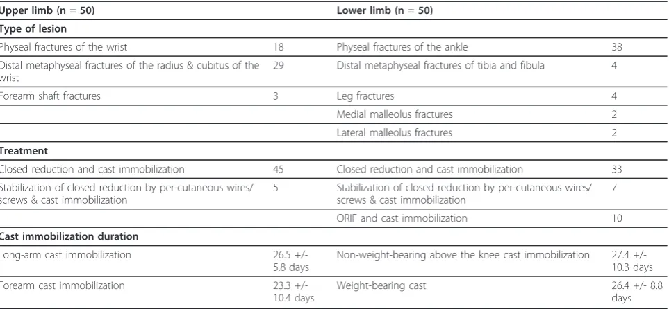
Table 1 Type of Lesion, Treatment, and Cast Immobilization Duration of Adolescents with Upper and Lower Limb Fractures
Upper limb (n = 50) Lower limb (n = 50)
Type of lesion
Physeal fractures of the wrist 18 Physeal fractures of the ankle 38
Distal metaphyseal fractures of the radius & cubitus of the wrist
29 Distal metaphyseal fractures of tibia and fibula 4
Forearm shaft fractures 3 Leg fractures 4
Medial malleolus fractures 2
Lateral malleolus fractures 2
Treatment
Closed reduction and cast immobilization 45 Closed reduction and cast immobilization 33
Stabilization of closed reduction by per-cutaneous wires/ screws & cast immobilization
5 Stabilization of closed reduction by per-cutaneous wires/ screws & cast immobilization
7
ORIF and cast immobilization 10
Cast immobilization duration
Long-arm cast immobilization 26.5
+/-5.8 days
Non-weight-bearing above the knee cast immobilization 27.4 +/-10.3 days
Forearm cast immobilization 23.3
+/-10.4 days
Weight-bearing cast 26.4 +/- 8.8
days
adolescents with fractures results in a decrease in energy expenditure and our clinical experience has shown that this decrease during the cast immobilization period may be the starting point of weight gain for children and adolescents [31].
An examination of time spent in vigorous PA is even more relevant as bone health is related to ground reac-tion forces [32]. It is well known that an intensificareac-tion
[image:4.595.58.540.111.326.2]of high impact and weight-bearing exercises has direct and positive consequences in relation to bone mass. Recent studies focus on the theory that high-intensity forces, especially if imposed rapidly, produce greater gains in bone mineral mass than low-to-moderate inten-sity forces [13,16-18]. Our study investigated specifically the time spent in vigorous PA, which reflects high-intensity forces beneficial to skeletal health, and Table 2 Characteristics and Physical Activity Measures of Adolescents with Upper Limb Fractures during Cast
Immobilization vs Healthy Controls
Injured adolescents (n = 35)
Healthy controls
(n = 35) p
value
Age (yr) 12.6 ± 1.7 12.6 ± 1.8 0.5244
Height (cm) 157.7 ± 13.4 157.7 ± 11.8 0.9989
Weight (kg) 47.3 ± 12.1 46.3 ± 11.4 0.4253
BMI (kg/m2) 18.7 ± 2.4 18.4 ± 2.5 0.2610
Number of valid monitored days (days) 8.7 +/- 2.4 8.1 +/- 2.4 0.2044
Daily duration of physical activity monitoring (min) 752.1 ± 26.8 740.9 ± 37.4 0.3177
Total activity (counts/min.day)
417.5 ± 179.1 597.1 ± 211.4 0.0003*
Time spent in sedentary activity (min/day) (% of total daily wearing time)
598.2 +/- 54.7 79.6
540.4 +/- 64.4 72.9
0.0001*
Time spent in light activity (min/day) (% of total daily wearing time)
111.8 +/- 41.1 14.9
136.5 +/- 40.1 18.4
0.0072*
Time spent in moderate (min/day) (% of total daily wearing time)
20.7 +/- 8.4 2.7
30.2 +/- 12 4.1
0.0003*
Time spent in vigorous activity (min/day) (% of total daily wearing time)
21.4 +/- 15.2 2.8
36.5 +/- 18.9 4.9
0.0008*
The results are expressed as mean ± SD.
*: statistically significant difference [please always place at foot of table].
Table 3 Characteristics and Physical Activity Measures of Adolescents with Lower Limb Fractures during Cast Immobilization vs Healthy Controls
Injured adolescents (n = 34)
Healthy controls
(n = 34) p
value
Age (yr) 13.6 ± 1.6 13.3 ± 1.6 0.2312
Height (cm) 161.9 ± 11.1 162 ± 12.9 0.9501
Weight (kg) 52.1 ± 11.8 51.8 ± 11.8 0.8128
BMI (kg/m2) 19.7 ± 2.5 19.5 ± 2.5 0.2614
Number of valid monitored days (days) 9.1 +/- 1.8 8.2 +/-2 0.0276*
Daily duration of physical activity monitoring (min) 746.7 ± 22.9 749.3 ± 33.3 0.8241
Total activity (counts/min.day)
200.9 ± 92.3 534.5 ± 207.3 <0.0001*
Time spent in sedentary activity (min/day) (% of total daily wearing time)
664.4 +/- 40.9 89
564.3 +/- 64.4 75.3
<0.0001*
Time spent in light activity (min/day) (% of total daily wearing time)
68.4 +/- 26.4 9.2
125.7 +/- 40.1 16.8
<0.0001*
Time spent in moderate (min/day) (% of total daily wearing time)
8.9 +/- 7.2 1.2
27.5 +/- 11.4 3.7
<0.0001*
Time spent in vigorous activity (min/day) (% of total daily wearing time)
5 +/- 6.8 0.6
31.8 +/- 18 4.2
<0.0001*
The results are expressed as mean ± SD.
[image:4.595.56.540.500.714.2]demonstrated a 41.4% and 84.4% decrease, respectively, in adolescents with upper and lower limb fractures. The significant reduction in high-intensity forces applied to the skeleton may provide also a valid explanation for disuse osteopenia [33]. Restoration of bone mineral mass may occur upon reaching pubertal maturity [33,34] and, above all, normal activity [35]. Nevertheless, there are currently no reported studies on the recovery of PA levels following limb fractures in children and adolescents.
The results of this study constitute a starting point for new investigations. The relationship between PA decrease and bone mineral loss in youth with fractures remains to be established in a prospective study. There are also some limitations to be considered when interpreting the findings of our study. First, our recruitment was relatively hetero-geneous, in particular with regard to the practice of sport. Even if we are unable to completely rule out the possibility of selection bias, there is no clear reason to believe that
[image:5.595.56.540.87.441.2]adolescents with markedly different PA profiles would have chosen to participate in the study. Second, there are activities during which accelerometers have to be removed (e.g., swimming) or do not accurately measure the inten-sity level (e.g., cycling). These “unmonitored”activities may result in an underestimation of PA in healthy con-trols. Nevertheless, Trost et al reported that children’s self-reported periods of“unmonitored”activity added to the registered accelerometer data led to no significant changes in calculated PA levels [36]. Third, to obtain 10 days of measurement recording, activity counts were aver-aged using a 1-min epoch to ensure that the accelerometer memory capacity was not exceeded. However, this method underestimates vigorous PA as such activity is rarely sustained for longer than 1 min [25]. Although previous studies have demonstrated that vigorous PA may be substantially underestimated [37], this is unlikely to be of importance in this study as we can hypothesize that it will be the same for the different groups.
Conclusions
PA measured by accelerometer is a useful and valid tool to assess the decrease of PA levels in adolescents with limb fractures. As cast immobilization and reduced PA are known to induce bone mineral loss, this study pro-vides important information to quantify the decrease of skeletal loading in adolescents with limb fractures. The significant reduction in time spent in vigorous PA, which reflects high-intensity forces beneficial to skeletal health, provides a valid explanation for disuse osteope-nia. These data are important and should be kept in mind by trauma practitioners to avoid an unnecessary prolongation of the cast immobilization period. This study gives an outline of the decrease of PA due to a sedentary behaviour and emphasizes the role of physical activity. Further research is needed to establish the rela-tionship between the PA level and bone mineral acquisi-tion during cast immobilizaacquisi-tion and after bone healing.
Acknowledgements
We thank the participants and their families for their willing cooperation in this study. We acknowledge Magali Oehrli and the staff of the pediatric orthopedic unit of the Department of Child and Adolescent, University of Geneva Hospitals, for their technical assistance. We acknowledge also Rosemary Sudan for editorial assistance. This work was supported by grants from the Swiss National Science Foundation (SNSF #405340-104611), and the funding source did not have any Funding sources: Supported by grants from the Swiss National Science Foundation (SNSF #405340-104611) role in the investigation.
Author details
1Pediatric Orthopedic Unit, University of Geneva Hospitals and University of
Geneva Faculty of Medicine, Geneva, Switzerland.2Clinical Epidemiology Service, University of Geneva Hospitals and University of Geneva Faculty of Medicine, Geneva, Switzerland.3Exercise Medicine, Pediatric Cardiology Unit, University of Geneva Hospitals and University of Geneva Faculty of Medicine, Geneva, Switzerland.4Department of Child and Adolescent, University of Geneva Hospitals and University of Geneva Faculty of Medicine, Geneva, Switzerland.
Authors’contributions
DC: participated in the design of the study, conceived and coordinated the study, collected and treated the patients, performed the CSA analysis, and drafted the manuscript. XM: conceived the study, participated in its coordination, collected the data, and performed the statistical analysis. CD: performed the statistical analysis and drafted the manuscript. NFL: participated in the design and conception of the study, and drafted the manuscript. All authors read and approved the final version of the manuscript.
Competing interests
The authors declare that they have no competing interests.
Received: 7 November 2010 Accepted: 4 May 2011 Published: 4 May 2011
References
1. Saggese G, Baroncelli GI, Bertelloni S:Osteoporosis in children and adolescents: diagnosis, risk factors, and prevention.J Pediatr Endocrinol Metab2001,14(7):833-59.
2. Barr SI, McKay HA:Nutrition, exercise, and bone status in youth.Int J Sport Nutr1998,8(2):124-42.
3. Craig CL, Marshall AL, Sjostrom M, Bauman AE, Booth ML, Ainsworth BE, Pratt M, Ekelund U, Yngve A, Sallis JF, Oja P:International physical activity
questionnaire: 12-country reliability and validity.Med Sci Sports Exerc
2003,35(8):1381-95.
4. Bailey DA, Martin AD, McKay HA, Whiting S, Mirwald R:Calcium accretion in girls and boys during puberty: a longitudinal analysis.J Bone Miner Res
2000,15(11):2245-50.
5. Kohrt WM, Bloomfield SA, Little KD, Nelson ME, Yingling VR:American College of Sports Medicine Position Stand: physical activity and bone health.Med Sci Sports Exerc2004,36(11):1985-96.
6. Heinonen A, Sievanen H, Kannus P, Oja P, Pasanen M, Vuori I:High-impact exercise and bones of growing girls: a 9-month controlled trial.
Osteoporos Int2000,11(12):1010-7.
7. Bailey DA:The Saskatchewan Pediatric Bone Mineral Accrual Study: bone mineral acquisition during the growing years.Int J Sports Med1997,
18(Suppl 3):S191-4.
8. Theintz G, Buchs B, Rizzoli R, Slosman D, Clavien H, Sizonenko PC, Bonjour JP:Longitudinal monitoring of bone mass accumulation in healthy adolescents: evidence for a marked reduction after 16 years of age at the levels of lumbar spine and femoral neck in female subjects.J Clin Endocrinol Metab1992,75(4):1060-5.
9. Slemenda CW, Miller JZ, Hui SL, Reister TK, Johnston CC Jr:Role of physical activity in the development of skeletal mass in children.J Bone Miner Res
1991,6(11):1227-33.
10. Cassell C, Benedict M, Specker B:Bone mineral density in elite 7- to 9-yr-old female gymnasts and swimmers.Med Sci Sports Exerc1996,
28(10):1243-6.
11. Courteix D, Lespessailles E, Peres SL, Obert P, Germain P, Benhamou CL:
Effect of physical training on bone mineral density in prepubertal girls: a comparative study between impact-loading and non-impact-loading sports.Osteoporos Int1998,8(2):152-8.
12. Khan KM, Bennell KL, Hopper JL, Flicker L, Nowson CA, Sherwin AJ, Crichton KJ, Harcourt PR, Wark JD:Self-reported ballet classes undertaken at age 10-12 years and hip bone mineral density in later life.Osteoporos Int1998,8(2):165-73.
13. Fuchs RK, Snow CM:Gains in hip bone mass from high-impact training are maintained: a randomized controlled trial in children.J Pediatr2002,
141(3):357-62.
14. MacKelvie KJ, Khan KM, Petit MA, Janssen PA, McKay HA:A school-based exercise intervention elicits substantial bone health benefits: a 2-year randomized controlled trial in girls.Pediatrics2003,112(6 Pt 1):e447. 15. MacKelvie KJ, McKay HA, Petit MA, Moran O, Khan KM:Bone mineral
response to a 7-month randomized controlled, school-based jumping intervention in 121 prepubertal boys: associations with ethnicity and body mass index.J Bone Miner Res2002,17(5):834-44.
16. McKay HA, Petit MA, Schutz RW, Prior JC, Barr SI, Khan KM:Augmented trochanteric bone mineral density after modified physical education classes: a randomized school-based exercise intervention study in prepubescent and early pubescent children.J Pediatr2000,136(2):156-62. 17. Morris FL, Naughton GA, Gibbs JL, Carlson JS, Wark JD:Prospective
ten-month exercise intervention in premenarcheal girls: positive effects on bone and lean mass.J Bone Miner Res1997,12(9):1453-62.
18. Petit MA, McKay HA, MacKelvie KJ, Heinonen A, Khan KM, Beck TJ:A randomized school-based jumping intervention confers site and maturity-specific benefits on bone structural properties in girls: a hip structural analysis study.J Bone Miner Res2002,17(3):363-72. 19. Vico L, Collet P, Guignandon A, Lafage-Proust MH, Thomas T, Rehaillia M,
Alexandre C:Effects of long-term microgravity exposure on cancellous and cortical weight-bearing bones of cosmonauts.Lancet2000,
355(9215):1607-11.
20. Frey-Rindova P, de Bruin ED, Stussi E, Dambacher MA, Dietz V:Bone mineral density in upper and lower extremities during 12 months after spinal cord injury measured by peripheral quantitative computed tomography.Spinal Cord2000,38(1):26-32.
21. Leblanc AD, Schneider VS, Evans HJ, Engelbretson DA, Krebs JM:Bone mineral loss and recovery after 17 weeks of bed rest.J Bone Miner Res
1990,5(8):843-50.
22. Szalay EA, Harriman D, Eastlund B, Mercer D:Quantifying postoperative bone loss in children.Journal of Pediatric Orthopedics2008,28:320-3. 23. Brage S, Wedderkopp N, Ekelund U, Franks PW, Wareham NJ, Andersen LB,
24. Ekelund U, Aman J, Westerterp K:Is the ArteACC index a valid indicator of free-living physical activity in adolescents?Obes Res2003,11(6):793-801. 25. Riddoch CJ, Bo Andersen L, Wedderkopp N, Harro M, Klasson-Heggebo L,
Sardinha LB, Cooper AR, Ekelund U:Physical activity levels and patterns of 9- and 15-yr-old European children.Med Sci Sports Exerc2004,36(1):86-92. 26. Schmidt MD, Freedson PS, Chasan-Taber L:Estimating physical activity
using the CSA accelerometer and a physical activity log.Med Sci Sports Exerc2003,35(9):1605-11.
27. Treuth MS, Sherwood NE, Butte NF, McClanahan B, Obarzanek E, Zhou A, Ayers C, Adolph A, Jordan J, Jacobs DR, Rochon J:Validity and reliability of activity measures in African-American girls for GEMS.Med Sci Sports Exerc
2003,35(3):532-9.
28. Hayden-Wade HA, Coleman KJ, Sallis JF, Armstrong C:Validation of the telephone and in-person interview versions of the 7-day PAR.Med Sci Sports Exerc2003,35(5):801-9.
29. Ekelund U, Sardinha LB, Anderssen SA, Harro M, Franks PW, Brage S, Cooper AR, Andersen LB, Riddoch C, Froberg K:Associations between objectively assessed physical activity and indicators of body fatness in 9- to 10-y-old European children: a population-based study from 4 distinct regions in Europe (the European Youth Heart Study).Am J Clin Nutr2004,80(3):584-90.
30. Strong WB, Malina RM, Blimkie CJR, Daniels SR, Dishman RK, Gutin B, Hergenroeder AC, Must A, Nixon PA, Pivarnik JM, Rowland T, Trost S, Trudeau F:Evidence Based Physical Activity for School-age Youth.The Journal of Pediatrics2005,146(6):732-737.
31. Jiménez-Pavón D, Kelly J, Reilly JJ:Associations between objectively measured habitual physical activity and adiposity in children and adolescents: Systematic review.Int J Pediatr Obes2010,5(1):3-18. 32. Freedson P, Pober D, Janz KF:Calibration of accelerometer output for
children.Med Sci Sports Exerc2005,37(11 Suppl):S523-30. 33. Ferrari SL, Chevalley T, Bonjour JP, Rizzoli R:Childhood fractures are
associated with decreased bone mass gain during puberty: an early marker of persistent bone fragility?J Bone Miner Res2006,21(4):501-7. 34. Jones IE, Williams SM, Dow N, Goulding A:How many children remain
fracture-free during growth? a longitudinal study of children and adolescents participating in the Dunedin Multidisciplinary Health and Development Study.Osteoporos Int2002,13(12):990-5.
35. Giangregorio L, Blimkie CJ:Skeletal adaptations to alterations in weight-bearing activity: a comparison of models of disuse osteoporosis.Sports Med2002,32(7):459-76.
36. Trost SG, Pate RR, Sallis JF, Freedson PS, Taylor WC, Dowda M, Sirard J:Age and gender differences in objectively measured physical activity in youth.Med Sci Sports Exerc2002,34(2):350-5.
37. Nilsson AEU, Yngve A, Sjöström M:Assessing physical activity among children with accelerometers using different time sampling intervals and placements.Pediatr Exerc Sci2002,14:87-96.
Pre-publication history
The pre-publication history for this paper can be accessed here: http://www.biomedcentral.com/1471-2474/12/87/prepub
doi:10.1186/1471-2474-12-87
Cite this article as:Ceroniet al.:Decrease of physical activity level in adolescents with limb fractures: an accelerometry-based activity monitor study.BMC Musculoskeletal Disorders201112:87.
Submit your next manuscript to BioMed Central and take full advantage of:
• Convenient online submission
• Thorough peer review
• No space constraints or color figure charges
• Immediate publication on acceptance
• Inclusion in PubMed, CAS, Scopus and Google Scholar
• Research which is freely available for redistribution