A Glanzmann’s mutation in
bb
3 integrin
specifically impairs osteoclast function
Xu Feng, … , F. Patrick Ross, Steven L. Teitelbaum
J Clin Invest.
2001;
107(9)
:1137-1144.
https://doi.org/10.1172/JCI12040
.
Osteoclastic bone resorption requires cell-matrix contact, an event mediated by the
a
v
b
3
integrin. The structural components of the integrin that mediate osteoclast function are,
however, not in hand. To address this issue, we generated mice lacking the
b
3 integrin
gene, which have dysfunctional osteoclasts. Here, we show the full rescue of
b
3
–/–osteoclast function following expression of a full-length
b
3 integrin. In contrast, truncated
b
3,
lacking a cytoplasmic domain (h
b
3Δc), is completely ineffective in restoring function to
b
3
–/–osteoclasts. To identify the components of the
b
3 cytoplasmic domain regulating osteoclast
function, we generated six point mutants known, in other circumstances, to mediate
b
integrin signaling. Of the six, only the S
752P substitution, which also characterizes a form of
the human bleeding disorder Glanzmann’s thrombasthenia, fails to rescue
b
3
–/–osteoclasts
or restore ligand-activated signaling in the form of c-src activation. Interestingly, the double
mutation Y
747F/Y
759F, which disrupts platelet function, does not affect the osteoclast. Thus
similarities and distinctions exist in the mechanisms by which the
b
3 integrin regulates
platelets and osteoclasts.
Article
Find the latest version:
Introduction
The osteoclast is a polykaryon of monocyte/macro-phage lineage (1, 2). It differs from other members of this family by its capacity to resorb bone, an event which necessitates contact between the osteoclast and bone matrix. Once this proximity is achieved, bone-derived signals induce the osteoclast to undergo dra-matic polarization eventuating in formation, at its interface with matrix, of a unique ruffled membrane which is the cell’s resorptive organelle. Thus, the means by which the osteoclast recognizes bone and transmits matrix-derived intracellular signals is critical to the cell’s capacity to resorb the skeleton.
Integrins are heterodimeric transmembrane proteins consisting of αand βsubunits, which not only mediate cell-cell and cell-matrix interaction, but also act as sig-naling receptors (3). The integrin αvβ3 is expressed by osteoclasts, and blocking studies establish that binding of this complex to bone is essential to the resorptive process (4, 5). Consistent with this posture, β3–/–mice
become progressively osteosclerotic with age, a phe-nomenon due to dysfunctional osteoclasts, which fail to adequately polarize and develop abnormal ruffled mem-branes (6, 7). In culture, these cells do not adequately organize their cytoskeleton, and thus fail to spread nor-mally. When placed on whale dentin, cultured β3–/–
osteoclasts only superficially excavate the surface and fail to generate normal resorptive lacunae (7).
Despite the critical role played by αvβ3 in skeletal resorption, the molecular mechanisms by which the integrin regulates osteoclast function are incomplete-ly understood. For example, the occupied integrin acti-vates c-src (8, 9), a molecule central to the osteoclast’s capacity to organize its cytoskeleton and resorb bone (10, 11), but the components of the integrin mediating this event are unknown.
Using a retroviral strategy, we demonstrate full rescue of β3–/–osteoclast function with a full-length β3cDNA. This
observation permitted us to ask if the β3 cytoplasmic domain, known to transmit intracellular signals in many cell types, is essential for osteoclast function. We find that, in contrast to the complete rescue achieved by full-length β3, deletion of the integrin subunit’s cytoplasmic domain renders it completely ineffective in β3–/–osteoclasts. To
identify the components of the β3 cytoplasmic domain regulating osteoclast function, we generated a series of point mutants known, in other circumstances, to mediate β3 integrin signaling. Of the six mutants, only the S752P substitution, which also characterizes a form of the human bleeding disorder Glanzmann’s thrombasthenia (12), fails to rescue the spreading and resorptive capacity of β3–/–osteoclasts or activate c-src upon ligation.
A Glanzmann’s mutation in
β
3 integrin
specifically impairs osteoclast function
Xu Feng,
1Deborah V. Novack,
1Roberta Faccio,
1,2Daniel S. Ory,
3Kunihiko Aya,
1Martin I. Boyer,
4Kevin P. McHugh,
1F. Patrick Ross,
1and Steven L. Teitelbaum
11Department of Pathology, Washington University School of Medicine, St. Louis, Missouri, USA 2Department of Human Anatomy, University of Bari, Bari, Italy
3Department of Medicine, and
4Department of Orthopedic Surgery, Washington University School of Medicine, St. Louis, Missouri, USA
Address correspondence to: Steven L. Teitelbaum, Washington University School of Medicine, Department of Pathology, Barnes-Jewish Hospital North, Mailstop #90-31-649, 216 South Kingshighway, St. Louis, Missouri 63110, USA. Phone: (314) 454-8463; Fax: (314) 454-5505; E-mail: [email protected].
Xu Feng and Deborah V. Novack contributed equally to this work.
Received for publication December 18, 2000, and accepted in revised form March 23, 2001.
Osteoclastic bone resorption requires cell-matrix contact, an event mediated by the αvβ3 integrin. The structural components of the integrin that mediate osteoclast function are, however, not in hand. To address this issue, we generated mice lacking theβ3 integringene, which have dysfunc-tional osteoclasts. Here, we show the full rescue of β3–/–osteoclast function following expression
of a full-length β3 integrin. In contrast, truncated β3, lacking a cytoplasmic domain (hβ3∆c), is completely ineffective in restoring function to β3–/–osteoclasts. To identify the components of the β3 cytoplasmic domain regulating osteoclast function, we generated six point mutants known, in other circumstances, to mediate βintegrin signaling. Of the six, only the S752P substitution, which
also characterizes a form of the human bleeding disorder Glanzmann’s thrombasthenia, fails to rescue β3–/–osteoclasts or restore ligand-activated signaling in the form of c-src activation.
Inter-estingly, the double mutation Y747F/Y759F, which disrupts platelet function, does not affect the
osteoclast. Thus similarities and distinctions exist in the mechanisms by which the β3 integrin reg-ulates platelets and osteoclasts.
Methods
Retrovirus vector construction. We used the ∆U3 retroviral vector to express the human β3 integrin (13). Using human β3 cDNA plasmid as a template, we performed PCR with the following primer pair: 5′- ATCCTCTAGACT-GCCATGCGAGCGCGGCCGCGGCCCCGGCCGCTC-3′and 5′- CTAGAGATCTTTAAGTGCCCCGGTACGTGATATTGGT-GAAGG-3′. The PCR product was digested with XbaI and BglII and subcloned into the shuttle vector pBluescriptI-ISK (Stratagene, La Jolla, California, USA). The pSK-hβ3 shuttle vector was digested with XbaI and BglII to gener-ate a 2.5-kb insert, which was cloned into the XbaI and BamHI cloning sites of ∆U3 to give rise to ∆U3-hβ3. The vector expressing human β3 lacking the cytoplasmic tail, ∆U3-hβ3∆c, was constructed using the primer pair: 5′- ATCCTCTAGACTGCCATGCGAGCGCGGCCGCGGCCC-CGGCCGCTC-3′ and 5′- CTAGAGATCTTTATTTCCATAT-GAGCAGGGCGGCAAGGCCAATGA-3′.
Mutagenesis. A 150-bp coding sequence from NdeI to BglII in pSK-hβ3 was used as a mutagenesis cassette. All mutations were generated using the QuickChange Site–directed Mutagenesis Kit (Stratagene). The mutated sites were confirmed by sequencing. The 150-bp fragment containing the desired mutation(s) was then released from the mutagenesis cassette by double digestion with NdeI and BglII, and then used to replace the 150-bp wild-type (WT) sequence of pSK-hβ3. The complete reconstituted full-length β3 cDNA with the desired mutation was subcloned into ∆U3 vector as described above.
Preparation of retrovirus. 293GPG packaging cells were cultured in DMEM with 10% heat-inactivated FBS sup-plemented with puromycin, G418, and tetracycline as described (13). ∆U3-hβ3 or its mutants were purified by CsCl gradient centrifugation. ∆U3-hβ3 and ∆U3-hβ3∆c were cotransfected with a plasmid encoding hygromycin into 293GPG cells using LipofectAmine Plus (Life Tech-nologies Inc., Rockville, Maryland, USA). Hygromycin-resistant stably transfected clones were selected for 2 weeks in media containing 100 µg/ml Hygromycin B (Sigma Chemical Co., St. Louis, Missouri, USA). The clones producing highest titer of virus, as determined by percent transduction of bone marrow macrophages (BMMs), were expanded, and virus-bearing supernatant was harvested under antibiotic-deficient conditions. Virus from the stable transfectants was used for ∆U3-hβ3 and ∆U3-hβ3∆c. The vectors encoding point mutants were transiently transfected into 293GPG cells using LipofectAmine Plus. Virus was collected at 48-, 72-, and 96-hour time points after transfection.
Infection of the BMMs. Macrophages were isolated from bone marrow of 4- to 8-week-old β3+/+or β3–/–mice,
cul-tured overnight in α-MEM containing 10% heat-inac-tivated FBS, and subjected to Ficoll-Hypaque (Ficoll; Sigma Chemical Co.; Hypaque 76; Nycomed, Prince-ton, New Jersey, USA) gradient purification as described (14). Cells at the gradient interface were col-lected and cultured in the presence of 10 ng/ml recom-binant M-CSF (R&D Systems Inc., Minneapolis,
Min-nesota, USA) in suspension in Teflon beakers (Fisher Scientific, Pittsburgh, Pennsylvania, USA) for 2 days. Cells were then transduced with virus for 24 hours in the presence of 20 ng/ml recombinant murine M-CSF and 8 µg/ml polybrene (Sigma Chemical Co.), without antibiotic selection. Transduced cells were grown an additional 2–3 days in suspension prior to analysis of expression or osteoclastogenesis.
In vitro generation of osteoclasts. Uninfected or infected marrow macrophages were cultured in α-MEM con-taining 10% heat-inactivated FBS with ST2 cells in pres-ence of 1 ×10⁻⁸M 1,25-(OH)
2vitamin D3and 1 ×10⁻⁶M dexamethasone in 24-well tissue culture plates (1 ×10⁵ BMMs and 1 ×10⁴ST2 cells per well). Under these con-ditions, osteoclasts begin to form at days 6–7. The cul-tures were stained for tartrate-resistant acid phosphatase (TRAP) activity at days 8–10. Percent area covered by spread osteoclasts was determined using OsteoMeasure software (Osteometrics, Decatur, Georgia, USA).
Bone resorption. Osteoclasts were generated on whale dentin slices from infected or uninfected marrow macrophages as described above. Dentin slices were har-vested at days 8–10. Cells were removed from the dentin slices with 0.25 M ammonium hydroxide and mechani-cal agitation. Dentin slices were then subjected to scan-ning electron microscopy (15). Maximum resorption lacunae depth was measured using a confocal micro-scope (Microradiance; Bio-Rad Laboratories Inc., Her-cules, California, USA) as described (7). For evaluation of pit number and resorbed area, dentin slices were stained with Coomassie brilliant blue and analyzed with light microscopy OsteoMeasure software (Osteometrics).
Immunostaining. In order to avoid background asso-ciated with ST2 stromal cells, osteoclasts were gener-ated from transduced and nontransduced precursors on glass coverslips in the presence of 25 ng/ml M-CSF and 40 ng/ml RANKL. After 8–10 days, cells were fixed in 4% paraformaldehyde, permeabilized in 0.1% Tri-tonX-100, rinsed in PBS, and immunostained with 1A2, an mAb against human β3 at 10 µg/ml in 0.1% BSA/PBS (a gift of S. Blystone, Department of Cell and Developmental Biology, State University of New York (SUNY) Upstate Medical University at Syracuse, Syra-cuse, New York, USA), followed by Cy3-conjugated goat anti-mouse Ab (Chemicon, Temecula, California, USA) and Alexa488-phalloidin (Molecular Probes Inc., Eugene, Oregon, USA).
HBSS and analyzed on a Becton-Dickinson FACScan (Becton-Dickinson Immunocytometry Systems, Moun-tain View, California, USA).
Src activation. Transduced BMMs were grown in α-MEM with 10% FBS, in the presence of M-CSF and RANKL for 3 days, then starved overnight in medium containing 1% FCS, without cytokines. On the fourth day, cells were lifted with 10 mM EDTA (37°C for 5 minutes) and pipetting, followed by two washes in α-MEM/0.5% BSA. Cells were either maintained in suspension or plated on vitronectin-coated (10 µg/ml, 4°C overnight) plates for 1 hour at 37°C. Suspension cultures or adherent cells were then lysed, as described (9). Cleared lysates (60 µg/condition) were subjected to immunoprecipitation with PY99-agarose beads (Santa Cruz Biotechnology Inc., Santa Cruz, Califor-nia, USA) at 4°C overnight, followed by immunoblot for p416-src (Cell Signaling Technology, Beverly, Mass-achusetts, USA). As a loading control, 10 µg cleared lysate was analyzed by immunoblot with a mono-clonal anti-src antibody (16).
Results
In vitro, osteoclasts generated from β3–/–BMMs develop
an abnormal cytoskeleton manifested by failure to spread on culture dishes. Furthermore, mutant osteo-clasts maintained on whale dentin slices excavate shal-low resorption lacunae (7). Thus, we asked if expressing human β3 integrin (hβ3) rescues the abnormal pheno-type of β3–/–murine osteoclasts. We chose the human
integrin because (a) high affinity mAb’s specific for hβ3 are available, and (b) the cytoplasmic domains of human and murine β3 are identical, making it very likely that hβ3 would be effective in murine cells.
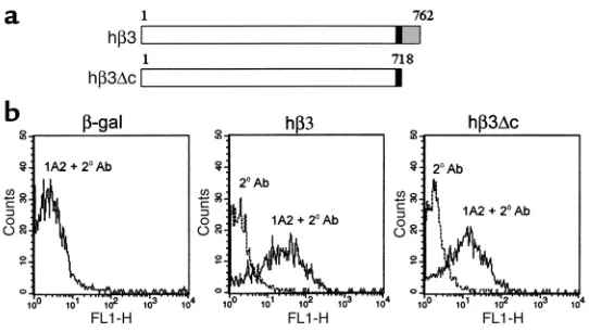
Because authentic osteoclast precursors, namely primary BMMs, are difficult to effi-ciently transfect, we used a retroviral approach (13). To this end, we cloned the full-length hβ3cDNA into the ∆U3 retroviral construct. To determine if integrin-transmit-ted intracellular signals are essential to osteo-clastic bone resorption, we also constructed a retrovirus vector encoding hβ3 lacking its cytoplasmic tail (hβ3∆c) (Figure 1a).
β3-deficient BMMs were transduced with ∆U3-hβ3 or ∆U3-hβ3∆c, and surface expres-sion of each was analyzed by flow cytometry with an antibody specific for the extracellu-lar domain of hβ3. ∆U3-β-gal served as neg-ative control. Retroviral transduction with either hβ3 construct yields equivalent sur-face expression of the WT and mutant inte-grin (Figure 1b). Following confirmation of hβ3 and hβ3∆c expression, transduced macrophages were cocultured with ST2 stromal cells in osteoclastogenic conditions. Nontransduced WT and β3–/–BMMs served
as controls. Eight days later, the cultures were analyzed for osteoclast expression of
the β3 external domain and stained for TRAP activity, a marker of osteoclast differentiation (Figure 2a).
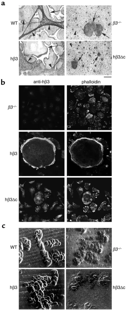
WT cultures consist of very large (150–400 µm diameter), well-spread TRAP-expressing, multinucle-ated cells. ST2 stromal cells either overlie or are pushed aside by these osteoclasts. β3–/–cultures also
contain numerous TRAP-expressing multinucleated cells, a manifestation of the fact that αvβ3 is not essential for osteoclastogenesis (7). These osteoclasts, however, spread poorly and therefore appear much smaller. Occasional larger osteoclasts are present but they also fail to spread well or push ST2 cells aside. Expression of full-length hβ3 completely restores the spreading capacity of the mutant osteoclasts. In con-trast, expression of the truncated hβ3∆c yields a cul-ture indistinguishable from that containing non-transduced β3–/–cells. Immunofluorescent staining
of mature osteoclasts derived from hβ3- and hβ3∆ c-transduced precursors confirms that retroviral-driv-en expression of these cDNAs persists and localizes with fibrillar actin as BMMs undergo osteoclast dif-ferentiation (Figure 2b).
We next turned to the functional implications of the morphological rescue of β3-deficient osteoclasts. To this end, we generated osteoclasts on whale dentin slices, and after 8 days assessed resorption lacunae formation by scanning electron microscopy. β3+/+
[image:4.576.267.538.454.607.2]osteoclasts form well-demarcated deep resorption pits, while those excavated by cells lacking the inte-grin are substantially fewer in number, shallow, and poorly defined (Figures 2c and 5b). Reflecting their recovered spreading capacity, the resorptive activity of β3–/–cells transduced with full-length hβ3 integrin is
Figure 1
Efficient hβ3 integrin surface expression is obtained after retroviral transduction of primary osteoclast precursors. (a) Schematic of full-length and truncated hβ3 proteins produced by retroviral transduction of macrophages with ∆U3-hβ3 and
∆U3-hβ3∆c constructs. Black boxes, transmembrane domain; hatched box, cyto-plasmic domain. (b) Flow cytometric analysis of β3–/– murine marrow
macrophages transduced with virus bearing β-gal, hβ3, or hβ3∆c. The cells were subjected to FACS analysis using a mAb (1A2) recognizing human but not murine
β3, and a FITC-conjugated secondary Ab (1A2 + 2oAb). An internal negative
con-trol using secondary antibody alone (2oAb) is shown in each panel. Cultures
indistinguishable from their WT counterparts. In contrast, deletion of its intracellular tail abrogates the capacity of the integrin to restore the resorptive activ-ity of β3-deficient cells.
Having established that the cytoplasmic domain of the β3 integrin is essential for osteoclast function, we turned to the individual amino acids mediating this event. On the basis of known regulatory capacity of αv-associated βintegrin subunits in other systems (17–22), we mutat-ed specific residues in the β3 cytoplasmic domain (Fig-ure 3a). The mutants were cloned into ∆U3 retroviral construct, which was used to generate retrovirus for transduction of β3–/–osteoclast precursors.
Once again, osteoclasts generated from uninfected β3–/–BMMs fail to spread (Figure 3, b and c). In
con-trast, infection of β3–/–osteoclast precursors with hβ3,
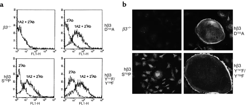
or any mutant, save one, restores the spreading capaci-ty of their osteoclast progeny, establishing that these altered amino acids are not essential for organization of the osteoclast cytoskeleton. hβ3(S752P) is the only mutant which obviates rescue of β3–/– osteoclasts’
capacity to spread. Flow cytometric analysis of trans-duced BMMs demonstrates that the failure of hβ3(S752P) to rescue spreading is not due to diminished surface expression of this mutant in osteoclast precur-sors (Figure 4a). Furthermore, immunofluorescent staining of mature osteoclasts demonstrates persistent expression of hβ3(S752P) and two other representative mutant β3 cDNAs (Figure 4b).
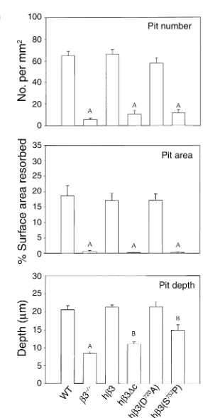
Mirroring spreading, the shallow, poorly-defined pits characteristic of β3–/–osteoclasts are completely
nor-malized by hβ3 and representative, nondisruptive mutants, hβ3(D723A) and hβ3(Y747F/Y759F) (Figure 5a). The lacunae formed by hβ3(S752P), in contrast, appear morphologically similar to those produced by β3–/–
osteoclasts. Quantitative analysis reveals that the num-ber of resorptive pits formed and the percent of dentin surface excavated by β3–/–osteoclasts transduced with
hβ3 or hβ3(D723A) are indistinguishable from those generated by WT cells (Figure 5b). Alternatively, β3–/–
cells bearing hβ3∆c, or the hβ3(S752P) mutant, mirror nontransduced β3–/–osteoclasts for these same indices
of resorptive activity. Pit depth is also completely res-cued in hβ3 and hβ3(D723A) cultures. Interestingly, while hβ3∆c and especially hβ3(S752P) transductants generate no more pits than do virgin β3–/–osteoclasts,
they partially normalize pit depth (P < 0.01 compared with both WT and β3–/–osteoclasts).
We next turned to c-src activation, an intracellular sig-nal mediated by αvβ3, in osteoclasts and asked if the event required the β3 cytoplasmic domain. Early osteoclasts were generated from β3–/–BMMs transduced with hβ3,
[image:5.576.65.287.52.590.2]hβ3∆c, or the hβ3(S752P) and hβ3(Y747F/Y759F) mutants. The transductants were lifted with EDTA and kept in sus-pension or plated on the αvβ3 ligand vitronectin. After 1 hour, phosphorylation of c-src at the activation-specific Y416site was determined by immunoblot (Figure 6). Adhe-sion to vitronectin activates c-src in osteoclasts generated from β3–/–marrow macrophages transduced with intact
Figure 2
The β3 integrin cytoplasmic domain is essential for osteoclast spreading and resorptive activity. (a) TRAP-stained osteoclast cul-tures derived from WT and β3–/–marrow macrophages, and from
β3–/–macrophages transduced with virus encoding hβ3 or hβ3∆c, in
coculture with ST2 stromal cells (arrows, osteoclasts; arrowheads, ST2 stromal cells; scale bar = 100 µm). (b) β3–/–BMMs, either
Figure 3
β3 integrin S752uniquely regulates osteoclast spreading. (a) Sequences of six point mutants of β3 integrin cytoplasmic domain. (b)
Osteoclasts derived from β3–/–macrophages infected with virus encoding WT hβ3 or mutations (detailed in a) were stained for TRAP
activity after 8 days of ST2 coculture. Scale bar = 100 µm. (c) Percent surface area of culture covered by spread osteoclasts for the experiment shown in b. Results are typical of those seen in four separate experiments. AP < 0.001 compared with hβ3 (without
muta-tion); error bars represent SEM.
Figure 4
hβ3(S752P) is effectively expressed by β3–/–osteoclasts and their precursors. (a) Flow cytometric analysis of β3–/–BMMs nontransduced
or transduced with virus encoding hβ3(D723A), hβ3(S752P), or hβ3(Y747F/Y759F), for hβ3 expression using 1A2 (1A2 + 2oAb). All mutants
are expressed at approximately equivalent levels. An internal negative control using secondary antibody alone (2oAb) is shown in each
panel. (b) Mature osteoclasts, generated from the same transduced β3–/–precursors shown in a, were analyzed for expression of the
[image:6.576.65.539.455.665.2]hβ3 or hβ3(Y747F/Y759F). In contrast, no such activation occurs in osteoclasts bearing hβ3∆c and hβ3(S752P), mir-roring the functional effects of these mutations.
Discussion
Osteoclastic bone resorption is initiated by matrix recognition, and formation, at the cell-bone interface, of an isolated, acidified microenvironment, which is the site of skeletal degradation (1). This physical intimacy between the cell and bone indicates that attachment molecules on the osteoclast are pivotal to skeletal remodeling. This posture is buttressed by experiments performed, in vitro and in vivo, demonstrating that αvβ3 blockade blunts the osteoclast’s ability to resorb bone (4–6). The clinical relevance of this observation is underscored by the capacity of soluble organic mimet-ics of the αvβ3 ligand to prevent experimental, post-menopausal osteoporosis (5).
With these experiments in mind, and the wish to determine the role of the αvβ3 integrin in skeletal development, we generated β3-deficient mice (7). Because the platelet integrin αIIbβ3 is not expressed in these animals, they serve as a model of the human bleeding dyscrasia Glanzmann’s thrombasthenia (23). Reflecting osteoclast dysfunction, β3–/– mice are
hypocalcemic and develop bone sclerosis as they age (7). While the bone phenotype in patients with Glanz-mann’s thrombasthenia is unknown, a reasonable pos-sibility holds that they too may have increased bone mass. A likely clinical consequence of this phenomenon would be protection against pathological bone loss such as that attending cessation of ovarian function.
The fact that β3–/–osteoclasts fail to normally
organ-ize their cytoskeleton, in vitro and in vivo, represents compelling evidence that the integrin transmits matrix-derived signals essential to the resorptive process. Given that the majority of known signaling events mediated by αvβ3 depend upon the β3 cytoplasmic domain, we asked if such was the case regarding the osteoclast. To address this issue, we first expressed full-length hβ3 in β3–/–osteoclasts. This undertaking was complicated by
[image:7.576.336.479.50.344.2] [image:7.576.71.291.58.197.2]the fact that primary macrophages, which are osteoclast precursors, cannot be transfected with high efficiency
Figure 5
β3 integrin S752P uniquely regulates osteoclast resorptive activity. (a)
β3–/–marrow macrophages, either nontransduced (β3–/–) or
trans-duced with retrovirus encoding hβ3 or its D723A, S752P, or Y747F/Y759F
mutants were cultured in osteoclastogenic conditions with ST2 cells on slices of whale dentin for 8 days. Resorption lacunae were examined by scanning electron microscopy. (b) Pit density and resorbed area were determined by examination of Coomassie brilliant blue–stained dentin slices using light microscopy. Pit depth was determined by confocal microscopy. AP < 0.001 compared with WT in all panels; BP < 0.01
com-pared with both WT and β3–/–); error bars represent SEM.
Figure 6
Activation of c-src requires the cytoplasmic tail of β3, and is abro-gated by the S752P mutation but not the Y747F/Y759F mutation. β3–/–
BMMs transduced with hβ3, hβ3∆c, or the S752P and Y747F/Y759F
mutants were grown in M-CSF and RANKL for 3 days to generate early (not fully spread) osteoclasts. Following overnight starvation, cells were lifted with EDTA, and either kept in suspension (S) or adhered to vitronectin-coated plates (A) for 1 hour. As a control, cleared lysates were analyzed by immunoblot for total c-src. Activa-tion of c-src was determined by immunoprecipitaActiva-tion of equal amounts of lysates with the anti-phosphotyrosine Ab PY99, followed by immunoblot with anti-pY416src. The ratio of pY416src band
[image:7.576.315.521.512.586.2]by traditional methods. Thus, we utilized a retroviral strategy (13) which permits effective expression of the transgene in virtually all osteoclast precursors. These transduced cells, when placed in osteoclastogenic con-ditions, differentiate into osteoclasts indistinguishable from WT, both in their capacity to spread and, most importantly, to resorb bone. In contrast, β3–/–
osteo-clasts are unaltered by the β3 integrin transgene lacking the cytoplasmic domain. Thus, signal transduction mediated by the β3 cytoplasmic tail is critical for inte-grin function in the osteoclast.
Previous studies have demonstrated that binding of RGD-containing peptides to the integrin triggers var-ious intracellular signals, including changes in intra-cellular calcium (24–26), and activation of c-src (8), PYK2 (9), p130cas (27), and PI3-kinase (28). Despite these observations, the structural components of the β3 cytoplasmic tail mediating osteoclast function have not been elucidated.
In an attempt to identify the amino acid residues in the β3 cytoplasmic tail critical to osteoclastic bone resorption, we generated single and double amino acid mutants based upon the demonstration, in other cells, that they alter βintegrin function. D723is implicated in forming a salt bridge with the αintegrin subunit, there-by stabilizing an activated conformation (20). P745, Y747, and Y759are located in two NPXY/NXXY motifs which are conserved among most βintegrin subunits and mediate many aspects of integrin function (17–19). Mutation of integrin β1A-P781, which corresponds to β3-P745, dampens expression and inactivates β1A in the mouse embryonic stem cell line GD25 (22).
Of the six point mutants known to impact βintegrin function, only S752P fails to rescue β3–/–osteoclasts.
This mutation has also been documented in Glanz-mann’s thrombasthenia (29), in which platelets fail to aggregate. Thus, the residue regulating human platelet function also regulates the cytoskeletal organization and bone resorptive activity of osteoclasts. Specifically, hβ3(S752P) fails to rescue both the impaired capacity of β3–/–osteoclasts to spread, and the frequency with
which they form resorptive lacunae. Similar to platelets (30), the effect of the mutation is specific for proline, as the more conservative mutation, S752A, is as effective as WT hβ3 in rescuing osteoclast spreading (data not shown). This result suggests that local secondary struc-ture, and not phosphorylation of S752, likely mediates its central role in osteoclast function.
It is of interest that, like S752, Y747and Y759are, in com-bination, also essential for platelet function (31). Both tyrosines, when phosphorylated, bind the signaling molecules SHC and GRB2, as well as the cytoskeletal protein myosin, in platelets (32, 33). Unlike S752, how-ever, these combined mutations fail to impact osteo-clast function. Thus, the osteoosteo-clast and platelet may share some β3-mediated signaling pathways, while oth-ers appear cell-specific.
c-src is a tyrosine kinase essential for osteoclast func-tion (10, 11, 34). The mechanism by which this
proto-oncogene activates the osteoclast is complex, but clear-ly involves both the kinase and protein docking domains (35, 36). We find that αvβ3-dependent activa-tion of c-src requires the cytoplasmic domain of β3, and is abrogated by the S752P, but not the Y747F/Y759F, muta-tion. Thus, the ability of β3 to activate c-src correlates with its capacity to stimulate osteoclast function.
While the S752P mutation affects both osteoclast and platelet function, the downstream signaling mechanisms appear to be distinct. Deletion of c-src has not been reported to cause platelet dysfunction (34), in contrast to osteoclasts, and therefore is unlikely to represent the relevant β3-mediated signal-ing pathway in platelets.
Integrin signaling is bidirectional, with inside-out signals affecting ligand binding affinity, and outside-in signals determoutside-inoutside-ing outside-intracellular events occurroutside-ing upon integrin-ligand interaction (37). In the context of overexpression in Chinese hamster ovary (CHO) cells, Chen et al. (29) showed that the S752P mutation disrupts binding of the ligand-induced binding site (LIBS) antibody PAC1, suggesting a defect in inside-out signaling. In contrast, we find that σosteoclasts expressing hβ3(S752P) bind AP5, another β3-specific LIBS antibody (R. Faccio, unpublished observations). Given that AP5 recognizes the activated form of αvβ3, this observation suggests that the β3(S752P) mutation, in osteoclasts, arrests outside-in signaling and thus, ligand-induced c-src activation.
Acknowledgments
This work is partially supported by NIH grants AR42404 (F.P. Ross); DE05413, AR32788, and AR45623 (S.L. Teitelbaum); 1F32AR08586 and DK07120 (D.V. Novack); a grant from Shriners Hospi-tal (S.L. Teitelbaum); a grant from Monsanto Corp. (S.L. Teitelbaum); and a Barnes-Jewish Hospital Foun-dation grant (X. Feng). We thank Scott Blystone for providing us with the 1A2 antibody.
1. Teitelbaum, S.L., Tondravi, M.M., and Ross, F.P. 1996. Osteoclast Biolo-gy. In Osteoporosis. R. Marcus, D. Feldman, and J. Kelsey, editors. Acade-mic Press. San Diego, California, USA. 61–94.
2. Suda, T., et al. 1999. Modulation of osteoclast differentiation and func-tion by the new members of the tumor necrosis factor receptor and lig-and families. Endocr. Rev.20:345–357.
3. Hynes, R.O. 1992. Integrins: versatility, modulation, and signaling in cell adhesion. Cell. 69:11–25.
4. Horton, M.A., Taylor, M.L., Arnett, T.R., and Helfrich, M.H. 1991. Arg-gly-asp (RGD) peptides and the anti-vitronectin receptor antibody 23C6 inhibit dentine resorption and cell spreading by osteoclasts. Exp. Cell Res.
195:368–375.
5. Engleman, V.W., et al. 1997. A peptidomimetic antagonist of the αvβ3 integrin inhibits bone resorption in vitro and prevents osteoporosis in vivo. J. Clin. Invest.99:2284–2292.
6. Nakamura, I., Tanaka, H., Rodan, G.A., and Duong, L.T. 1998. Echistatin inhibits the migration of murine prefusion osteoclasts and the formation of multinucleated osteoclast-like cells. Endocrinology.139:5182–5193. 7. McHugh, K.P., et al. 2000. Mice lacking β3 integrins are osteosclerotic
because of dysfunctional osteoclasts. J. Clin. Invest.105:433–440. 8. Sanjay, A., et al. 2001. Cbl associates with Pyk2 and Src to regulate Src
kinase activity, αvβ3 integrin-mediated signaling, cell adhesion, and osteoclast motility. J. Cell Biol.152:181–196.
cel-lular activity and osteoclast-mediated resorption. Bone. 28:54–64. 11. Missbach, M., et al. 1999. A novel inhibitor of the tyrosine kinase Src
suppresses phosphorylation of its major cellular substrates and reduces bone resorption in vitro and in rodent models in vivo. Bone. 24:437–449. 12. Chen, Y.P., et al. 1992. Ser-752—>Pro mutation in the cytoplasmic domain of integrin beta 3 subunit and defective activation of platelet integrin alpha IIb beta 3 (glycoprotein IIb-IIIa) in a variant of Glanz-mann thrombasthenia. Proc. Natl. Acad. Sci. USA.89:10169–10173. 13. Ory, D.S., Neugeboren, B.A., and Mulligan, R.C. 1996. A stable
human-derived packaging cell line for production of high titer retrovirus/vesic-ular stomatitis virus G pseudotypes. Proc. Natl. Acad. Sci. USA.
93:11400–11406.
14. Abu-Amer, Y., et al. 1998. Tumor necrosis factor-αactivation of nuclear transcription factor-κB in marrow macrophages is mediated by c-Src tyrosine phosphorylation of IκBα. J. Biol. Chem.273:29417–29423. 15. Greenfield, E.M., et al. 1992. Avian osteoblast conditioned media
stim-ulate bone resorption by targeting multinucleating osteoclast precur-sors. Calcif. Tissue Int. 51:317–323.
16. Lipsich, L.A., Lewis, A.J., and Brugge, J.S. 1983. Isolation of monoclonal antibodies that recognize the transforming proteins of avian sarcoma viruses. J. Virol. 48:352–360.
17. Filardo, E.J., Brooks, P.C., Deming, S.L., Damsky, C., and Cheresh, D.A. 1995. Requirement of the NPXY motif in the integrin beta 3 subunit cytoplasmic tail for melanoma cell migration in vitro and in vivo. J. Cell. Biol.130:441–450.
18. Ylanne, J., et al. 1995. Mutation of the cytoplasmic domain of the inte-grin beta 3 subunit. Differential effects on cell spreading, recruitment to adhesion plaques, endocytosis, and phagocytosis. J. Biol. Chem.
270:9550–9557.
19. O’Toole, T.E., Ylanne, J., and Culley, B.M. 1995. Regulation of integrin affinity states through an NPXY motif in the beta subunit cytoplasmic domain. J. Biol. Chem. 270:8553–8558.
20. Hughes, P.E., et al. 1996. Breaking the integrin hinge. A defined structural constraint regulates integrin signaling. J. Biol. Chem.271:6571–6574. 21. Schaffner-Reckinger, E., Gouon, V., Melchior, C., Plancon, S., and
Kief-fer, N. 1998. Distinct involvement of beta3 integrin cytoplasmic domain tyrosine residues 747 and 759 in integrin-mediated cytoskeletal assem-bly and phosphotyrosine signaling. J. Biol. Chem. 273:12623–12632. 22. Sakai, T., Peyruchaud, O., Fassler, R., and Mosher, D.F. 1998.
Restora-tion of Beta(1)A integrins is required for lysophosphatidic acid-induced migration of Beta(1)-null mouse fibroblastic cells. J. Biol. Chem.
273:19378–19382.
23. Hodivala-Dilke, K.M., et al. 1999. β3-integrin-deficient mice are a model for Glanzmann thrombasthenia showing placental defects and reduced
survival. J. Clin. Invest.103:229–238.
24. Shankar, G., Davison, I., Helfrich, M.H., Mason, W.T., and Horton, M.A. 1993. Integrin receptor-mediated mobilisation of intranuclear calcium in rat osteoclasts. J. Cell Sci. 105:61–68.
25. Zimolo, Z., et al. 1994. Soluble αvβ3-integrin ligands raise [Ca2+] in rat
osteoclasts and mouse-derived osteoclast-like cells. Am. J. Physiol.
266:C376–C381.
26. Paniccia, R., et al. 1995. Calcitonin down-regulates immediate cell sig-nals induced in human osteoclast-like cells by the bone sialoprotein-IIA fragment through a postintegrin receptor mechanism. Endocrinology.
136:1177–1186.
27. Nakamura, I., et al. 1998. Tyrosine phosphorylation of p130Cas is involved in actin organization in osteoclasts. J. Biol. Chem.
273:11144–11149.
28. Hruska, K.A., Rolnick, F., Huskey, M., Alvarez, U., and Cheresh, D. 1995. Engagement of the osteoclast integrin alpha v beta 3 by osteopontin stimulates phosphatidylinositol 3-hydroxyl kinase activity. Endocrinolo-gy.136:2984–2992.
29. Chen, Y.P., O’Toole, T.E., Ylanne, J., Rosa, J.P., and Ginsberg, M.H. 1994. A point mutation in the integrin beta 3 cytoplasmic domain (S752-->P) impairs bidirectional signaling through alpha IIb beta 3 (platelet glyco-protein IIb-IIIa). Blood. 84:1857–1865.
30. Kieffer, N., et al. 1996. Serine 752 in the cytoplasmic domain of the beta 3 integrin subunit is not required for αvβ3 postreceptor signaling events.
Cell Adhes. Commun. 4:25–39.
31. Law, D.A., et al. 1999. Integrin cytoplasmic tyrosine motif is required for outside-in alphaIIbbeta3 signalling and platelet function. Nature.
401:808–811.
32. Law, D.A., Nannizzi-Alaimo, L., and Phillips, D.R. 1996. Outside-in inte-grin signal transduction. Alpha IIb beta 3-(GP IIb IIIa) tyrosine phos-phorylation induced by platelet aggregation. J. Biol. Chem.
271:10811–10815.
33. Jenkins, A.L., et al. 1998. Tyrosine phosphorylation of the beta3 cyto-plasmic domain mediates integrin-cytoskeletal interactions. J. Biol. Chem.
273:13878–13885.
34. Soriano, P., Montgomery, C., Geske, R., and Bradley, A. 1991. Targeted disruption of the c-src proto-oncogene leads to osteopetrosis in mice.
Cell. 64:693–702.
35. Schwartzberg, P.L., et al. 1997. Rescue of osteoclast function by trans-genic expression of kinase-deficient Src in src–/– mutant mice. Genes Dev.
11:2835–2844.