Original Article
Long non-coding RNA AFAP1-AS1 was up-regulated
in triple-negative breast cancer and regulated
proliferation and invasion
Fan Yang*, Si-Yang Dong*, Lin Lv, Ye-Huan Liu, Zhi-Han Yao, Xiao-Hua Zhang, Ou-Chen Wang
Department of Surgical Oncology, The First Affiliated Hospital of Wenzhou Medical University, Wenzhou, Zhejiang,
PR China. *Equal contributors.
Received February 10, 2015; Accepted April 28, 2016; Epub June 1, 2016; Published June 15, 2016
Abstract: Triple-negative breast cancer (TNBC) is usually associated with an unfavorable prognosis and lacks effec-tive therapeutic targets. Long non-coding RNAs (lncRNAs) contribute to the initiation and progression of a variety of human cancers. The identification of dysregulated lncRNAs involved in TNBC might provide additional insights into its aggressive biological behavior. Here, we reported one lncRNA, actin filament associated protein 1 anti-sense RNA1 (AFAP1-AS1), was significantly overexpressed in TNBC and associated with lymph node metastasis, distant metastasis and stage. In vitro experiments demonstrated that the proliferation and invasion potential was significantly reduced in AFAP1-AS1-silenced TNBC cells. To the best of our knowledge, it was firstly reported that AFAP1-AS1 was involved in breast cancer. Our results suggested that AFAP1-AS1 represents a novel and a potential therapeutic target for TNBC. Furthermore, increased understanding of such onco-lncRNAs could provide additional insights into cancer tumorigenesis and progress.
Keywords: lncRNA, AFAP1-AS1, triple-negative breast cancer, proliferation, migration, invasion
Introduction
Breast cancer (BC) is the most common type of female cancer worldwide [1]. Triple-negative breast cancer (TNBC) is characterized by a lack of expression of estrogen receptor (ER) and progesterone receptor (PR) as well as human epidermal growth factor receptor 2 (HER-2). Overall, the prevalence of TNBC accounts for 11%-20% of the BC patient cohort [2]. TNBC is associated with the worst prognosis among all BC subtypes, partly because of a lack of effec-tive targeted drugs such as tamoxifen in ER-positive BC and trastuzumab in HER2-positive BC [3, 4]. Although remarkable prog-ress has been made during recent decades, the molecular mechanisms underlying the aggressive biological behavior of TNBC remain to be elucidated.
Long non-coding RNAs (lncRNAs) are a class of transcripts longer than 200 bp that lack a pro-tein-coding capacity [5]. An increasing number of studies indicate that lncRNAs are involved in
a variety of biological processes including epi-genetics, transcription, post-transcription, and translation [6-8]. The dysregulation of lncRNAs has also been shown to contribute to the initia-tion and progression of several human cancers including BC [9-14]. Hence, identification of cancer-associated lncRNAs and investigation of their biological functions might help identify novel therapeutic targets.
vali-datedthe biological function of AFAP1-AS1 in TNBC.
Materials and methods
Patient samples
BC patients undergoing modified radical mas-tectomy were included in this study. All samples were confirmed as TNBC by postoperative his-topathological examination and immunohisto-chemistry. Primary cancer tissues and non-tumorous tissues were snap-frozen in liquid nitrogen immediately after resection and then stored at -80°C before RNA extraction. Informed consent was obtained from all individual par-ticipants included in the study. The study was approved by the Ethics Committee of the First Affiliated Hospital of Wenzhou Medical University.
RNA extraction and quantitative reverse tran -scription PCR (qRT-PCR)
Total RNA was extracted from tissue samples using TRIzol (Life Technologies, Carlsbad, CA) according to the manufacturer’s protocol. qRT-PCR was performed using a Thunderbird SYBR qPCR Mix (Toyobo, Osaka, Japan) in the Applied Biosystems 7300 Real Time PCR System (Applied Biosystems, Foster City, CA). The prim-er sequences for PCR are as follows: AFAP1-AS1, forward 5’-AATGGTGGTAGGAGGGAGGA-3’ and reverse 5’-CACACAGGGGAATGAAGAGG-3’. β-Actin was used as an internal control and the-comparative CT method (∆∆CT) method was used to evaluate the relative quantification of AFAP1-AS1. Each sample was in triplicate.
Cell lines and small interfering RNA (siRNA)
TNBC cancer cell lines BT-549 and MBA-MD- 231 were cultured in RPMI 1640 medium (Gibco, Grand Island, NY) supplemented with 10% fetal bovine serum (FBS; Gibco) in a humidified incubator with 5% CO2 at 37°C. Cells were seeded overnight and transfected with either 100 nM siRNA or nontarget scramble control siRNA (GenePharma, Shanghai, China) using Lipofectamine RNAiMAX Reagent (Invi- trogen, Carlsbad, CA) in OptiMEM medium (Gibco) for gene knockdown.
Colony formation assay and MTT assay
For colony formation assay, 5000 cells were plated in each well of a six-well plate. When
there was visible colony by naked eye, cells were fixed with 4% formaldehyde and were stained with crystal violet (0.25%). Colonies were then counted. For MTT assay, 2000 cells from each group were plated in each well of five 96-well plates. To analyze cell proliferation, 20 μL of MTT substrate at a concentration of 2.5 mg/mL in PBS was added to each well. The plates were then returned to a standard tissue incubator for an additional 4 h. The medium was then removed, and the cells were solubi-lized in 150 μL of dimethylsulfoxide for the colo-rimetric analysis (wavelength, 490 nm). One plate was analyzed immediately after the cells adhered. Then, one plate per day was exam-ined for the next 4 consecutive days.
Wound healing assay
Cells were grown to 90% confluency in six-well culture plates. A p200 pipette tip was used to scratch the cell monolayer. Images were cap-tured immediately and 24 h and 48 h after wounding, and wound closure was monitored by microscopy. Wound sizes were verified with an ocular ruler.
Transwell assay
Cell migration and invasion were measured on Transwell plates (Costar, New York, NY) and Matrigel chamber plates (BD Biosciences, Bedford, MA), respectively. A total of 1 × 10⁵ cells were seeded onto Transwell or Matrigel insert membranes on day 2 following transfec-tion. Growth medium containing 20% FBS was used as a chemoattractant. After incubation at 37°C for 24 h, cells that did not migrate or invade through the pores of the Transwell inserts were manually removed with a cotton swab. Cells that had invaded through the filter pores were fixed with methanol, stained with 0.1% crystal violet, and observed under a microscope. The number of invasive tumor cells was counted from five randomly selected 20 × fields for each experiment and averaged. Statistical analysis
compari-sons in cellular experiments. A P value less than 0.05 was considered statistically signifi-cant. All statistical analyses were performed using SPSS version 22.0 (Chicago, IL).
Results
LncRNA AFAP1-AS1 was up-regulated in TNBC
The expression of AFAP1-AS1 was detected in 102 TNBC tissues and 95 non-cancerous
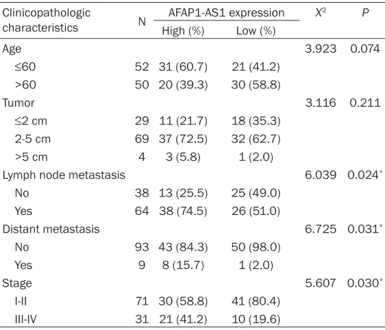
tis-the control cells (P<0.01, Figure 2B). Similarly, MTT assay revealed that cells transfected with siRNA1 and siRNA2 and not control cells, had significantly inhibited growth and proliferation of TNBC cells (P<0.001, Figure 2C).
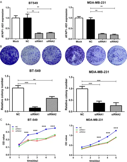
Inhibition of AFAP1-AS1 leads to reduced migration and invasion
[image:3.612.91.372.70.253.2]Furthermore, the role of AFAP1-AS1 in TNBC cell metastasis was demonstrated. Wound Figure 1. AFAP1-AS1 is overexpressed in TNBC. AFAP1-AS1 expression
lev-els in 102 TNBC tissues and 95 non-cancerous tissues were confirmed by qRT-PCR. ***P<0.001; Student t-test.
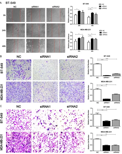
Table 1. Relationship between AFAP1-AS1 expression and patient clinicopathological characteristics
Clinicopathologic
characteristics N AFAP1-AS1 expression X
2 P
High (%) Low (%)
Age 3.923 0.074
≤60 52 31 (60.7) 21 (41.2)
>60 50 20 (39.3) 30 (58.8)
Tumor 3.116 0.211
≤2 cm 29 11 (21.7) 18 (35.3)
2-5 cm 69 37 (72.5) 32 (62.7)
>5 cm 4 3 (5.8) 1 (2.0)
Lymph node metastasis 6.039 0.024*
No 38 13 (25.5) 25 (49.0)
Yes 64 38 (74.5) 26 (51.0)
Distant metastasis 6.725 0.031*
No 93 43 (84.3) 50 (98.0)
Yes 9 8 (15.7) 1 (2.0)
Stage 5.607 0.030*
I-II 71 30 (58.8) 41 (80.4)
III-IV 31 21 (41.2) 10 (19.6)
*P<0.05; Chi-square test.
sues by qRT-PCR. As showed in Figure 1, the expression of AFAP1-AS1 in TNBC tissues was almost 5 folds of non-can-cerous tissues (P<0.001). The relationship between
AFAP1-AS1 expression and
clinical features
To analyze whether AFAP1-AS1 was associated with the devel-opment and progression of TNBC, we investigated itsrela-tionship with clinical features. In qPCR cohort, patients were divided into low and high AFAP1-AS1 expression groups according to the median value. The results revealed lymph no- de metastasis (P=0.024), dis-tant metastasis was (P=0.031) and stage (P=0.030) were significant related with the expression of AFAP1-AS1 posi-tively (Table 1).
Inhibition of AFAP1-AS1 in
TNBC cells leads to reduced
proliferation
[image:3.612.91.372.338.579.2]recovery was significantly delayed when AFAP1-AS1 was knocked down compared with control cells (P<0.05, Figure 3A). Besides, cell migra-tion and invasion were also significantly reduced, as assessed by a transwell assay. As
[image:4.612.93.521.72.614.2]Discussion
TNBC patients usually have significantly lower rates of disease-free and overall survival, and
[image:5.612.94.522.70.609.2]drawn extra attention from oncologists. Growing numbers of lncRNAs are proven to have a crucial effect in the biological regulation of a range of cancers [20]. However, consider-ing the high number of lncRNAs, knowledge of lncRNAs and their mechanisms of action is sur-prisingly limited, especially with respect to TNBC. Here, we proved that lncRNA AFAP1-AS1 is significantly overexpressed in TNBC and associated with lymph node metastasis, dis-tant metastasis and stage. In vitro experiments demonstrated that the proliferation and metas-tasis potential was significantly reduced in AFAP1-AS1-silenced TNBC cells. To the best of our knowledge, it was firstly reported that lncRNA AFAP1-AS1 was involved in breast cancer.
Wu previously reported that AFAP1-AS1 is upregulated in esophageal cancer because of extreme hypomethylation in its promoter [21]. AFAP1-AS1 silencing was shown to inhibit prolif-eration and colony-forming ability, induce apop-tosis, and reduce migration and invasion of esophageal cancer cells [21]. Zeng reported the upregulation of AFAP1-AS1 in lung cancer, and showed it to be associated with poor prog-nosis [22], while Bo similarly revealed its over-expression in nasopharyngeal carcinoma and association with metastasis and poor progno-sis [16]. Taken together, this indicated that AFAP1-AS1 was involved in multiple cancers and it might act as an onco-lncRNA.
The exact mechanism of AFAP1-AS1 action remains unclear. Previous studies have report-ed widespread sense-antisense transcripts in mammalian cells, and shown that perturbation of antisense RNA can alter the expression of the sense gene [23, 24]. AFAP1, AFAP1-AS1’s coding counterpart, has been found to promote focal adhesion formation in MDA-MB-231 BC cells [15]. We therefore hypothesize that AFAP1-AS1 might exert its function by regulating AFAP1. Further studies should investigate the mechanisms by which AFAP1-AS1 modulates its sense counterpart.
In conclusion, our results suggest that AFAP1-AS1 represents a novel therapeutic target for TNBC. Furthermore, increased understanding of suchonco-lncRNAs in TNBC could provide additional insights into cancer tumorigenesis and progress.
Acknowledgements
This study was funded by the Key Project of Science and Technology Innovation Team of Zhejiang Province (2013TD10) and the National Natural Science Foundation of China (No. 81372380).
Disclosure of conflict of interest
None.
Address correspondence to: Xiao-Hua Zhang and Ou-Chen Wang, Department of Surgical Oncology, The First Affiliated Hospital of Wenzhou Medical University, Wenzhou, Zhejiang, PR China. Tel: +86 577-55579462; E-mail: xiaohuazhang2015@sina. com (XHZ); Tel: +86 577-55578462; E-mail: [email protected] (OCW)
References
[1] Siegel RL, Miller KD and Jemal A. Cancer sta-tistics, 2015. CA Cancer J Clin 2015; 65: 5-29. [2] Lin NU, Vanderplas A, Hughes ME, Theriault
RL, Edge SB, Wong Y, Blayney DW, Niland JC, Winer EP and Weeks JC. Clinicopathological features and sites of recurrence according to breast cancer subtype in the National Compre-hensive Cancer Network (NCCN). J Clin Oncol 2009; 27.
[3] Carey L, Winer E, Viale G, Cameron D and Gi-anni L. Triple-negative breast cancer: disease entity or title of convenience? Nat Rev Clin On-col 2010; 7: 683-692.
[4] Di Cosimo S and Baselga J. Management of breast cancer with targeted agents: impor-tance of heterogenicity. Nature Reviews Clini-cal Oncology 2010; 7: 139-147.
[5] Ponting CP, Oliver PL and Reik W. Evolution and functions of long noncoding RNAs. Cell 2009; 136: 629-641.
[6] Huarte M, Guttman M, Feldser D, Garber M, Koziol MJ, Kenzelmann-Broz D, Khalil AM, Zuk O, Amit I, Rabani M, Attardi LD, Regev A, Land-er ES, Jacks T and Rinn JL. A large intLand-ergenic noncoding RNA induced by p53 mediates glob-al gene repression in the p53 response. Cell 2010; 142: 409-419.
[7] Mercer TR and Mattick JS. Structure and func-tion of long noncoding RNAs in epigenetic reg-ulation. Nat Struct Mol Biol 2013; 20: 300-307.
[8] Qiu MT, Hu JW, Yin R and Xu L. Long noncoding RNA: an emerging paradigm of cancer re-search. Tumor Biol 2013; 34: 613-620. [9] Pandey GK, Mitra S, Subhash S, Hertwig F,
A, Mondal T and Bandaru S. The risk-associat-ed long noncoding RNA NBAT-1 controls neuro-blastoma progression by regulating cell prolif-eration and neuronal differentiation. Cancer Cell 2014; 26: 722-737.
[10] Martens-Uzunova ES, Böttcher R, Croce CM, Jenster G, Visakorpi T and Calin GA. Long non-coding RNA in prostate, bladder, and kidney cancer. Eur Urol 2014; 65: 1140-1151. [11] Wang Y, He L, Du Y, Zhu P, Huang G, Luo J, Yan
X, Ye B, Li C and Xia P. The Long Noncoding RNA lncTCF7 Promotes Self-Renewal of Hu-man Liver Cancer Stem Cells through Activa-tion of Wnt Signaling. Cell Stem Cell 2015; 16: 413-425.
[12] Sun M, Gadad SS, Kim DS and Kraus WL. Dis-covery, Annotation, and Functional Analysis of Long Noncoding RNAs Controlling Cell-Cycle Gene Expression and Proliferation in Breast Cancer Cells. Mol Cell 2015; 59: 698-711. [13] Liu B, Sun L, Liu Q, Gong C, Yao Y, Lv X, Lin L,
Yao H, Su F and Li D. A Cytoplasmic NF-κB In-teracting Long Noncoding RNA Blocks IκB Phosphorylation and Suppresses Breast Can-cer Metastasis. CanCan-cer Cell 2015; 27: 370-381.
[14] Xing Z, Lin A, Li C, Liang K, Wang S, Liu Y, Park PK, Qin L, Wei Y and Hawke DH. lncRNA directs cooperative epigenetic regulation downstream of chemokine signals. Cell 2014; 159: 1110-1125.
[15] Dorfleutner A, Stehlik C, Zhang J, Gallick GE and Flynn DC. AFAP-110 is required for actin stress fiber formation and cell adhesion in MDA-MB-231 breast cancer cells. J Cell Physiol 2007; 213: 740-749.
[16] Bo H, Gong Z, Zhang W, Li X, Zeng Y, Liao Q, Chen P, Shi L, Lian Y, Jing Y, Tang K, Li Z, Zhou Y, Zhou M, Xiang B, Li X, Yang J, Xiong W, Li G and Zeng Z. Upregulated long non-coding RNA AFAP1-AS1 expression is associated with pro-gression and poor prognosis of nasopharyn-geal carcinoma. Oncotarget 2015; 6: 20404-20418.
[17] Deng J, Liang Y, Liu C, He S and Wang S. The up-regulation of long non-coding RNA AFAP1-AS1 is associated with the poor prognosis of NSCLC patients. Biomed Pharmacother 2015; 75: 8-11.
[18] Ye Y, Chen J, Zhou Y, Fu Z, Zhou Q, Wang Y, Gao W, Zheng S, Zhao X, Chen T and Chen R. High expression of AFAP1-AS1 is associated with poor survival and short-term recurrence in pancreatic ductal adenocarcinoma. J Transl Med 2015; 13: 137.
[19] Foulkes WD, Smith IE and Reis-Filho JS. Triple-negative breast cancer. New Engl J Med 2010; 363: 1938-1948.
[20] Kung JT, Colognori D and Lee JT. Long noncod-ing RNAs: past, present, and future. Genetics 2013; 193: 651-669.
[21] Wu W, Bhagat TD, Yang X, Song JH, Cheng Y, Agarwal R, Abraham JM, Ibrahim S, Bartens-tein M, Hussain Z, Suzuki M, Yu Y, Chen W, Eng C, Greally J, Verma A and Meltzer SJ. Hypo-methylation of noncoding DNA regions and overexpression of the long noncoding RNA, AFAP1-AS1, in Barrett’s esophagus and eso- phageal adenocarcinoma. Gastroenterology 2013; 144: 956-966, e954.
[22] Zeng Z, Bo H, Gong Z, Lian Y, Li X, Li X, Zhang W, Deng H, Zhou M, Peng S, Li G and Xiong W. AFAP1-AS1, a long noncoding RNA upregulat-ed in lung cancer and promotes invasion and metastasis. Tumour Biol 2015; 1-9.
[23] Katayama S, Tomaru Y, Kasukawa T, Waki K, Nakanishi M, Nakamura M, Nishida H, Yap C, Suzuki M and Kawai J. Antisense transcription in the mammalian transcriptome. Science 2005; 309: 1564-1566.