T O P I C A L R E V I E W
Voltage-gated proton channels: what’s next?
Thomas E. DeCoursey
Department of Molecular Biophysics and Physiology, Rush University Medical Center, 1750 W. Harrison, Chicago, IL 60612 USA
This review is an attempt to identify and place in context some of the many questions about voltage-gated proton channels that remain unsolved. As the gene was identified only 2 years ago, the situation is very different than in fields where the gene has been known for decades. For the proton channel, most of the obvious and less obvious structure–function questions are still wide open. Remarkably, the proton channel protein strongly resembles the voltage-sensing domain of many voltage-gated ion channels, and thus offers a novel approach to study gating mechanisms. Another surprise is that the proton channel appears to function as a dimer, with two separate conduction pathways. A number of significant biological questions remain in dispute, unanswered, or in some cases, not yet asked. This latter deficit is ascribable to the intrinsic difficulty in evaluating the importance of one component in a complex system, and in addition, to the lack, until recently, of a means of performing an unambiguous lesion experiment, that is, of selectively eliminating the molecule in question. We still lack a potent, selective pharmacological inhibitor, but the identification of the gene has allowed the development of powerful new tools including proton channel antibodies, siRNA and knockout mice.
(Received 15 August 2008; accepted after revision 17 September 2008; first published online 18 September 2008)
Corresponding authorT. E. DeCoursey: Department of Molecular Biophysics and Physiology, Rush University Medical Center, 1750 W. Harrison, Chicago, IL 60612 USA. Email: [email protected]
A few years ago, a H+-ATPase researcher made the astonishing remark that his field was finished – all the major questions had been answered! It had never even occurred to me that this was possible – I have worked mainly in areas that are rife with problems to solve. The voltage-gated proton channel is a poster child of unsolved problems! Before I begin to list several important unanswered questions, I will briefly introduce this little-known channel.
First, the voltage-gated proton channel is an ion channel. For reasons that escape me, some people seem to want to call it a ‘proton pump.’ It is nothing like a pump. The proton channel cannot move protons against an electrochemical gradient – it is a passive pathway across the membrane. It does not need or use ATP. Proton channels open and close just like other ion channels, generating noise and single-channel currents. They open with depolarization, like the classical Na+, K+ and Ca2+ channels of excitable cells. Although these properties define the proton channel as an ion channel, it does have a number of distinctive, if not unique properties. Its unitary conductance is∼103smaller than most other channels, roughly 15 fS at room temperature and physiological pH (Chernyet al.2003). Many channels are selective, but imperfectly so; the proton channel
has apparently perfect selectivity. Proton channels have stronger temperature dependence, both of conductance and gating kinetics, than almost any other ion channel. Reminiscent of inward rectifier K+ channels, the voltage dependence of proton channel gating is not absolute, but varies with the permeant ion concentration. Inward rectifiers conduct mainly inward K+ current; proton channels conduct mainly outward H+current. Finally, the proton channel may be unique in lacking a water-filled pore that acts as the conduction pathway. Protons can travel in ways that other ions cannot, such as Grotthuss conduction in water (de Grotthuss, 1806; Pom`es, 2006) and hydrogen-bonded chain conduction through proteins (Nagle & Morowitz, 1978). It is virtually certain that the conducted species is the proton, H+, not the hydronium ion, H3O+ (DeCoursey & Cherny, 1997, 1998). The lack of a continuous aqueous pore could explain the extreme selectivity of the proton channel.
these cells engulf microbes, the enzyme NADPH oxidase begins to produce superoxide anion, the precursor to a host of bactericidal reactive oxygen species. NADPH oxidase is electrogenic (Henderson et al. 1987; Schrenzel et al.
1998), and proton channels provide the bulk of charge compensation (Henderson et al. 1988a,b; Murphy & DeCoursey, 2006), which prevents extreme depolarization that would stop enzyme function (DeCourseyet al.2003). Recent evidence that proton channels are required for histamine release by human basophils (Musset et al.
2008b) might be explained by proton extrusion, charge compensation or some other mechanism.
Molecular and genetic properties
Has the right gene been identified? The historical controversies over whether the gp91phox component of NADPH oxidase might function as a voltage-gated proton channel (Henderson et al. 1995; Henderson & Meech, 1999; B´anfi et al. 2000; DeCoursey et al. 2000, 2001b; Maturana et al. 2001) have been thoroughly discussed (Touret & Grinstein, 2002; DeCoursey et al.
[image:2.595.45.279.425.574.2]2002; Henderson & Meech, 2002; Maturanaet al. 2002; DeCoursey, 2003, 2008; Musset et al. 2008a) and little new can be added. What can be stated without any
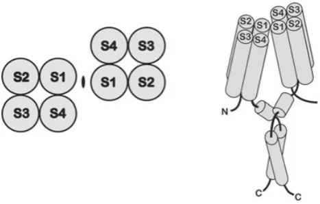
Figure 1. Topology of the voltage-gated proton channel
Voltage-gated proton channels resemble the voltage-sensing domain (VSD) of ordinary voltage-gated cation channels. Hydropathy plots of the protein coded for by the proton channel gene indicate four membrane-spanning regions that resemble S1–S4 of voltage-gated K+ channels (Ramseyet al.2006; Sasakiet al.2006). The proton channel appears to assemble as a dimer (Kochet al.2008; Leeet al.2008; Tombolaet al.2008). The top view of the channel (with the
membrane in the plane of the page) is on the left, the side view on the right. In the latter, the S1–S4 cylinders represent alpha helices spanning the membrane and both the N and C termini are intracellular. Proposed interaction sites are just external to S1 and within a predicted coiled-coil region in the C terminus. (From Leeet al.
2008, with permission.)
doubt is that bona fide voltage-gated proton channel genes were identified in 2006. The Clapham lab identified and characterized the human proton channel gene, HV1 (Ramsey et al. 2006). The Okamura lab (Sasaki et al.
2006) identified a voltage-gated proton channel, CiVSOP, inCiona intestinalis, a sea squirt, in a project to classify the genome of this creature, and found a homologue in the mouse, mVSOP. A gene coding for a voltage-sensitive phosphatase had been identified previously (Murataet al.
2005); the proton channel was a fortuitous discovery in an effort to catalogue other homologues. The proton channel gene products have several remarkable and unexpected properties. As shown in Fig. 1, the proton channel has four putative membrane-spanning regions, S1–S4, which are generally similar to the S1–S4 regions of many voltage-gated ion channels (Sasaki et al. 2006; Ramsey
et al.2006) as well as the voltage-sensitive phosphatase in
Ciona(Murataet al.2005). However, the protein lacks the S5–S6 regions, which in ordinary ion channels form the conduction pathway. The proton channel is thus a voltage sensor without a conventional pore.
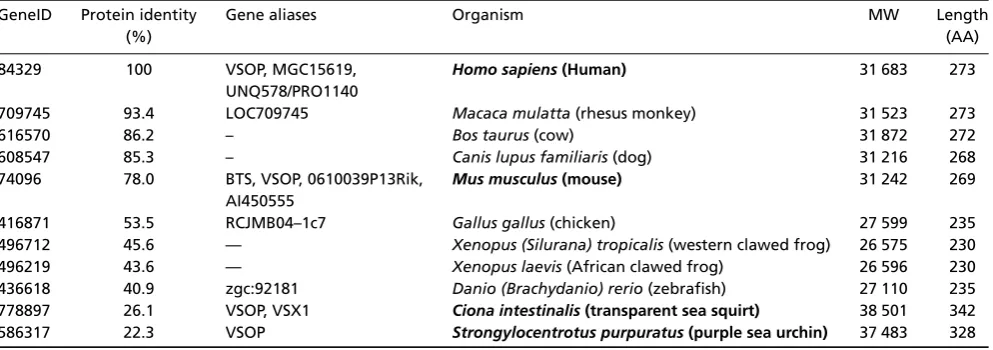
Genes that are highly homologous to the proton channel are present in at least two dozen species, with selected species given in Table 1. Other species with similar genes, listed in descending order of predicted protein similarity (to the human HVCN1) include:
Pan troglodytes (chimpanzee), Sus scrofa (pig), Equus caballus(horse),Monodelphis domestica(grey, short-tailed opossum), Rattus norvegicus (rat), Ornithorhynchus anatinus (platypus), Trichoplax adhaerens (placozoan),
Nematostella vectensis (starlet sea anemone), Tetraodon nigroviridis(puffer fish),Macaca fascicularis(crab-eating macaque), Laccaria bicolor (basidiomycete fungus),
Aspergillus niger (black mold) and Physcomitrella patens
(moss) which is 23% identical to HV1 (NCBI BLAST). Each protein in Table 1 has four predicted transmembrane domains that have substantial homology, fairly long C and N terminal domains, and short linkers with the highest homology in the intracellular S2–S3 linker. In most species, the C terminus contains a long predicted intra-cellular coiled-coil region, where intersubunit interaction may occur (Kochet al.2008; Leeet al.2008; Tombolaet al.
2008).
Is the functional channel really a dimer, and how independent are its pathways? This year, three groups presented diverse evidence indicating that the voltage-gated proton channel functions as a dimer, and each subunit has its own conduction pathway (Koch
Table 1. Voltage-gated proton channel (HVCN1) family
GeneID Protein identity Gene aliases Organism MW Length
(%) (AA)
84329 100 VSOP, MGC15619, Homo sapiens(Human) 31 683 273
UNQ578/PRO1140
709745 93.4 LOC709745 Macaca mulatta(rhesus monkey) 31 523 273
616570 86.2 – Bos taurus(cow) 31 872 272
608547 85.3 – Canis lupus familiaris(dog) 31 216 268
74096 78.0 BTS, VSOP, 0610039P13Rik, Mus musculus(mouse) 31 242 269 AI450555
416871 53.5 RCJMB04–1c7 Gallus gallus(chicken) 27 599 235
496712 45.6 — Xenopus (Silurana) tropicalis(western clawed frog) 26 575 230 496219 43.6 — Xenopus laevis(African clawed frog) 26 596 230 436618 40.9 zgc:92181 Danio (Brachydanio) rerio(zebrafish) 27 110 235 778897 26.1 VSOP, VSX1 Ciona intestinalis(transparent sea squirt) 38 501 342 586317 22.3 VSOP Strongylocentrotus purpuratus(purple sea urchin) 37 483 328
Characteristics of proteins coded by selected proton channel genes. Species for which the expressed gene product has been demonstrated by voltage clamp to function as a proton channel are in bold: human (Ramsey et al. 2006), mouse and Ciona (Sasakiet al.2006), and sea urchin (personal communication, Y. Okamura). VSOP, voltage sensor domain-only protein. Identity with the human protein is from EMBL-EBI (http://www.ebi.ac.uk/Tools/es/cgi-bin/jobresults.cgi/needle). Some aliases are from UniProt (http://www.uniprot.org/). The isotopically averaged molecular weight (MW) of a monomer is given using Protein Calculator v. 3.3 (www.scripps.edu).
2008), a clear demonstration of the architecture of the assembled native proton channel, especially in phagocytes, would be most welcome.
Kochet al.(2008) tagged the C terminus of the mouse channel, mVSOP, with either HA or Myc, co-expressed the two constructs and then fished with antibodies to the tags. Each antibody detected the other tag, showing that the proton channel comprised multimers that contain both tags. They then used fluorescence resonance energy transfer (FRET) on theCiona intestinalisproton channel, CiVSOP, engineering donor and acceptor fluorophores on the extracellular S3–S4 linker (at S242C), and determined a distance of 42.2 ˚A between them, a proximity consistent with multimerization. To determine the number of sub-units, they generated tandem dimers of CiVSOP in both sequences: WT-S242C and S242C-WT. Co-expression should lead to a FRET signal if tetramers formed, but there was no FRET, speaking against tetramerization, although not unequivocally ruling out a trimer, for example. Finally, Western blots of mVSOP revealed mainly monomers but with a faint band at the dimer position. After treatment with the cross-linker DSS (disuccinimidyl suberate), a strong dimer band was observed. There is evidently weak interaction in the cytoplasmic regions. This interaction was demonstrated by truncating N or C termini, or both. The channel still functioned (electrophysiologically) as a monomer with both C and N termini removed, although gating kinetics was altered. Evidently, the channel can function as a monomer, but usually occurs as a dimer. Clearly, each monomer contains an independent conduction pathway. The authors speculated that a
trans-ition between monomer and dimer might underlie the ‘activation’ of proton channels in phagocytes.
MacKinnon’s group (Lee et al. 2008) found that the human proton channel HV1 migrated as a monomer on Western blots, but with increasing concentrations of the crosslinking agent DSS, predominantly occurred in dimer form. They introduced Cys residues to identify putative interaction sites. All mutants migrated as monomers in reducing conditions but mild to strong oxidation led to dimer formation, with key residues identified as C249 (one of two naturally occurring Cys in HV1) in the C terminus (where coiled-coil interaction was proposed) and I127near the external end of S1. The proposed interaction regions are shown in Fig. 1.
MTS reagents only partially reduced the current of a common pore. Dimerization was found to involve the cytoplasmic domain, using chimeras between HV1 and CiVSP, a related phosphatase that is thought to exist as a monomer (Murataet al.2005; Kohoutet al.2008). The N and C termini of HV1 were required for dimer formation, assessed by photobleaching. Finally, enforcing monomeric expression by substituting both N and C termini from CiVSP into HV1 resulted in proton currents that, like those reported by Kochet al.(2008), activated substantially more rapidly than the WT dimer.
In summary, the diversity of evidence indicating that the expressed proton channel exists as a dimer lends strong support to this conclusion. It also seems clear that each monomer forms a separate conduction pathway. As mentioned above, all of these studies were done on expressed proton channels, which in the next section are shown to have subtle but distinct differences from native channels. Furthermore, there is little or no evidence regarding whether the two protomers function independently or in a concerted manner (as speculated by Leeet al.2008). Concerted gating might help explain the sigmoidal activation kinetics and certain peculiarities of gating charge measurements (below), and might conceivably be involved in the conversion of the resting channel to its ‘enhanced gating mode’ during phagocyte activation (as speculated by Kochet al.2008).
Why are there differences between expressed and native proton channels? In almost every respect, the electrophysiological properties of the proton channel gene products resemble those of native proton channels. Features common to both include: (1) a depolarization-activated conductance; (2) sigmoidal activation kinetics; (3) generally slow activation kinetics; (4) potent inhibition by Zn2+ that, (a) slows current turn-on, (b) shifts the gH–V relationship positively, and (c) exhibits profound pH dependence reflecting competition with H+ for an external binding site; (5) a 40 mV shift of the gH–V relationship when pHo is increased by one unit or pHi is decreased by one unit; (6) extraordinarily strong temperature dependence (Kuno
et al. 1997; DeCoursey & Cherny, 1998; Ramsey et al.
2006); and (7) perfect selectivity for protons over all other ions. However, in a detailed study (Musset et al.
2008a), one difference was observed between expressed and native proton currents. In heterologous expression systems, expressed human and murine proton channels, HV1 and mVSOP, both opened at potentials about 30 mV more negative at any given pH gradient,pH, than did native proton channels in over a dozen cell types. As it turns out, this deviant behaviour affects a central characteristic of proton channels – the regulation of their gating by pH that results in only outward current in the steady state (ergo acid extrusion), as discussed below (Sensitivity
of gating to pH). This aberrant voltage dependence may reflect a requirement for an additional co-factor or accessory protein, or a difference between the expression systems (COS-7 and HEK-293) and native cells, although the small endogenous proton currents in HEK-293 cells exhibit normal voltage dependence. Rather than speculate further, I will sit back and wait to see how this is resolved.
Permeation
How is perfect selectivity achieved? A hallmark of the voltage-gated proton channel is its extremely high selectivity. Selectivity determined from measurement of the reversal potential, Vrev, provides two clear results. First, when pH is changed, Vrev changes by an amount that approaches the Nernst potential for protons, EH (DeCoursey, 2003). Second, when the predominant cation or anion in the bath is substituted, there is no detectable change in Vrev (Barish & Baud, 1984; Mahaut-Smith, 1989a; Bernheim et al. 1993; Demaurex et al. 1993; Kapus et al. 1993; DeCoursey & Cherny, 1993, 1994a, 1996a; Qu et al. 1994; Eder et al. 1995; Gordienko
et al. 1996; Kuno et al. 1997) provided that liquid junction potentials are corrected (Neher, 1992) and that one avoids creating conditions in which Na+−H+ anti-port activity changes pHi(DeCoursey & Cherny, 1994a; Demaurexet al.1995; Klee et al.1999). Calculated with the Goldman–Hodgkin–Katz voltage equation (Goldman, 1943; Hodgkin & Katz, 1949; Hille, 2001), the relative permeability of H+ is 106−108 greater than that of any other ion (Demaurex et al. 1993; Kapus et al. 1993; Bernheim et al. 1993; DeCoursey & Cherny, 1994a,b, 1996a, 1997; Chernyet al.1995, 2001b; Gordienkoet al.
1996; DeCourseyet al.2001b; Schillinget al.2002), even under the improbable worst-case assumption that all deviation from Nernst is due to permeation of the other cation. This high relative permeability results in part from the extremely low proton concentration, typically ∼106 lower than the predominant cation. However, ion sub-stitution experiments reveal that ion species other than H+ have no detectable effect onVrevand hence, the selectivity of the proton channel is effectively perfect.
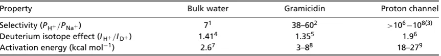
Table 2. Proton conduction in water and through gramicidin and voltage-gated proton channels
Property Bulk water Gramicidin Proton channel
Selectivity (PH+/PNa+) 71 38–602 >106−108(3)
Deuterium isotope effect (IH+/ID+) 1.414 1.355 1.96
Activation energy (kcal mol−1) 2.67 3–88 18–279
The activation energies listed are for proton permeation through open channels. The temperature dependence of gramicidin includes measurements of several modified dioxolane-linked channels.1Robinson & Stokes, 1959;2Myers
& Haydon (1972);3DeCoursey (2003);4Lewis & Doody (1933); 5Akeson & Deamer (1991); Chernyshevet al.2003; 6DeCoursey & Cherny (1997);7Robinson & Stokes (1959);8Akeson & Deamer (1991); Chernyshev & Cukierman, 2002; 9DeCoursey & Cherny (1998).
acids as well as intervening water molecules. An HBC mechanism that included side chains would enable perfect proton selectivity; other ions are excluded (Nagle & Tristram-Nagle, 1983).
In Table 2, proton conduction through the voltage-gated proton channel is compared with proton conduction through the water-filled gramicidin channel, as well as with proton conduction in bulk water. The gramicidin channel has been useful as a model ion channel – it is relatively small, structurally simple and robust – it still functions at 5–6M HCl (Eisenman et al. 1980;
Cukierman et al. 1997)! The gramicidin channel has a narrow cylindrical pore that is filled with a single-file row of a dozen water molecules (Levitt et al. 1978). Gramicidin is a non-selective cation channel, but it conducts protons much better than any other ion, because protons can hop through a water wire without displacing the water molecules. In contrast a Na+ or K+ ion must wait patiently for the water molecules to diffuse through before it can permeate. For roughly the same reason, protons diffuse in bulk water 5 times faster than K+ (Danneel, 1905; Robinson & Stokes, 1959). Gramicidin conducts up to 2×10⁹H+s−1(Cukierman, 2000), faster than any other narrow-pore channel conducts any other ion. The impression that any proton that appears at the mouth of gramicidin permeates is supported by a nearly direct proportionality between proton current and proton concentration over 5 orders of magnitude (from pH 4.5 to pH−0.5) (DeCoursey, 2003). Proton conductivity of bulk water is similarly proportional to concentration up to pH∼0 (Owen & Sweeton, 1941). In contrast, although the single-channel conductance of voltage-gated proton channels increases at lower pHi, the increase is only 4-fold between pHi6.5 and 5.5 (Chernyet al.2003). It would be intriguing to obtain single-channel currents at lower pH, but biological membranes seem unwilling to cooperate in this endeavour.
The impression from Table 2 is that proton permeation through gramicidin is not too different from conduction in bulk water, but permeation through voltage-gated proton channels is something completely different. Deuterons permeate both gramicidin and voltage-gated proton channels, but the isotope effect is substantially greater
for permeation through voltage-gated proton channels. The temperature dependence of the open-channel conductance is stronger for the voltage-gated proton channel than for almost any other ion channel. Together with these factors, the concentration dependence mentioned above supports the conclusion that traversing voltage-gated proton channels is challenging for protons. The contrast between proton permeation through gramicidin, a water-filled ion channel, and voltage-gated proton channels has led to the proposal that the conduction pathway in the latter is not likely to comprise a simple water wire, but instead may comprise a HBC that includes at least one titratable group (DeCoursey & Cherny, 1994a,b, 1997, 1998; Henderson & Meech, 1999; Cherny et al. 2001b; Maturana et al. 2001; DeCoursey, 2003; Demaurex & Pethe¨o, 2005).
Other proton selectivity mechanisms may exist, however. The perfect proton selectivity of the M2 viral proton channel (Chizhmakov et al. 1996; Lin & Schroeder, 2001; Mould et al.2000) has been explained by two different mechanisms. One is a traditional HBC mechanism in which protonation/deprotonation of a ring of His residues ensures selectivity (Pinto et al.
1997; Schweighofer & Pohorille, 2000; Shuck et al.
2000; Lear, 2003). Alternatively, channel opening might complete a water wire that experiences a constriction that is sufficiently narrow to prevent cation (or water) permeation, but still permits proton transfer between adjacent waters (Sansom et al. 1997; Kukol et al. 1999; Smondyrev & Voth, 2002; Kass & Arkin, 2005; Chenet al.
2007; Stoufferet al.2008).
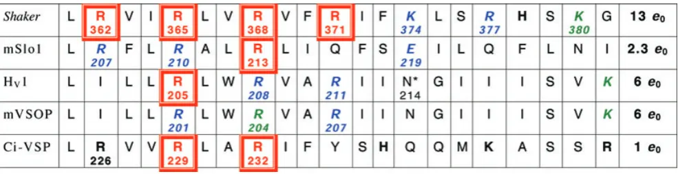
Table 3. S4 regions of voltage-gated K+and H+channels and a voltage-sensing phosphatase (VSP)
A sequence of 21 amino acids in the putative S4 regions of theShakerK+channel, the maxi-K calcium-activated K+channel (mSlo), the human proton channel (HV1), the mouse proton channel (mVSOP), and a voltage-sensing phosphatase inCiona(Ci-VSP). Potentially
charged amino acids are indicated in bold. Single neutralization mutations at the numbered positions in red boxes reduce the gating charge; those in italics (in blue) do not affect gating charge, or were inconclusive (in green) (Aggarwal & MacKinnon, 1996; Seohet al. 1996; Bezanilla, 2000; Murataet al.2005; Ramseyet al.2006; Sasakiet al.2006; Maet al.2006; Mussetet al.2008a; Hossainet al. 2008). Total gating charge (e0) estimates are listed in the final column; for HV1 and mVSOP, these are from the limiting slope method
(Mussetet al.2008a). The mouse R204Q mutant did not express well; R207Q was identical to wild type, and for R201Q activation was faster and thegH–Vrelationship was shifted by−50 mV and had a slightly steeper slope (z=1.9versus1.4 for wt) (Sasakiet al.2006).
In human proton channels, gating was faster for all three Arg mutants, and for R205A the midpoint was shifted positively, and the slope (fromgH–V relationships) was less steep by 1/3 (zδ=0.57versus0.90 for wt) (Ramseyet al.2006). The VSD of the Shaker K+
channel can be transformed into a proton channel by the R362H mutation (Starace & Bezanilla, 2004), and R362X where X=Cys, Ala, Ser or Val produces non-selective cationic ‘omega’ current through the voltage sensor (Tombolaet al.2005).∗The N214R mutation greatly attenuates conduction (Tombolaet al.2008). A ClustalW alignment of proteins containing S4 regions homologous to HV1 was
manually adjusted to reflect predicted transmembrane regions. In some cases, plausible alternative alignments can be obtained by shifting a sequence by three residues. These previously unpublished alignments were generously provided by S. M. E. Smith (Emory University).
Arg→His are alternately exposed to internal or external solutions during gating, whereas Arg→His positioned on either end apparently forms a solitary constriction that has simultaneous access to both sides of the membrane, one in the closed (R362H) and the other in the open (R371H) conformation of the central K+ pore. If R362 is mutated into any of several other amino acids, the result is non-selective cation current through the VSD (Tombolaet al.2005, 2007). At two other locations in the VSD, Ile→His mutations (I241H in S1 and I287H in S2) produce proton channels (Camposet al. 2007). Finally, in the VSD of voltage-gated Na+ channels, Arg→His mutation of one of the S4 residues (R663H) produces a proton selective channel (Struyk & Cannon, 2007), whereas Arg→X mutation produces non-selective cation current (Sokolov et al. 2005). It is difficult to imagine that the proton selectivity that is uniquely achieved by positioning a His residue at these locations does not result from protonation/deprotonation of the His during conduction. Unfortunately, it is also difficult to conceive an experiment that would reveal with certainty whether this is the case.
Finally, there are several recent examples of non-selective channels that conduct protons paradoxically well, including TRPV1 (Hellwiget al. 2004), TRP-ML1 (Soyombo et al. 2006), TRPM7 (Jiang et al. 2005; Numata & Okada, 2008) and colicin A (Slatin et al.
2008). The relative proton permeability, PH+/PCs+>1000 for TRPV1 and PH+/PCs+=104−106 for TRP-ML1, is astonishing, and that PH+/PK+=12 000 for colicin A is truly astounding in view of this channel having a lumen ∼10 ˚A in diameter (Slatin et al. 2008). Whether the proton permeability of these channels can be explained by conventional paradigms remains to be determined.
Where is the permeation pathway? Answering this question would be facilitated by knowing the structure. Although many multimeric channels form their conduction pathway at the interface between subunits, as discussed above, the proton channel appears to be a dimer and each subunit has its own pore (Koch et al.
2008; Tombola et al.2008; Lee et al.2008). At present, no published data exist that indicate which amino acids form the conduction pathway, although the N214R mutation greatly attenuates conduction, and thus may be near the inner mouth of the ‘pore’ (Tombolaet al.2008). The general similarity of the proton channel molecule to the voltage-sensing domain of a K+channel (Sasakiet al.
or internal solutions depending on protein movements that occur during gating (channel opening and closing). If the architecture of the proton channel follows this pattern, then the general framework for a proton pathway may reasonably occur in each monomer.
What is the concentration dependence of the proton conductance? Examination of macroscopic gH data in many studies indicated that gH,max – the limiting gH – increased only 2-fold per unit decrease in pHi(DeCoursey, 1998). The prediction of the Goldman–Hodgkin–Katz equation (Goldman, 1943; Hodgkin & Katz, 1949; Hille, 2001) is a 10-fold increase, which presumes that entry into the channel is rate determining. This discrepancy suggested that permeation, rather than entry, was rate determining. The implicit assumption is that the same number of channels is open during a large depolarization at any pHi. Surprisingly however, when determined at various pHi values, the single-channel conductance increased from 38 fS to 140 fS at pHi 6.5 and 5.5, respectively, nearly a 4-fold increase (Chernyet al.2003). The open probability was 75% and 95%, respectively, in these conditions. The macroscopic and microscopic conductances can be reconciled if there is an inhibitory process at low pHithat removes functional channels from the fray. Systematic study of this issue would be welcome, but has been hampered because biological membranes do not tolerate extremely low pH. Perhaps incorporation of proton channels into artificial lipid bilayers would enable direct observation over a wider pH range.
At the other extreme, it is curious at how high pHi one could still detect proton currents. At high pH, free protons are exceedingly scarce and buffer (used at its pKa) holds on to its proton tightly. At sufficiently high pH, deprotonation of buffer must become rate determining, at which point proton current should decrease in direct proportion to [H+] (Br¨onsted & Pedersen, 1923; Eigen & Hammes, 1963), assuming that buffer protonation is diffusion limited. Our attempts to probe this question have been limited by two factors: (1) cells do not survive well at extremely high pH; and (2) it is necessary to monitorVrev to determine how well one has established the intended pH. Credible outward proton currents with Nernstian (i.e. reasonable)Vrev have been seen in excised patches at pHi8.5 with pHo7.5 (V. V. Cherny & T. E. DeCoursey, unpublished data), where only 3 nMpermeant ion (H+) was present.
Voltage gating
Is S4 the voltage sensor? It is well established that the S4 region contributes substantially to the ability of several voltage-gated ion channels to sense membrane potential (Noda et al. 1984; St¨uhmer et al. 1989). This said, it
should be emphasized that charged residues elsewhere in K+ channels also contribute (Seoh et al. 1996), and the mechanism of gating remains controversial. As shown in Table 3, every third amino acid in S4 of the Shaker
K+ channel is a potentially charged Arg or Lys and each of the first four Arg residues appears to contribute to voltage sensing (Aggarwal & MacKinnon, 1996; Seohet al.
1996; Bezanilla, 2000). Three of these Arg residues are conserved in the voltage-gated proton channel (Table 3). However, when these were mutated individually to neutral amino acids, only one appeared to reduce the effective charge moved during gating (channel opening) in the human channel (Ramseyet al.2006), and none of them contributed in the mouse channel (Sasaki et al. 2006). One limitation of these studies is that they were based on Boltzmann fits togH–V relationships, which may not capture the entire gating charge moved (Sigworth, 1993). Preferable estimates are from the limiting slope of theg–V
relationship (Almers, 1978; Sigworth, 1993). The gating charge movement estimated by this method is 12–14e0for Na+or K+channels (Hirschberget al.1995; Schoppaet al.
1992; Aggarwal & MacKinnon, 1996; Seohet al.1996) and 5.4–8e0 for voltage-gated proton channels (DeCoursey & Cherny, 1996b, 1997; Mussetet al.2008a). Even more direct estimates of gating charge movement come from gating current measurements (Sigworth, 1993). Gating current occurs when channels open or close, but must be isolated from ionic current, for example by blockers. However, this measurement is nearly impossible for voltage-gated proton channels, because: (a) it is impossible to abolish ionic current by eliminating the permeant ion; (b) reducing the permeant ion concentration shifts the
gH–V relationship (see Sensitivity of gating to pH) rather than permitting gating in the absence of ionic current; (c) attempts to measure gating currents at
EH (the Nernst potential for H+), which would pre-clude ionic current, still did not reveal detectable gating current, probably because gating is very slow (V. V. Cherny, V. Sokolov & T. E. DeCoursey, unpublished data).
major participant in voltage sensing in proton channels. A similar conclusion was reached for BK channels, although their total gating charge is relatively petite (Maet al.2006). Proton current kinetics changed fairly dramatically even in the Arg mutations that did not affect total charge, however (Sasaki et al. 2006; Ramsey et al. 2006), suggesting that these groups do participate in gating. In summary, the voltage-sensing apparatus and its mechanism in proton channels are largely unknown.
How does gating work? Voltage-gated proton channels open and close like other ion channels. Single channel currents of∼10 fA can just barely be resolved under highly favourable conditions: in excised patches, at low pHi, with seal resistances in the teraohm range, and with no other conductances present (Chernyet al.2003). Proton channel gating generates current fluctuations that exhibit archetypal stochastic behaviour. Distinct fluctuations (noise) appear at voltages at which thegHis activated, and the variance of this noise increases to a maximum near the midpoint of the gH–V relationship, precisely where one would expect the frequency of opening and closing transitions to be maximal. Beyond this, almost nothing is known about the gating mechanism. Given the likelihood of HBC conduction, a proton conduction pathway might appear or disappear in ways that would not be effective for gating conduction of other ions, so we need to keep an open mind. For example, subtle changes in the length or orientation of hydrogen bonds (Pauling, 1939; Scheiner, 1981) or in the pKaof groups in the pathway could enable or disable a HBC.
The similarity between the proton channel and the VSD, combined with the demonstration that the K+ channel VSD can be converted into a proton-selective channel by single mutations of Arg residues in S4 (Starace & Bezanilla, 2001, 2004) raises the obvious possibility that movement analogous to that which is thought to occur during VSD gating also occurs in the proton channel and results in channel ‘opening.’ Changes in accessibility of the four key Arg residues during K+channel gating led to the conclusion that they ratchet outwards past a solitary constriction, with the first and fourth Arg at the narrow point when the K+channel is closed or open, respectively (Larssonet al.1996; Bezanilla, 2000; Horn, 2005; Tombola
et al.2006). Tombolaet al.(2008) found that in HV1, the N214R mutation abolished conduction and proposed that the proton channel opens when the polar Asn214 moves into this hypothetical constriction.
Some constraint on the gating process is placed by the extraordinarily strong temperature dependence of gating. Three parameters describing proton channel gating kinetics – the delay and activation time constant τact obtained from fitting proton current turn-on with a delay followed by a rising exponential, and τtail obtained by fitting tail current decay with a single exponential – all
had activation energies of 30–38 kcal mol−1 (Q
10 values of 6–9) in six types of cells (DeCoursey & Cherny, 1998). Activation energies for the opening and closing of most ion channels, including K+ channels (Bezanilla, 2000), are only about half this large. That all kinetic parameters for proton channel gating had similar large activation energies suggests that both opening and closing involve a single rate-limiting, energetically demanding transition that occurs in multiple independent subunits. Whether the two pores of the proton channel dimer gate independently or cooperatively has not been established.
What is the real kinetics of proton channels? From the beginning, it has always been evident that proton currents are prone to manifestations of depletion of the permeant ion. Since in many situations the purpose of proton currents is to change pH, this property should not come as a surprise. Nevertheless, someone who is familiar with K+ currents for which the cellular concentration of permeant ion is 10−1M is in for a rude awakening when studying channels for which the cellular concentration of permeant ion is 10−7M. One should first of all be impressed that proton currents are as large as they are – in many cells they are as large or larger than any other conductance present, including K+ currents (Byerly & Suen, 1989; DeCoursey, 1991; Demaurexet al.1993; DeCoursey & Cherny, 1994b, 1996a; Femlinget al.2006). The conjunction of large currents and minuscule permeant ion concentration leads to depletion of the permeant ion. The most obvious manifestation of depletion is decay or droop of proton currents. Proton channels do not inactivate in any tissue studied to date (p. 532, DeCoursey, 2003). However, proton currents often decay during sustained depolarization. That the cause is proton depletion-induced pH changes has been verified by measuring pHiby pH electrode (Thomas & Meech, 1982; Meech & Thomas, 1987), by fluorescent dye (Demaurex
et al.1993; Kapuset al.1993; Schwiening & Willoughby, 2002), or by using the proton channel as a pH meter and measuringVrev of proton currents (Barish & Baud, 1984; DeCoursey, 1991; Kapuset al.1993; DeCoursey & Cherny, 1994b; Humezet al.1995; Gordienkoet al.1996; Morihataet al.2000; Schrenzelet al.1996). There seems to be a spontaneous tendency for researchers to feel guilty about proton depletion, and to attempt to conceal droopy currents, for example. On the one hand, it makes sense to try to minimize depletion, because it compromises the measurements. On the other hand, it is dangerous to pretend that depletion is not occurring, because this can lead to gross misinterpretation of data.
to buffer. A 10μm diameter spherical cell dialysed with 100 mMbuffer at its pKa contains only 315 000 free H+ at pH 6 (DeCoursey, 2003). These protons can carry a 100 pA H+ current for only 0.5 ms. Although there are 1.6×10¹⁰ protonated buffer molecules in the cell, 6.25×10⁸(4%) are consumed each second of the 100 pA H+ current. If they are not replenished by diffusion from the pipette solution, pHiwill increase. (3) Finally, proton channels in most mammalian cells open quite slowly, with activation time constants,τact, in the range of seconds, in contrast with a few milliseconds in snail neurones (Byerlyet al.1984). By the time what appears to be a steady-state level of activation is achieved during the pulse, depletion has already occurred. Figure 2Aillustrates this phenomenon. In order to determine the voltage dependence of ‘steady-state activation’ of the gH, one typically applies a family of pulses. The current at the end of the pulse can be used to calculate the conductance, usingVrevmeasured previously by the tail-current method (Hodgkin & Huxley, 1952). These values are plotted as blue circles, and they appear to saturate and could be fitted by a Boltzmann function, as we are wont to do. However, the gH ought to be identical at the start of the tail current, and many purists prefer measuring tail current amplitude, because this precludes worries about open-channel rectification. These values are plotted as red diamonds and they show little inclination to saturate. What has happened, of course, is that each pulse removed protons from the cytoplasm, so that pHi was higher at the end of the pulse, and probably also at the start of the next pulse than it was for the previous pulse. The estimates ofVrevin Fig. 2Bconfirm a 9 mV depolarizing shift ofVrevby the end of the pulse family. Consequently, calculatinggHusing a singleVrevvalue does not reflect the true situation. We can attack this problem by realizing that we could use these interpolatedVrevvalues for each pulse, and obtain the truegHat that instant (green squares). The only problem is that our goal was to determine thegH at one particular pHi, not over a range of continuously changing pHithat spans 0.15 pH units. This distinction is not trivial, because as we will see in the next section, the position of thegH–V relationship depends strongly on pHi.
Obviously, increasing the buffer concentration reduces the rate and extent of depletion (DeCoursey, 1991; Kapus
et al.1993; Demaurexet al.1993; DeCoursey & Cherny, 1994b, 1996b; Kunoet al.1997). Most labs routinely use ≥100 mM buffer, which reduces but does not prevent
depletion; in the experiment in Fig. 2 there was 100 mM
buffer in all solutions. Figure 3 illustrates the effects of buffer concentration on proton currents in an excised inside-out patch of membrane. Several features are remarkable. First, it is clear that depletion occurs even in excised patches. One can easily imagine that in whole-cell configuration depletion might be a problem, but both H+
[image:9.595.312.546.209.555.2]currents and unstirred compartment volumes ought to be much smaller in patches. Still, the membrane migrates some distance up from the tip of the pipette (Milton & Caldwell, 1990; Ruknudinet al.1991), so there may be a substantial unstirred volume from which protons will be removed by depolarizing pulses (in inside-out patches). The impression from Fig. 3Ais that the pulses begin well, but increasingly, the current at lower [buffer] sags below the one at higher [buffer]. Although frank droop is seen only during the pulse to+60 mV, it is glaringly obvious
Figure 2. Proton depletion complicates measurement of the voltage dependence of the proton conductance,gH
A, thegH–Vrelationship estimated by three methods at each of three
pHovalues in a COS-7 cell transfected with the mouse proton channel
gene, mVSOP. The pipette contained pHi6.5 with 100 mMBisTris
buffer. At pHo8.0, 2 s pulses were applied in 5 mV increments every
20 s fromVhold= −100 mV. At pHo7.0 or 6.0,Vholdwas−60 or −40 mV, respectively, and 6 s pulses were applied every 20 s. As indicated in the inset,gHwas estimated from proton current
measured at the end of each pulse (Iend, blue circles) or at the start of
the tail current (Itail, red diamonds) assuming a single constant
measured value forVrev. Alternatively,gHwas calculated as the slope
conductance (green squares) betweenIendandItail, which does not
require an estimate ofVrev.B, actualVrevestimated by theXaxis
intercept of a line connectingIendandItailfor the indicated pulses in
that the time course of the current is radically different at low [buffer], even at+20 mV where the current is much smaller. Clearly, lack of droop does not signify that the time course of the current reflects pure gating kinetics. At lower [buffer],τact will be artificially fast. In Fig. 3B, we see that 10 mMbuffer was not enough, despite the normal
appearance of the family in Fig. 3A, because the currents are larger and ‘activate’ even more slowly at 100 mMbuffer.
We sometimes use 200 mMbuffer, after Gabor Pethe¨o and
Nic Demaurex showed us this was possible, but even then, can we be sure that the time course of the current reflects the true time course of gating? The deconvolution of the true gating kinetics of proton channels from diffusional effects on the time course of recorded currents may require serious modelling. In the meanwhile, it is best to remember that much of what you see is not a direct reflection of gating kinetics, and that one must guard against being seduced into over-interpreting data.
[image:10.595.45.284.438.574.2]Depletion is more pronounced in whole-cell configuration. Proton accumulation outside the cell (Zifarelliet al.2008b) and proton depletion inside the cell (Swietachet al.2003) during constant proton efflux have been modelled. Certainly depletion during proton efflux is more severe inside the cell, and replenishment depends on diffusion of protonated buffer from the pipette solution, whose rate of equilibration has also been evaluated (Pusch & Neher, 1988). The effective diffusion coefficient for protons in cells is slowed by the effects of fixed and mobile intrinsic buffers (Junge & McLaughlin, 1987) by at least
Figure 3. High buffer concentration reduces but does not eliminate depletion
These records were obtained in inside-out patches of membrane from two rat alveolar epithelial cells, at pHo7.5 and pHi6.5. As one might
expect, diffusion limitation is less severe in patches than in whole-cell configuration. However, depletion still occurs in patches.A, the currents in were recorded at the indicated voltages during pulses from a holding potential of−40 mV with [buffer] in the bath
(corresponding to intracellular) either 10 mM(continuous lines) or 1 mM(dotted lines).B, proton currents in another patch during pulses to+60 mV at the indicated BisTris concentrations. Note the radically different kinetics at different buffer concentration. Pulses were applied at 100 mMbuffer before and after the other concentrations were applied. (From DeCoursey & Cherny, 1996b.)
two (Vaughan-Jones et al. 2002; Swietach et al. 2003) or even three orders of magnitude (Stewartet al.1999), particularly at low pH (Swietach & Vaughan-Jones, 2005; Swietachet al.2007) where the intrinsic buffering capacity is increased (p. 483, DeCoursey, 2003). In practical terms, establishing control of pHi by 100 mM buffer requires 8–10 min in large cells of diameter 120μm (Byerly & Moody, 1986), and 1–3 min in small cells of diameter 10–20μm (Demaurex et al. 1993; Kapus et al. 1993), depending on pipette geometry. Adequate control is never achieved at low (<20 mM) buffer concentration (Byerly &
Moody, 1986; DeCoursey, 1991; Demaurexet al.1993). The inevitability of depletion has several consequences. As mentioned, one must be alert to this phenomenon and not over-interpret data. With depletion in mind, we often use the threshold for channel activation,Vthreshold, as a rough indicator of the voltage dependence of gating. This parameter is arbitrary, because if gH decreases exponentially with hyperpolarization, no true threshold exists; the Vthreshold detected will depend on gH,max and noise levels. However, Vthreshold is a useful and practical compromise for several reasons. It is easy to determine. It precludes the need for large depolarizations that cause extensive depletion. By definition, currents nearVthreshold are very small and produce little depletion. It does not assume any specific gating model, nor does it require force-fitting data to a Boltzmann function. In practice,
Vthresholdgave results similar to more traditional methods of analysis (Mussetet al.2008a). We suspect thatVthreshold is a more reliable indication of the voltage dependence of proton channel gating than parameters obtained from Boltzmann fits, which are strongly influenced by depletion and the specific conditions of the measurements.
Sensitivity of gating topH
Which groups sense pHo and pHi? A quintessential
feature of voltage-gated proton channels is that their voltage dependence is strongly regulated by the pH gradient, pH (defined as pHo– pHi) (Cherny et al. 1995). ThegH–Vrelationship shifts 40 mV per unit change in pH over a wide range encompassing all physio-logically attainable values. The result of this extraordinary regulation by pH is that proton channels open only when the electrochemical proton gradient is outward, when opening will result in acid extrusion from the cell. Consequently, it is universally accepted that acid extrusion, especially in situations of excessive metabolic activity, is a major function of voltage-gated proton channels. All native voltage-gated proton channels share the property of pH-dependent gating, and the voltage at which the gH first turns on, Vthreshold, can be predicted by:
that protonation of sites on the channel accessible to the external solution stabilizes the closed channel and protonation from the inside stabilizes the open channel. The model works only if the regulatory protonation sites are accessible to just one side of the membrane at a time, and accessibility is transferred by an unspecified conformational change in the protein (Chernyet al.1995). At this moment, we have no idea what or where these protonation sites are, or whether these hypothetical sites exist at all.
An informal survey of amino acid mutations that significantly alter pH sensing in 35 membrane proteins revealed the following: 20 involved His residues, singly or in clusters of up to six, 15 involved Glu, 7 Asp, 6 Arg, 6 Lys, and 3 Gly. The apparent preference for Glu over Asp is surprising, but may be fortuitous. In some cases, a single residue seems to provide the entire pH sensitivity. In many others, the pH sensitivity seems to involve a complex inter-play between groups of nearby amino acids; sometimes mutation of only one residue of the group has no effect. The ClC-0 chloride channel is sensitive to pHi, but when mutation of 22 candidate amino acids failed to reveal a sensor, the idea of a proton sensor was abandoned in favour of a different mechanism involving OH− (Zifarelliet al.
2008a).
How do the pH sensors communicate with the voltage sensor? The mechanism of this coupling has not even been imagined. One can hardly speculate on this mechanism, despite its obvious importance, because we do not know what or where the sensors are or how gating works.
Pharmacology
Zinc’s secrets revealed! One structure–function question has been solved, namely the location of the external Zn2+ binding site on the human proton channel. Zn2+is among the most potent inhibitors of voltage-gated proton channels (Mahaut-Smith, 1989b). The effects of Zn2+ on proton currents closely resemble the effects of poly-valent cations on nearly all voltage-gated ion channels; namely, thegH–V relationship is shifted positively and the turn-on of current during depolarizing pulses is slowed (largerτact). Qualitatively, these effects correspond to the electrostatic effects of positioning two positive charges at the outer end of the channel – with the result that the voltage sensor is tricked into thinking the membrane potential is more negative than it really is (Frankenhaeuser & Hodgkin, 1957). The efficacy of Zn2+in inhibiting the proton channel is greatly reduced at low pHo, and the details of the competition between H+and Zn2+could be explained only by assuming that the external Zn2+binding site comprised 2 or 3 (but not 1) titratable groups with pKa
6.2–7.0, suggesting His residues (Cherny & DeCoursey, 1999). The human proton channel turns out to have two His residues that are accessible to the external solution, and their mutation individually to Ala lowers Zn2+ sensitivity by an order of magnitude; the double His→Ala mutant has only weak Zn2+ sensitivity (Ramsey et al. 2006).
To a rough approximation, mouse and human proton channels have similar Zn2+sensitivity (Ramseyet al.2006; Sasakiet al.2006), but the Cionaproton channel is less sensitive (Sasakiet al.2006). There are several His residues at suggestive locations in the mouse proton channel, and fewer in theCionachannel (Sasakiet al.2006; DeCoursey & Cherny, 2007), but whether this explains the weaker Zn2+binding remains to be determined.
The internal Zn2+binding site has not been identified. Applied intracellularly, Zn2+has less dramatic effects that are qualitatively consistent with surface charge effects, namely shifting thegH–V relationship negatively, slowing tail current decay (largerτtail) and decreasing the limiting
gH, gH,max (Cherny & DeCoursey, 1999; Pethe¨o et al. 2003).
What about a potent, selective inhibitor? Unfortunately, Zn2+ is not a very selective inhibitor, which greatly hampers attempts to evaluate possible functions of proton channels. A potent and selective toxin or venom would be very handy, but so far, none has been discovered.
How are H+channels regulated?
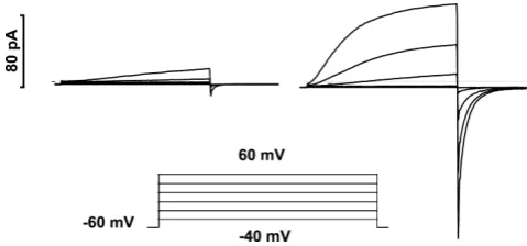
[image:11.595.309.549.521.633.2]Where is(are) the phosphorylation site(s)? In phagocytes, voltage-gated proton channels can be transformed into an ‘enhanced gating mode’ that seems to coincide with NADPH oxidase activity (B´anfiet al.1999). As illustrated in Fig. 4, agents that activate NADPH oxidase, such as PMA
Figure 4. Agents that activate NADPH oxidase in phagocytes also greatly enhance the opening of proton channels
Identical families of pulses (inset) were applied to a human eosinophil before (left) and after (right) addition of PMA to the bathing solution. The enhanced gating response can be observed in perforated-patch configuration, but not whole-cell. This cell was exposed to a pH 7.0 bath solution with 50 mMNH4+on both sides of the membrane to
or arachidonic acid, profoundly alter the gating of proton channels. PMA shifts thegH–V relationship by−40 mV, speeds activation, slows deactivation and increasesgH,max (DeCourseyet al.2000, 2001a,b; Bankers-Fulbrightet al.
2001; Cherny et al. 2001a). These effects are prevented or largely reversed by the PKC inhibitors GF109203X (GFX) or staurosporine (Bankers-Fulbright et al. 2001; Mori et al. 2003; Morgan et al. 2007), which suggests that phosphorylation by PKC of either the channel or a regulatory molecule creates the enhanced gating mode. On a more phenomenological level, PKC stimulates both NADPH oxidase activity and proton efflux in intact human neutrophils (Hendersonet al.1987; Nanda & Grinstein, 1991; Kapuset al.1992). Accepting that phosphorylation is a major mechanism of proton channel regulation, where is(are) the phosphorylation site(s)? The next question is, by what mechanism do these sites exert such a strong influence on proton channel gating? Does the level of ‘activation’ of proton channels reflect a balance between phosphorylation and dephosphorylation? If so, which kinases and phosphatases are involved?
[image:12.595.44.372.513.720.2]Does phosphorylation account entirely for proton channel ‘activation’ in phagocytes? When GFX, a fairly selective PKC inhibitor, is applied to an activated eosinophil, the electron current turns off completely (Fig. 5), indicating that sustained NADPH oxidase activity requires ongoing PKC activity. At the same time, the enhanced features of proton currents are largely, but not completely reversed (Morgan et al. 2007). Two explanations for the less complete reversal of proton channel modulation are possible. First, it might be that deactivation reflects the activity of phosphatases, and those that dephosphorylate proton channels are less active then those that turn off NADPH oxidase. The phosphatase inhibitor, okadaic acid, does slow the reversal
Figure 5. The enhanced gating mode is partially reversed by the PKC inhibitor, GFX
From a holding potential of−60 mV, test pulses were applied every 30 s to+60 mV, resulting in the upward deflections. Where indicated, 60 nMPMA was added, stimulating
increased proton current as well as inward current at−60 mV. This inward current (shown at amplified gain below) is electron current that is generated by the activity of NADPH oxidase, and can be inhibited by the NADPH oxidase inhibitor DPI (diphenylene iodonium). Examples of proton currents during test pulses at right are labelled with lower case letters that show the time each was recorded. At the second arrow, 3μMGFX was added. (From Morgan
et al.2007.)
of proton current enhancement by GFX (Morgan et al.
2007). Alternatively, the activation of proton channels may involve something more than PKC phosphorylation. If there are additional or alternative pathways of activating proton channels, what are they?
What is the relationship between proton channels and NADPH oxidase? This is one of the most intriguing mysteries regarding the activity of proton channels. As mentioned in ‘Has the right gene been identified?’ the preponderance of evidence speaks against gp91phox functioning as a proton channel. However, there remain several inexplicable interactions between these two molecules. The existence of these interactions contributed substantially to the attraction of the idea that gp91phox might be a proton channel.
2. Proton currents are converted into the enhanced gating mode by numerous activators of NADPH oxidase (Figs 4 and 5). NADPH oxidase activity was first associated with proton channel ‘activation’ in a whole-cell study in which ingredients supportive of NADPH oxidase activity were included in the pipette solution (B´anfiet al. 1999). In perforated-patch studies, agonists that activate both NADPH oxidase and proton channels include: PMA, AA (arachidonic acid), oleic acid, LTB4 (leukotriene B4), IL-5 (interleukin-5), fMLF (formyl-methionyl-leucyl-phenylalanine, a chemo-tactic peptide) and spontaneous activation presumably due to adherence (DeCoursey et al. 2000, 2001a,b; Bankers-Fulbrightet al.2001; Chernyet al.2001a; Mori
et al.2003; Morganet al.2007).
3. Proton channels can sense whether NADPH oxidase is working or not. One of the characteristics of proton channels in the enhanced gating mode is profound slowing of tail current decay, withτtail increasing by 4- to 6-fold (B´anfiet al.1999; DeCourseyet al. 2000, 2001a, 2001b; Morganet al.2007). When the NADPH oxidase inhibitor DPI is introduced, the electron current turns off and at the same time,τtail returns toward its original rapid kinetics (DeCourseyet al.2000, 2001a). DPI only affects ‘activated’ proton channels, not resting channels in unstimulated cells (DeCourseyet al.2000). However, there is no immediate effect of DPI on the other enhanced gating properties of proton channels. The association of slowing of τtail with electron currents is uncanny. In experiments on differentiated HL-60 cells, a fraction of cells responded to PMA stimulation with characteristic changes in proton currents seen in other phagocytes;τtailslowed appreciably only in cells in which electron current was detected (V. V. Cherny, D. Morgan and T. E. DeCoursey, unpublished data).
4. On the other hand, all of the features of enhanced gating except forτtail(including the−40 mV shift of the
gH–Vrelationship that produces inward currents) remain enhanced when NADPH oxidase is inhibited by DPI or by removal of substrate O2 (B´anfi et al. 1999). Thus, most enhanced gating features do not require NADPH oxidase activity (electron current).
5. An extension of this phenomenon is observed in granulocytes from patients with CGD. Stimulation of neutrophils or eosinophils from CGD patients by PMA produces most of the enhanced gating features of proton channels that occur in normal cells, except the negative shift of thegH–V relationship is smaller and τtail does not slow appreciably (DeCourseyet al.2001b; DeCoursey, 2003). By definition, there is no electron current in these cells. Presumably the only difference between these and normal cells is the dysfunction of NADPH oxidase, yet the proton channel response is different.
6. A similar phenomenon occurs in human baso-phils. These leucocytes are developmentally related to
eosinophils and, more distantly, to neutrophils, but they do not express detectable levels of NADPH oxidase (de Boer & Roos, 1986). In a recent study, we found that baso-phils respond to PMA almost identically to CGD cells: the shift ofVthresholdis only−20 mV instead of−40 mV, and τtailis slowed negligibly (Mussetet al.2008b).
7. In non-leukocytic cells that do not express high levels of NADPH oxidase, there is little evidence of the enhanced gating mode. So far, this has been tested only in rat alveolar epithelial cells (DeCourseyet al.2000), and in HEK-293 and COS-7 cell lines (Mussetet al.2008a). Nevertheless, the coincidence of proton channel enhancement with NADPH oxidase expression is striking.
In the case of proton channels sensing NADPH oxidase activity (no. 3 above), one proposed mechanism is via local pH (DeCourseyet al.2000). NADPH oxidase activity in phagocytes generates large quantities of cytoplasmic protons (van Zwieten et al. 1981; Gabig et al. 1984; Borregaard et al. 1984), and according to the model of proton channel gating, the effect of intracellular protons is to stabilize the open proton channel (Cherny et al.
1995). If sufficient protons were generated in the vicinity of proton channels, this could account forτtail slowing, at least qualitatively. Rough calculations do not support the likelihood of sufficiently large pH changes if random distribution of both molecules in the plasma membrane is assumed (p. 553–554, DeCoursey, 2003), but any number ofad hoc explanations could be offered. In summary, no mechanistic explanation for the cross-talk between proton channels and NADPH oxidase has been established.
How about PI(3,4)P2? The stability of the enhanced
gating mode of proton channels in inside-out patches appears to be supported by a combination of ATP, GTPγS and PI(3,4)P2 (phosphoinositide 3,4-bisphosphate). The mechanism was hypothesized to include interaction with the p47phox component of NADPH oxidase (Pethe¨o et al. 2006), adding to the intriguing question of the nature of the relationship between proton channels and NADPH oxidase. The details of the interactions of these regulatory molecules with proton channels need further clarification.
What are the functions of proton channels?
The identification of the proton channel gene now enables several new approaches to this question. Both groups who identified the gene (Ramseyet al.2006; Sasakiet al.
2006) have generated knockout mice. We eagerly await reports of the manifestations of this genetic loss. Of course, in many knockouts, the functions performed by the missing protein are compensated by other molecules that may not normally act in this capacity. But this puts the apologetics before the horse. Antibodies to external epitopes on the proton channel that alter function in intact cells would be very useful, but to my knowledge do not yet exist. It is now also possible to use siRNA (small interfering RNA, or silencing RNA) to knock down the activity of the channel. The cautionary note for this approach is that the knockdown must be nearly complete. Ion channels are activated (i.e. they open) in a probabilistic manner. Even if 63% (e−1) of all functional proton channels were eliminated, the cell would need only 4.2 mV additional depolarization – the limiting slope of thegH–V relationship (Mussetet al.2008a) – to activate the same proton conductance. Whether this small extra depolarization would significantly affect anything else in the cell is not clear.
The converse of eliminating proton channel function is increased proton conductance. In brown fat cells, a proton leak across mitochondrial inner membrane generates heat. The inappropriate appearance of proton current occurs with certain mutations that insert His into the VSD of a Na+ channel, and may be responsible for some of the consequences of hypokalaemic periodic paralysis (Struyk & Cannon, 2007).
In general, any of the consequences of proton channel activity may serve a useful function in a cell. These consequences include: (1) electrical effects, such as hyper-polarization of the membrane or charge compensation; (2) increasing pHi; (3) decreasing pHo; (4) osmotic effects; and (5) other specialized consequences. Specific situations have been identified in which each consequence of proton channel activity plays an important role.
Charge compensation. Opening proton channels will drive the membrane potential toward the Nernst potential for H+. Since proton channels open mainly when the electrochemical gradient for protons is outward, their activity tends to hyperpolarize the membrane. The importance of electrical effects has been shown abundantly in phagocytes, where proton efflux balances the electrical consequences of the electrogenic electron flux through NADPH oxidase. NADPH oxidase works by translocating electrons from cytoplasmic NADPH across the membrane to reduce O2to superoxide anion, O2·−(Hendersonet al. 1987). As soon as NADPH oxidase is activated, there is rapid and profound depolarization (Seligmann & Gallin, 1980; Whitinet al.1980; Lazzariet al.1986; Di Virgilioet al.
1987; Hendersonet al.1987; Puginet al.1997; Geisztet al.
1997; Jankowski & Grinstein, 1999; Bankers-Fulbright
et al.2003; Radaet al.2004; Demaurex & Pethe¨o, 2005), which is exacerbated by Zn2+(Hendersonet al.1987; B´anfi
et al.1999; Bankers-Fulbrightet al.2003; Radaet al.2004; Demaurex & Pethe¨o, 2005). Zn2+has no direct effect on NADPH oxidase (Yatsuyanagi & Ogiso, 1988; Schrenzel
et al.1998; DeCourseyet al.2003), but potently inhibits proton channels (Thomas & Meech, 1982; Mahaut-Smith, 1989b; Cherny & DeCoursey, 1999; DeCoursey & Cherny, 2007). When enough proton channels are open, H+efflux balances electron extrusion, and there is no further change in membrane potential. If the electronic charge were not compensated, depolarization would reach levels that directly inhibit NADPH oxidase by opposing electron translocation (DeCourseyet al.2003).
A recent speculative explanation for the need for proton channels in basophils is to compensate electrically for Ca2+ influx, which is required for anti-IgE-mediated histamine release (Mussetet al.2008b).
Acid extrusion (increasing pHi). The classical way to
demonstrate involvement of proton channels in acid extrusion is to load a cell with acid, either by the NH4+ pre-pulse method (Roos & Boron, 1981) or by direct injection of HCl (Thomas & Meech, 1982), and then observe the recovery. Under conditions in which other acid extrusion mechanisms are precluded, Zn2+ inhibits pHirecovery in many cells (Nandaet al.1992; Nordstr¨om
et al.1994, 1997; Demaurexet al.1996; Kunoet al.1997; Sheldon & Church, 2002; Murphyet al.2005; Chenget al.
2008). A complementary result is that transfecting mVSOP into HEK-293 cells greatly accelerated their recovery from an acid load (Sasakiet al.2006).
Acid secretion (lowering pHo). The distinction from acid
extrusion is mainly teleological – the former has a ‘goal’ of maintaining pHiin an optimal range for cellular functions, whereas the ‘intent’ of acid secretion is to regulate the acidity of the extracellular environment. In the case of respiratory epithelium, proton channel activity may serve to maintain the airway surface liquid at a moderately low pH (Fischeret al.2002; Schwarzeret al.2004), although additional functions have been proposed (Schwarzeret al.
2004; Fischer, 2007).
swelling is much less (Reeveset al.2002), it is clear that most of the compensation occurs by H+flux. The osmotic consequences of H+ flux into the phagosome are mild because most of the products of reactions involving H+ that occur in the phagosome (e.g. H2O, H2O2, HOCl) are membrane permeable.
Other consequences. Another important function of proton flux in phagocytes is to provide the H+ needed in large quantities as a substrate for the production of several reactive oxygen species (H2O2, HOCl, etc.) that kill microbes (Klebanoff, 2005; Nauseef, 2007; Radaet al.
2008). Proton flux into the phagosome delivers this sub-strate to the site of its utilization.
Conclusion
During the interval between the discovery of voltage-gated proton channels and the identification of their gene, many of the fundamental properties of these channels were determined. Now that the gene has been identified, a host of questions can be addressed, including the whole gamut of structure–function questions. Genetic approaches may facilitate further understanding of the functions of proton channels in the many cells that express them. The surprising similarity of the proton channel architecture to the VSD of other voltage-gated ion channels has the serendipitous effect that progress in understanding one type of channel has already and will continue to contribute in unpredictable ways to the understanding of other channels.
References
Aggarwal SK & MacKinnon R (1996). Contribution of the S4 segment to gating charge in theShakerK+channel.Neuron
16, 1169–1177.
Akeson M & Deamer DW (1991). Proton conductance by the gramicidin water wire: model for proton conductance in the F1F0ATPases?Biophys J60, 101–109.
Almers W (1978). Gating currents and charge movements in excitable membranes.Rev Physiol Biochem Pharmacol82, 96–190.
Babior BM (1991). The respiratory burst oxidase and the molecular basis of chronic granulomatous disease.Am J
Hematol37, 263–266.
B´anfi B, Maturana A, Jaconi S, Arnaudeau S, Laforge T, Sinha B, Ligeti E, Demaurex N & Krause K-H (2000). A mammalian H+channel generated through alternative splicing of the NADPH oxidase homolog NOH-1.Science287,
138–142.
B´anfi B, Schrenzel J, N¨usse O, Lew DP, Ligeti E, Krause K-H & Demaurex N (1999). A novel H+conductance in
eosinophils: unique characteristics and absence in chronic granulomatous disease.J Exp Med190, 183–194.
Bankers-Fulbright JL, Gleich GJ, Kephart GM, Kita H & O’Grady SM (2003). Regulation of eosinophil membrane depolarization during NADPH oxidase activation.J Cell Sci
116, 3221–3226.
Bankers-Fulbright JL, Kita H, Gleich GJ & O’Grady SM (2001). Regulation of human eosinophil NADPH oxidase activity: a central role for PKCδ.J Cell Physiol189, 306–315.
Barish ME & Baud C (1984). A voltage-gated hydrogen ion current in the oocyte membrane of the axolotlAmbystoma.J
Physiol352, 243–263.
Bernheim L, Krause RM, Baroffio A, Hamann M, Kaelin A & Bader C-R (1993). A voltage-dependent proton current in cultured human skeletal muscle myotubes.J Physiol470, 313–333.
Bezanilla F (2000). The voltage sensor in voltage-dependent ion channels.Physiol Rev80, 555–592.
Borregaard N, Schwartz JH & Tauber AI (1984). Proton secretion by stimulated neutrophils: significance of hexose monophosphate shunt activity as source of electrons and protons for the respiratory burst.J Clin Invest74, 455–459. Br¨onsted JN & Pedersen K (1923). Die katalytische Zersetzung
des Nitramids und ihre physikalisch-chemische Bedeutung.
Z Phys Chem108, 185–235.
Byerly L, Meech R & Moody W Jr (1984). Rapidly activating hydrogen ion currents in perfused neurones of the snail,
Lymnaea stagnalis.J Physiol351, 199–216.
Byerly L & Moody WJ (1986). Membrane currents of internally perfused neurones of the snail,Lymnaea stagnalis, at low intracellular pH.J Physiol376, 477–491.
Byerly L & Suen Y (1989). Characterization of proton currents in neurones of the snail,Lymnaea stagnalis.J Physiol413, 75–89.
Campos FV, Chanda B, Roux B & Bezanilla F (2007). Two atomic constraints unambiguously position the S4 segment relative to S1 and S2 segments in the closed state of Shaker K channel.Proc Natl Acad Sci U S A104, 7904–7909.
Chen H, Wu Y & Voth GA (2007). Proton transport behavior through the influenza A M2 channel: insights from molecular simulation.Biophys J93, 3470–3479.
Cheng YM, Kelly T & Church J (2008). Potential contribution of a voltage-activated proton conductance to acid extrusion from rat hippocampal neurons.Neurosci151,
1084–1098.
Cherny VV & DeCoursey TE (1999). pH-dependent inhibition of voltage-gated H+currents in rat alveolar epithelial cells by Zn2+and other divalent cations.J Gen Physiol114,
819–838.
Cherny VV, Henderson LM, Xu W, Thomas LL & DeCoursey TE (2001a). Activation of NADPH oxidase-related proton and electron currents in human eosinophils by arachidonic acid.J Physiol535, 783–794.
Cherny VV, Markin VS & DeCoursey TE (1995). The voltage-activated hydrogen ion conductance in rat alveolar epithelial cells is determined by the pH gradient.J Gen
Physiol105, 861–896.