Polymer Separation by Size Exclusion
Chromatography
See II/CHROMATOGRAPHY/Size Exclusion Chromatography of Polymers
Protein Separation
R. K. Scopes, La Trobe University,
Melbourne, Victoria, Australia
Copyright^ 2000 Academic Press
Introduction
Proteins are the essence of life processes. DNA con-tains the coded information for life, and is analogous to computer software, but it is the proteins that are analogous to the hardware, that actually carry out the job. Proteins have many roles, from catalysts (enzymes), through proteins that bind and interact with other molecules to control their behaviour, to structural and storage proteins which, although less functional, are nevertheless just as essential. Some proteins are ‘solid’, e.g. the proteins in our skin; others are soluble, such as those in our blood.
As with most separation procedures, those de-signed for proteins are dede-signed to deal with a com-plex mixture of similar components, and separation often depends on slight and subtle differences between these components. Moreover, whereas some proteins may comprise a substantial proportion of the starting mixture, others may make up only a tiny fraction. The situation is very much like mining for minerals: Rrst, select a source that is particularly enriched in the component you want, then work on removing all those you do not want. However, pro-tein puriRers have one advantage over miners: no-body has succeeded inRnding the philosopher’s stone to turn base metals into gold, but molecular biologists do have the equivalent}the ability to greatly enrich the starting material with the desired protein. Conse-quently when talking about protein puriRcation today, it is necessary to include a discussion of tech-niques for production of the protein by recombinant procedures. We also refer to the overall separation processes as ‘puriRcation’, since the object is usually to obtain a homogeneous preparation of one single protein type, a ‘pure’ protein (even if this aim is not always quite achieved).
Separation procedures depend on differences in properties between the components, and fortunate-ly proteins do come in a wide range of shapes and sizes. The properties that are exploited are solubility, ionic charge, size and shape, surface features and natural biological interactions. With recombinant techniques it is possible to modify the protein’s struc-ture so as to greatly simplify its puriRcation.
The Starting Material
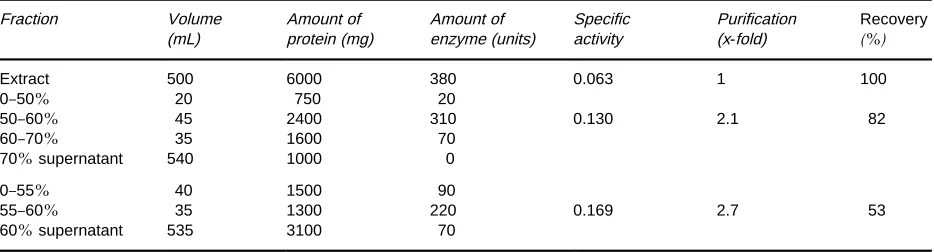
Table 1 Results of a typical ammonium sulfate fractionation procedure in purifying an enzyme. Percentages are given as a percent-age of saturation with ammonium sulfate. Specific activity is in units of enzyme activity per milligram of protein. Although the degree of purification is not high (2.1-fold), it is useful to note that much of the nonprotein material stays in the supernatant, and the precipitated fraction containing the enzyme can be dissolved in a much smaller volume than the starting extract. Results are also given for a second trial, in which recovery of activity has been sacrificed for a higher degree of purification
Fraction Volume (mL)
Amount of protein (mg)
Amount of enzyme (units)
Specific activity
Purification (x-fold)
Recovery
(%)
Extract 500 6000 380 0.063 1 100
0}50% 20 750 20
50}60% 45 2400 310 0.130 2.1 82
60}70% 35 1600 70
70%supernatant 540 1000 0
0}55% 40 1500 90
55}60% 35 1300 220 0.169 2.7 53
60%supernatant 535 3100 70
In most cases the desired protein will be soluble in aqueous media. If not, there are both advantages and disadvantages: the advantages include the ability to remove all the water-soluble proteins by simple ex-traction, which is a major separation process in itself. But the disadvantages include the problems of deciding what to do next, and how to separate the many insoluble proteins from each other. The answer is to get them into solution, and often this involves detergents, or other agents that may actually disrupt the natural biological state of the proteins. The natural state is called ‘native’ protein, whereas the disrupted state is called ‘denatured’. It is sometimes possible to ‘renature’ a protein from the denatured state, and this may be necessary when recombinant expression of otherwise soluble proteins results in an insoluble, denatured product (inclusion bodies).
Solubility
Separation by solubility characteristics is the oldest technique in protein puriRcation. Changes that result in proteins becoming less soluble in aqueous media include addition of salts, miscible organic solvents and organic polymers, and adjustment of pH. The aim is to make some proteins less soluble than others by varying these conditions, so the proteins can be separated from each other by centrifugation. It is not possible to predict which proteins will be affec-ted, so it is essential to have a method for detecting the protein that is to be isolated, so that its position is known after any separation procedure. Typical precipitants include ammonium sulfate as a salt, ethyl alcohol and acetone as miscible solvents, polyethy-lene glycol as an organic polymer, and pH adjustment with a weak acid to about pH 5, which coagulates complex proteins.
After centrifuging the precipitate, it can be re-dissolved in a buffer that lacks the precipitant.
Table 1illustrates an idealized analysis of the results
of an ammonium sulfate fractionation of an extract containing an enzyme. Before the next separation procedure, it may be necessary to remove excess precipitant by dialysis or gelRltration.
Ionic Charge
All proteins have charges on them as a result of amino acid side chains such as aspartate, glutamate, histidine, lysine and arginine. The net charge on a given protein depends on its exact composition, and on the pH. Consequently at a given pH dif-ferent proteins will have difdif-ferent net charges, and a shift in pH will change this value for each protein, though for all them it will become more negative for a higher pH, and more positive for a lower pH.
Figure 1 Example of protein separation using anion exchange chromatography. (A) A diagram of the basic principles of the equipment. The amount of protein emerging from the column is monitored by detecting the absorbance of light in the ultraviolet region (280 nm or 215 nm) due to proteins, and separate fractions are collected automatically. (B) The elution pattern. Some proteins did not bind to this adsorbent, being positively charged at this pH. The elution position of a specific protein is indicated by the shaded peak.
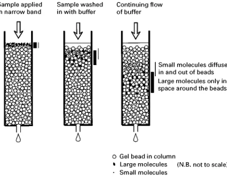
Figure 2 Principles of gel filtration. A mixture of proteins is applied to the column. Only the smallest molecules can penetrate the beads, while the larger molecules pass round the outside of the beads. Intermediate-sized molecules can partially pen-etrate the beads. As the separation continues, the larger molecu-les run out ahead of the smaller ones.
[image:3.568.294.517.469.645.2]strength), and proteins of the opposite charge to the column are adsorbed. Proteins are generally eluted by increasing the ionic strength with gradual salt addi-tion to the buffer, so that proteins of successive adsorbing strengths elute as the salt concentration goes up. This enables relatively high resolution of separate protein components, giving a high degree of puriRcation. There are many commercial adsorbents and equipment to use them with. The type to be used will depend very much on the purpose; ‘high perfor-mance’ systems are expensive to operate and may not be suitable for commercial production of the protein, but most suited to research and development. Speed may be desirable, or may be of little concern; there are materials for all needs. An example of the separ-ation of a fairly complex mixture of proteins on a ‘moderate performance’ adsorbent is shown in
Figure 1.
Size and Shape
The sizes of protein molecules vary over a large range, but most have a molecular weight 20 000}200 000 Da (native protein). This translates to a size, assuming a perfect sphere, of roughly 3}8 nm in diameter. Many proteins are not spherical, however, so their longer dimensions will be greater than theseRgures. Separation by size is carried out by a process called gel Rltration or size exclusion chromatography. The principle of gel Rltration is shown inFigure 2. Beads that are porous, with pores of similar size to the proteins, are packed into a col-umn. The largest proteins cannot penetrate the beads because the pores are too small, so theySow quickly around the outside of the beads, and emerge Rrst.
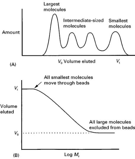
They do not emerge until after a volume (the void volume,V0) that is equivalent to the volume of liquid outside the beads has passed through the column. This volume is typically about 30% of the total col-umn volume, Vt. The smallest proteins are able to
Figure 3 (A) The elution pattern of a mixture of proteins from a gel filtration column. (B) Plot of the ‘elution volume’ against the logarithm of the molecular weight. All small molecules emerge at Vt, all very large molecules earlier at V0, and the values for
intermediate-sized molecules fall on a straight line over about a 10-fold increase in molecular weight range. By comparing the results with the elution volumes of proteins of known molecular weight, the molecular weight of an unknown protein can be deter-mined.
beads have a range of pore sizes, so that intermediate-sized proteins can spend some time inside the beads, but not as much as the smallest proteins. Conse-quently the emergence volume of each protein is re-lated to size, as shown inFigure 3.
This is also the principle used to separate large molecules from small molecules in general, and is very useful for removing salts from protein solutions. In this case the beads have pores so small that even the smallest proteins cannot penetrate the beads, but salts can.
GelRltration is a very gentle method; the proteins remain in solution at all times, and need not be exposed to any extremes of pH or salt concentrations. Resolution depends on the relative sizes of the pro-teins in the starting mixture. GelRltration will com-monly be used at a later stage of puriRcation, when the quantities of sample have been reduced.
There are also membranes available which have pores of suitable size to separate proteins. A force, such as gas pressure, is used to push the liquid, plus smaller molecules through the membrane. The largest molecules are retained, and concentrated
behind the membrane. This has application in the relatively crude separation of large proteins from small ones.
Separation by size is the commonest technique in the analysis of proteins, and is described in a later section. Using slab gels, which have pore sizes similar to the beads described previously, proteins are forced by electrophoresis to separate according to their size, the larger ones having more difRculty in Rnding their way through the gel, and so moving more slow-ly. This method has been applied preparatively, but only on a relatively small scale and mainly with unfol-ded, denatured proteins. Normally, one wants to iso-late the protein in its native state, as not all proteins can be re-foldedin vitro.
Other Surface Features of Proteins
The charged amino acids in a protein are responsible for the properties that allow separation on the basis of overall electrical charge. Other amino acids, espe-cially those exposed on the surface of the molecule, confer other properties that can be exploited in separ-ation methods. In particular, hydrophobic side chains would prefer not to be in contact with water. These amino acid side chains like to stick to each other, or to some surface that is also hydrophobic, and will do so in preference to being surrounded by water.
An adsorbent that consists of fat-like molecules attached to the insoluble beads is called a hydropho-bic adsorbent, and will attract those proteins that have a larger proportion of hydrophobic side chains on their surface. Hydrophobic interactions are strengthened by inclusion in the solution of high con-centrations of certain salts such as sulfates. A typical procedure would be to add up to 1Msodium sulfate
to the protein mixture, then run it through a column of hydrophobic adsorbent. Many proteins will not bind; these have few hydrophobic side chains. Then the salt concentration is gradually decreased, and proteins successively elute. The most hydrophobic proteins remain even a low salt concentration, and need other solutes such as chaotropic salts (e.g. thiocyanates), nonionic detergents, or high concen-trations of glycols to elute them.
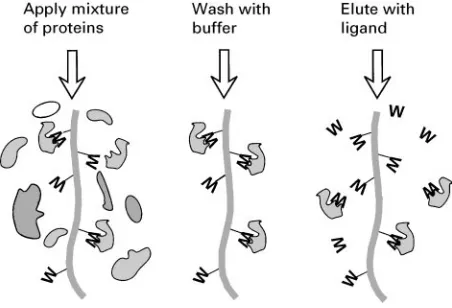
Figure 4 Principles of affinity chromatography. A ligand W that the desired protein binds to naturally is immobilized by attachment to column material. The protein mixture is applied, and the desired protein sticks to the immobilized ligand, while other proteins pass through and are washed away. Ideally only the proteins that bind to ligand W are left, though in practice there are usually others. Consequently a second ‘affinity’ step, involving washing the col-umn with the natural, free ligand, is used to displace the specific proteins from the column.
in a narrow and very reproducible range of solvent concentrations, and provided that the proteins refold rapidly as they are eluted, this gives a very high resolution of components. This has been highly suc-cessful with small proteins and peptides, but is less useful for larger and more unstable protein components.
Bioaf
\
nity
One surface feature that most proteins have is a bind-ing site that corresponds to their natural physiolo-gical role. Enzymes, for example, bind to substrates known as ligands, which may be small molecules or parts of large ones. This binding is often very tight and speciRc, and can be made use of in sepa-ration procedures. The method is called ‘afRnity chromatography’, and exploits the afRnity of the protein for its natural ligand. The principle is illus-trated inFigure 4. The adsorbent is designed so that it selects out only those proteins that bind to that par-ticular ligand. The proteins may subsequently be eluted using some solute that disrupts the interaction, often just salt or more speciRcally with the natural ligand, free in solution.
Af
\
nity Methods Associated with
Recombinant Proteins
The most signiRcant development in the past decade for purifying proteins has been the use of molecular
biology techniques not only to express the desired protein in large amounts, but also to modify it in a way that makes its puriRcation easy. This is achieved by making the protein longer with extra amino acid residues (polypeptide) that can be recog-nized by an afRnity column. The product is called a fusion protein. There are many systems available, and the process is made easy by commercially avail-able expression systems that automatically add the desired polypeptide. In some cases, the addition may be fewer than ten amino acids. In other cases it is a whole extra protein, which folds into an active shape while linked to the desired protein being ex-pressed. Often the overall expression level is even higher with these ‘fusion products’ than with the unmodiRed protein, because the expression of the fusion portion has been optimized in designing the vector.
After purifying the fusion protein, it may be neces-sary to remove the fusion portion. This can be done with selective proteases, though these usually still leave a few extra amino acids on the end. One system involving an intein, or self-splicing protein, enables release of exactly the desired protein with no gain or loss of amino acid.
Integrated Puri
\
cation Schemes
PuriRcation of proteins, even when using the highly selective afRnity methods, usually requires more than one step, with the extra steps carried out sequen-tially. The starting material is the most bulky stage, the crudest and least pure sample of the desired pro-tein, and theRrst separation step needs to be able to deal with such material. A traditional scheme is to use salt (ammonium sulfate) fractionation Rrst, then an ion exchange column, followed by gelRltration. Each of these steps exploits a different property of the proteins, so when combined they give a good chance of high selectivity. Repeated use of one particular procedure, e.g. ion exchange chromatography, will produce diminishing returns in terms of separation and recovery of protein.
An alternative Rrst stage is to use an adsorbent that acts as a pseudo-afRnity material and selects out the desired protein (along with many others), allowing nonprotein material to be removed. Any such adsorbent must be cheap to produce, because the crude extract can cause changes to its properties after a few uses, such as clogging or degradation. Dye adsorbents have been used for this purpose: brightly-coloured columns bind certain proteins se-lectively, and these can be washed off, then put through other steps such as ion exchange and/or gel
Table 2 Purification of an enzyme using three different procedures: dye adsorbent chromatography exploiting surface character-istics; ion exchange chromatography exploiting charge differences; and gel filtration to separate by size. The enzyme was xylulokinase from a thermophilicBacillus sp. (author’s unpublished results). Specific activity is in units of enzyme activity per milligram of protein. Nearly 200-fold purification was achieved, and the final enzyme was judged to be about 95%pure. This implies that the xylulokinase enzyme made up just 0.5%of the total protein in the original extract.
Purification step Volume (mL)
Amount of protein (mg)
Amount of enzyme (units)
Specific activity
Purification (x-fold)
Recovery
(%)
Extract 50 650 290 0.45 1 100
Off dye adsorbent 29 36 208 5.8 13 72
Off anion exchange 15 7.5 150 20 44 52
(NH4)2SO4precipitated 0.65 2.2 150 66 147 52
Gel filtration 2.0 1.4 120 86 190 41
TheRnal stage in a protein puriRcation will often be a high resolution step such as HPLC. Any asym-metry of theRnal peak might indicate that impurities are still present.
A puriRcation procedure ideally results in a good yield of a ‘pure’ protein. If the Rnal product is an enzyme it is relatively easy to determine how much remains at theRnal stage, compared with the amount in the crude extract, by measuring its activity.
SacriRces of recovery for purity (or vice versa) during the procedure will need to be made, depending on the relative importance of these two factors.
Table 2illustrates a typical puriRcation of an enzyme,
with a respectable recovery of activity and an end product that was analytically close to homogeneous.
Analytical Separations
At various stages, but more particularly at the end, one needs to know how close to purity the protein fraction is. Of many different analytical tech-niques that have been used, one is universally em-ployed. This is known as SDS-PAGE, which stands for sodium dodecyl sulfate-polyacrylamide gel elec-trophoresis. The gel (polyacrylamide) has pores that are similar in size to the protein molecules, and the mixture of proteins is forced through the gel by an electricReld. First the proteins are fully denatured by treatment with the anionic detergent sodium dodecyl sulfate, which binds to all the proteins and confers on them a negative charge proportional to their size. The smaller proteins move through the gel relatively eas-ily, but the larger ones are retarded by the lack of pores large enough to move through. Consequently there is a separation of components according to size, in a logarithmic way similar to that observed for gel
Rltration (see Figure 3). The individual components remain in tight bands; as a consequence proteins that differ in size by only 3}5% can be completely separated. This sharp resolution is thus able to detect small amounts of impurity, and a judgement can be
made on the absolute purity of the sample. One further feature is that, as with gelRltration, the dis-tance moved by an unknown protein can be com-pared with known standards and the unknown mo-lecular weight determined to within about 5% accu-racy. The amount of protein required is only about 1g or less, depending on the detection (staining) method. Capillary electrophoresis systems can be even more sensitive and accurate for analytical work.
See also: II/Affinity Separation. Theory and Develop-ment of Affinity Chromatography. Chromatography: Pro-tein Separation. Chromatography: Liquid: Column Technology; Large-Scale Liquid Chromatography; Mecha-nisms: Ion Chromatography. Electrophoresis: One-dimensional Sodium Dodecyl Sulphate Polyacrylamide Gel Electrophoresis; Proteins, Detection of; Two-dimen-sional Polyacrylamide Gel Electrophoresis.
Further Reading
Coligan J, Dunn B, Ploegh H, Speicher D and WingReld P (eds) (1995) Current Protocols in Protein Science. New York: John Wiley.
Deutscher MP (ed.) (1990) Methods in Enzymology, vol. 182: Guide to Protein PuriTcation. San Diego: Academic Press.
Harris ELV and Angal S (eds) (1990)Protein PuriTcation Applications;A Practical Approach. Oxford: IRL Press. Janson J-C and Ryden L (eds) (1989)Protein PuriTcation: Principles,High Resolution Methods,and Applications. New York: VCH Publishers.
Kenney A and Fowell S (eds) (1992) Practical Protein Chromatography. New Jersey: Humana Press.
Price NC (ed.) (1995)Proteins Labfax. Oxford: Bios Scien-tiRc Publishers. San Diego: Academic Press.
Scopes RK (1993)Protein PuriTcation,Principles and Prac-tice, 3rd edn. New York: Springer-Verlag.
Scopes RK (1997) Protein puriRcation in the nineties. Biotechnology and Applied Biochemistry23:197}204. Wheelwright SM (1991)Protein PuriTcation;Design and