EVALUATION OF ETCH PATTERNS AND BOND STRENGTH AFTER USING DIFFERENT ENAMEL
CONDITIONERS WITH AND WITHOUT DEPROTEINIZING
1
Dr. Rohini Sharma,
2Dr. Seema Gupta,
2, *
Dr. Surbhi Sharma,
7Dr. Raghu Pratap Thapa,
1Department of Orthodontics and Dentofacial Orthopaedics, Swami Devi Dayal Hospital and Dental College,
2Department of Orthodontics and Dentofacial Orthopaedics, Surendera Dental College and Research Institute,
3Department of Orthodontics and Dentofacial Orthopaedics, Institute of Dental Sciences, Sehora, India 4Department of Public Health Dentistry, Swami Devi Dayal Hospital and Dental College, Barwala,
5Department of Pedodontics, Institute of Dental Sciences, Seho
6Department of Oral Medicine and Radiology, Institute of dental Sciences, Sehora, Jammu and Kashmir, India 7F234 Raipur, Satwari,
ARTICLE INFO ABSTRACT
Objective
scores using different enamel conditioners with and without deproteinizing agent.
Study Design
A, B1, C1 i
20 seconds respectively. In Groups B2 and C2, etchants were same as B1 and C1 but enamel deproteinization with 5.25% sodium hypochlorite was done for 1 minute before en
Etching patterns were then evaluated in SEM, bond strength was examined with universal mechanical test machine and ARI scores were evaluated to check the bracket debonding interface. Data was analyzed statistically.
Results
bracket adhesive interface compared to the control group (A), whereas the Group C1showed lowest mean SBS, type 4 etching patterns and debonding occurring at the enamel adhesive interfa
B2 and C2 showed higher SBS and better etching patterns compared to the Group B1 and C1 where deproteinizing agent not was used.
Conclusions
etching patterns and incre acids to etch the enamel surface. Copyright © 2018, Rohini Sharma et al. This is an open
use, distribution, and reproduction in any medium, provided
INTRODUCTION
It was a cutting edge in dentistry which came up at the end of the 20th century with the advent of aesthetic adhesive dental materials. Buonocore demonstrated an increased adhesion of acrylic resins on enamel treated with 85% phosphoric acid (H3PO4). Further research was fundamental to the understanding and acceptance of enamel etching and the adhesion system in dentistry (Espinosa
Phosphoric acid remained the principal enamel etchant since it was first introduced by Buonocorein 1955
Newman in 1965.
*Corresponding author: Dr. Surbhi Sharma,
Department of Orthodontics and Dentofacial Orthopaedics, Surendera Dental College and Research Institute, Sriganganagar, Rajasthan, India
ISSN: 0975-833X
Article History: Received 20th June, 2018
Received in revised form 27th July, 2018
Accepted 20th August, 2018
Published online 30th September, 2018
Citation: Dr. Rohini Sharma, Dr. Seema Gupta, Dr. Sachin Ahuja
conditioners with and without deproteinizing agent: an in vitro study Key Words:
Shear Strength, Bonding, Acids,
Scanning electron Microscopy.
RESEARCH ARTICLE
EVALUATION OF ETCH PATTERNS AND BOND STRENGTH AFTER USING DIFFERENT ENAMEL
CONDITIONERS WITH AND WITHOUT DEPROTEINIZING AGENT: AN IN VITRO STUDY
Dr. Seema Gupta,
2Dr. Sachin Ahuja,
2Dr. Eenal Bhambri,
Dr. Raghu Pratap Thapa,
4Dr. Aditi Sharma,
5Dr. Alpna and
Department of Orthodontics and Dentofacial Orthopaedics, Swami Devi Dayal Hospital and Dental College, Barwala, Panchkula, Harayana, India
Department of Orthodontics and Dentofacial Orthopaedics, Surendera Dental College and Research Institute, Sriganganagar, Rajasthan, India
Department of Orthodontics and Dentofacial Orthopaedics, Institute of Dental Sciences, Sehora, India Department of Public Health Dentistry, Swami Devi Dayal Hospital and Dental College, Barwala,
Panchkula, Harayana, India
Department of Pedodontics, Institute of Dental Sciences, Sehora, Jammu and Kashmir, India
Department of Oral Medicine and Radiology, Institute of dental Sciences, Sehora, Jammu and Kashmir, India F234 Raipur, Satwari, Jammu Cantt, Jammu, Jammu and Kashmir, India
ABSTRACT
Objective: The objective of this study was to compare etch patterns, shear bond strength and ARI scores using different enamel conditioners with and without deproteinizing agent.
Study Design: A total of 125 human premolars were divided into 5 groups (n = 25) as follows: Group A, B1, C1 in which teeth are etched with 37% phosphoric acid, 10% maleic acid, 20% lactic acid for 20 seconds respectively. In Groups B2 and C2, etchants were same as B1 and C1 but enamel deproteinization with 5.25% sodium hypochlorite was done for 1 minute before en
Etching patterns were then evaluated in SEM, bond strength was examined with universal mechanical test machine and ARI scores were evaluated to check the bracket debonding interface. Data was analyzed statistically.
Results: Group B2 showed superior SBS, more of type 1 etching patterns and debonding occurring at bracket adhesive interface compared to the control group (A), whereas the Group C1showed lowest mean SBS, type 4 etching patterns and debonding occurring at the enamel adhesive interfa
B2 and C2 showed higher SBS and better etching patterns compared to the Group B1 and C1 where deproteinizing agent not was used.
Conclusions: Enamel deproteinization with 5.25% sodium hypochlorite showed improvement in etching patterns and increased shear bond strength of orthodontic brackets bonded with alternative acids to etch the enamel surface.
open access article distributed under the Creative Commons Attribution provided the original work is properly cited.
dentistry which came up at the end of century with the advent of aesthetic adhesive dental materials. Buonocore demonstrated an increased adhesion of acrylic resins on enamel treated with 85% phosphoric acid ). Further research was fundamental to the understanding and acceptance of enamel etching and the
Espinosa et al., 2008). Phosphoric acid remained the principal enamel etchant since it was first introduced by Buonocorein 1955 and used by
Department of Orthodontics and Dentofacial Orthopaedics, Surendera Dental College and Research Institute, Sriganganagar, Rajasthan, India
Phosphoric acid concentration between 30% in the most retentive etching pattern
Current clinical application of phosphoric acid uses a 37% acid concentration (Olsen et al., 1997
between enamel surfaces and restorative dental material is affected by several factors that include acid etching, etching time, pH, acid composition (organic inorganic), acid concentration, enamel morphology, calcium phosphate concentration, enamel site, and rinsing time. In the process of bonding, the microporous enamel surface created by acid etching with phosphoric acid plays a main role in the bond strength of restorative dental materials for primary and permanent teeth (Loyola-Rodriguez
speculated that37% phosphoric acid is very acidic
al., 2006). Acid etching has been reported to causeenamel loss.
International Journal of Current Research
Vol. 10, Issue, 09, pp.73885-73890, September, 2018
DOI: https://doi.org/10.24941/ijcr.32338.09.2018
Dr. Rohini Sharma, Dr. Seema Gupta, Dr. Sachin Ahuja et al., 2018. “Evaluation of etch patterns and bond strength after using
conditioners with and without deproteinizing agent: an in vitro study”, International Journal of Current Research, 10, (09
EVALUATION OF ETCH PATTERNS AND BOND STRENGTH AFTER USING DIFFERENT ENAMEL
AGENT: AN IN VITRO STUDY
Dr. Eenal Bhambri,
3Dr. Amit Choudhary,
Dr. Alpna and
6Dr. Dheeraj Sharma
Department of Orthodontics and Dentofacial Orthopaedics, Swami Devi Dayal Hospital and Dental College,
Department of Orthodontics and Dentofacial Orthopaedics, Surendera Dental College and Research Institute,
Department of Orthodontics and Dentofacial Orthopaedics, Institute of Dental Sciences, Sehora, India Department of Public Health Dentistry, Swami Devi Dayal Hospital and Dental College, Barwala,
ra, Jammu and Kashmir, India
Department of Oral Medicine and Radiology, Institute of dental Sciences, Sehora, Jammu and Kashmir, India India- 180003
study was to compare etch patterns, shear bond strength and ARI scores using different enamel conditioners with and without deproteinizing agent.
: A total of 125 human premolars were divided into 5 groups (n = 25) as follows: Group n which teeth are etched with 37% phosphoric acid, 10% maleic acid, 20% lactic acid for 20 seconds respectively. In Groups B2 and C2, etchants were same as B1 and C1 but enamel deproteinization with 5.25% sodium hypochlorite was done for 1 minute before enamel etching. Etching patterns were then evaluated in SEM, bond strength was examined with universal mechanical test machine and ARI scores were evaluated to check the bracket debonding interface. Data was
uperior SBS, more of type 1 etching patterns and debonding occurring at bracket adhesive interface compared to the control group (A), whereas the Group C1showed lowest mean SBS, type 4 etching patterns and debonding occurring at the enamel adhesive interface. Group B2 and C2 showed higher SBS and better etching patterns compared to the Group B1 and C1 where
: Enamel deproteinization with 5.25% sodium hypochlorite showed improvement in ased shear bond strength of orthodontic brackets bonded with alternative
ribution License, which permits unrestricted
Phosphoric acid concentration between 30% and 40% results in the most retentive etching pattern (Alsulaimani, 2014). Current clinical application of phosphoric acid uses a 37% acid
1997). The tensile bond strength between enamel surfaces and restorative dental material is affected by several factors that include acid etching, etching time, pH, acid composition (organic inorganic), acid concentration, enamel morphology, calcium phosphate centration, enamel site, and rinsing time. In the process of bonding, the microporous enamel surface created by acid etching with phosphoric acid plays a main role in the bond strength of restorative dental materials for primary and
Rodriguez et al., 2009). It is speculated that37% phosphoric acid is very acidic (Mazzoni et
Acid etching has been reported to causeenamel loss. OF CURRENT RESEARCH
The permanent loss of enamel calcium during the acid etching procedure may render the enamel surface more susceptible to demineralization during and after the orthodontic treatment. Cehreli and Altay observed that regardless of treatment time, etching with 37% phosphoric acid results in irreversible damage of the enamel surface (Cal-Neto and Miguel, 2006). Therefore, to minimize the extent of enamel surface damage, alternate conditioners, such as lactic acid, maleic acid, nitric acid, polyacrylic acid, and ethylenediaminetetraacetic acid, have been used to obtain clinically useful bond strengths by decreasing the depth of enamel dissolution (Alsulaimani, 2014). Espinosa et al. (2008) showed that when 5.25% of NaOCl was used as deproteinizing agent before acid etching with alternative etchants, increased the bond strength and produced more of type 1 and type 2 etching patterns while without the use of NaOCl, more of type 3 etch patterns were found. The present study was conducted to test the following hypothesis:
a) Different enamel etchants will provide different etching patterns and different shear bond strength to enamel. b) Improvement in etching patterns, shear bond strength
and debonding interface will be seen after the use of deproteinizing agent for 1 minute before the acid etching.
MATERIALS AND METHODS
One hundred and twenty-five extracted human premolars were collected from the Department of Oral and Maxillofacial Surgery. The teeth extracted were from the patients whose treatment plan needed orthodontic extractions and were collected with the informed consent of the patients. The sample size was estimated and this study was designed to have power of 90%. The exclusion criteria for selection of the samples were the teeth with caries, cracks, erosion, fluorosis orhypo-calcification, and restored teeth. A total of 125 human premolars were, divided into 5 groups (n = 25), and denominated as follows:
Group A(n=25):The enamel surface was etched with 37%
Phosphoric acid (3M Unitek) applied with a microbrush for 20 seconds, washed with water and air spray for 20 seconds, then dried with oil free compressed air.
Group B1 (n=25): The enamel surface was etched with 10% Maleic acid (99.9% From Loba Chemie, Diluted in the Pharmacology Lab at Surendera Dental College and Research Institute)applied with a microbrush for 20 seconds, washed with water and air spray for 20 seconds, then dried with oil free compressed air.
Group B2 (n=25): The enamel surface was treated with 5.25%
sodium hypochlorite applied with sterile cotton pellet for one minute, washed, then dried with water for 10 seconds and etched with 10% Maleic acid (99.9% From LobaChemie, Diluted in the Pharmacology Lab at Surendera Dental College and Research Institute) applied with a microbrush for 20 seconds, washed with water and air spray for 20 seconds, then dried with oil free compressed air.
Group C1 (n=25): The enamel surface was etched with 20% Lactic acid (99.9% From Loba Chemie, Diluted in the Pharmacology Lab at Surendera Dental College and Research Institute) applied with a microbrush for 20 seconds, washed
with water and air spray for 20 seconds, then dried with oil free compressed air.
Group C2 (n=25):The enamel surface was treated with 5.25%
sodium hypochlorite applied with sterile cotton pellet for one minute,washed, then dried with water for 10 seconds and etched with 20% Lactic acid (99.9% From LobaChemie, Diluted in the Pharmacology Lab at Surendera Dental College and Research Institute) applied with a microbrush for 20 seconds, washed with water and air spray for 20 seconds, then dried with oil free compressed air.
The orthodontic brackets (MBT brackets, 0.022” slot, American Orthodontics) for premolars were fixed in the centre of the crown using Transbond XT Adhesive (3M Unitek). Excess adhesive was removed from the teeth with a probe, and each bracket was then lightpolymerized with a LED appliance (Woodpecker, Guilin Woodpecker Medical Instrument company Ltd, Guangxi, China) for 20 s (5 s on each side). After bonding, all samples were stored in distilled water with 0.1% thymol solution. Each tooth was mounted in acrylic resin in the respective mould with roots embedded upto cervical line in the acrylic portion to enhance the control and manipulation of the specimens.
Thermocycling and Shear bond strength testing
The test samples were subjected to 500 thermocycles at temperature (5°C to 55°C) before testing. The dwell time was 30 seconds for each bath as advised by International Organisation for Standardization before testing to mimic thermally stressed accelerated aging. 20 samples from each group were subjected to a shear load in a universal testing machine model (Star testing systems model no. 9036 TD) with a knife-edged blade at a crosshead speed of 1 mm/min. The force was applied parallel to the long axis of the tooth on top of each orthodontic bracket base, and the shear load was recorded at the point of failure. The force per unit area required to dislodge the bracket was recorded in Newtons for each specimen and divided by the surface area of the bracket base to estimate the SBS in megapascals (MPa).
Assessment of adhesive remnant index
The enamel surfaces were examined with a stereomicroscope under40×magnifications to determine the amount of composite remaining, and then they were classified according to adhesive remnant index (ARI). The ARI scores ranged from 1 to 5 with 1 indicating all the composite remained on the tooth as well as the impression of the bracket base, 2 indicating more than 90% of the composite remained, 3 indicating more than 10% but less than 90% of the composite remained on the tooth, 4 indicating less than 10% of composite remained on the tooth surface and 5 indicating that no composite remained on the enamel.
Scanning electron microscope examination
Five premolars from each group were used for ultra-structural examination of the etched enamel surfaces using a scanning electron microscope (JEOL, JSM-6100 LV, USA). Each etched buccal surface was observed under SEM and photomicrograph was taken in the centre of the test surface at 2000X magnification. The teeth were pre-treated with the respective acid, then rinsed with water for 10 s, and dried. The
teeth were separated from the acrylic moulds and gold sputtering was done separately for each group. A total of 25 photomicrographs (5 for each experimental group) were taken. Under the scanning electron microscope, all
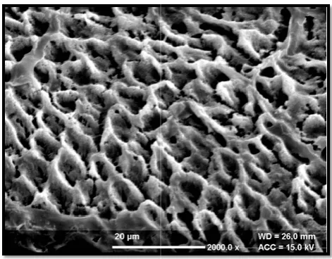
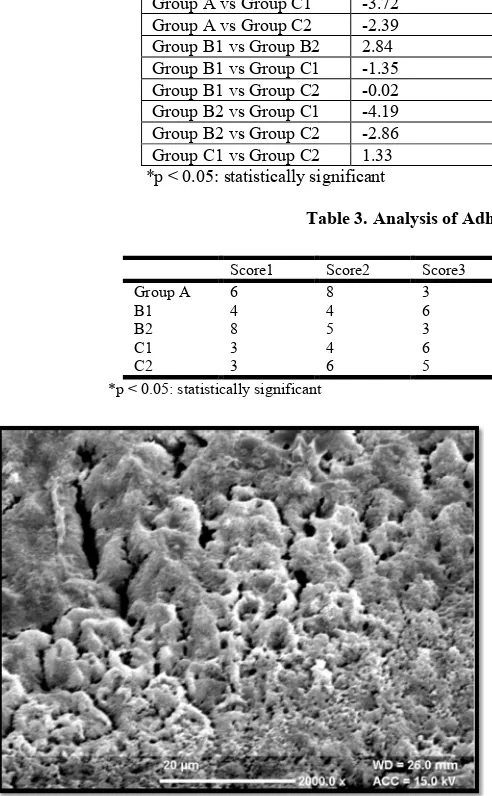
surfaces resulted in different etch patterns where enamel prism cores preferentially removed with peripheries relatively intact shows type 1 etching patterns , peripheral regions of the prisms removed leaving relatively unaffected prism cores sh
2 etching patterns, regions in which the pattern of etching could not be related to prism morphology shows type 3 etching patterns, prismless enamel displaying no rod or prism patterns shows type 4 etching patterns and flat and smooth lacking micro irregularities shows the type 5 etching patterns. second invigilator who was experienced in SEM analysis analyzed the etching patterns seen on the buccal surface and they were recorded. Hence both the assessing invigilators were blinded. After recording the pictures, they were evaluated.
Statistical analysis
Statistical analyses were performed with
Version 23 for window, release 7.5.1, Chicago, USA). Descriptive statistics that included mean, standard deviation, median, minimum and maximum values were calculated for all five groups. Analysis of variance was applied to determine whether significant differences existed among the groups. Post hoc Tukey's test was used to compare the mean between the individual groups. Chi-square test was used for assessing ARI scores. The level of significance adopted was 5% (p<0.05).
RESULTS
Scanning Electron Microscope
Group A showed both type 1 and type 2 etching patterns with more of type 2 etching patterns. Group B1 showed more of type 3 etching patterns. Group C1 showed less well
patterns indicating more of type 4 etching patterns. With t use of deproteinizing agent for one minute before acid etching, Group B2 showed type 1 etching patterns and Group C2showedtype 2 etching patterns. On comparing the SEM images after etching, Group B2 showed the best etching patters, followed by Group A, Group C2, Group B1 and Group C1 showed the etch patterns which were lessfavourable (Figure 1-5).
Universal Testing Machine
The highest SBS was recorded for Group B2 (10.09±0.47 MPa), followed by Group A (9.62±0.41 MPa) and then Group B1 (7.25±0.56MPa) which was almost similar to Group C2 (7.23±0.28 MPa). Group C1 had the least bond strength with the values (5.9±0.36MPa). The ANOVA test showed significant difference between the groups with respect to the median load (p< 0.01) (Table 1).To find out the diff between the groups, Turkey HSD Post-hoc Test was carried out and results were given in (Table 2). The significant differences were found between all the groups except B1 and Group C2which showed non-significant difference between the bond strength.
Analysis of ARI Scores: The ARI was used to determine the bond failure location between the different adhesives and etching techniques. The results of the Chi Square test comparing the scores indicated the presence of significant teeth were separated from the acrylic moulds and gold sputtering was done separately for each group. A total of 25 photomicrographs (5 for each experimental group) were taken. Under the scanning electron microscope, all the etched surfaces resulted in different etch patterns where enamel prism cores preferentially removed with peripheries relatively intact shows type 1 etching patterns , peripheral regions of the prisms removed leaving relatively unaffected prism cores shows type regions in which the pattern of etching could not be related to prism morphology shows type 3 etching patterns, prismless enamel displaying no rod or prism patterns shows type 4 etching patterns and flat and smooth lacking o irregularities shows the type 5 etching patterns. The second invigilator who was experienced in SEM analysis analyzed the etching patterns seen on the buccal surface and they were recorded. Hence both the assessing invigilators were
ing the pictures, they were evaluated.
Statistical analyses were performed with the SPSS (SPSS Version 23 for window, release 7.5.1, Chicago, USA). Descriptive statistics that included mean, standard deviation, minimum and maximum values were calculated for all five groups. Analysis of variance was applied to determine significant differences existed among the groups. Post hoc Tukey's test was used to compare the mean between the square test was used for assessing ARI scores. The level of significance adopted was 5% (p<0.05).
Group A showed both type 1 and type 2 etching patterns with Group B1 showed more of Group C1 showed less well-defined patterns indicating more of type 4 etching patterns. With the use of deproteinizing agent for one minute before acid etching, Group B2 showed type 1 etching patterns and Group C2showedtype 2 etching patterns. On comparing the SEM images after etching, Group B2 showed the best etching Group C2, Group B1 and Group C1 showed the etch patterns which were lessfavourable (Figure
The highest SBS was recorded for Group B2 (10.09±0.47 MPa), followed by Group A (9.62±0.41 MPa) and then Group ich was almost similar to Group C2 (7.23±0.28 MPa). Group C1 had the least bond strength with the values (5.9±0.36MPa). The ANOVA test showed significant difference between the groups with respect to the median load (p< 0.01) (Table 1).To find out the difference hoc Test was carried out and results were given in (Table 2). The significant differences were found between all the groups except Group significant difference
The ARI was used to determine the bond failure location between the different adhesives and etching techniques. The results of the Chi Square test comparing the scores indicated the presence of significant
differences between the various groups (p<0.03). Group B2, A, C2 had bond failure at the composite
[image:3.595.309.545.109.305.2]whereas Group B1 and C1 revealed bond failure primarily at the enamel-composite interface (Table 3).
Figure 1. Type 1 and Type 2 etching patterns, wi etching patterns seen after etching the enamel s
[image:3.595.314.556.362.544.2]phosphoric acid
Figure 2. Type 3 etching patterns seen after etching the enamel surface with 10% maleic
Figure 3. Type 1 etching patterns seen after treating the enamel surface with deproteinizing agent before enamel etching with 10%
maleic acid
n the various groups (p<0.03). Group B2, A, C2 had bond failure at the composite-bracket interface, whereas Group B1 and C1 revealed bond failure primarily at
[image:3.595.316.552.578.762.2]composite interface (Table 3).
Figure 1. Type 1 and Type 2 etching patterns, with more of type 1 etching patterns seen after etching the enamel surface with 37%
phosphoric acid
Figure 2. Type 3 etching patterns seen after etching the enamel surface with 10% maleic acid
Figure 3. Type 1 etching patterns seen after treating the enamel surface with deproteinizing agent before enamel etching with 10%
Figure 4. Type 4 etching patterns seen after etching the enamel surface with 20% lactic acid
DISCUSSION
The use of lactic acid as an alternative to phosphoric acid for enamel etching has been documented in the literature (Alsulaimani, 2014). 10 % maleic acid has also been used as a conditioner that could etch dentine as well as be used for enamel bonding. Therefore, maleic acid has been suggested as alternative conditioner that can maintain a clinically useful bond strength while decreasing the depth of enamel dissolution, thus decreasing enamel surface damage (Sinhoreti
[image:4.595.113.486.199.338.2]et al., 1998; Hermsen et al., 1993).
Figure 5. Type 2 etching patterns seen after treating the enamel surface with deproteinizing agent before enamel etching with 20% lactic acid
However, the etch patterns created by these alternative acids have been found to be inconsistent. So, elimination of organic substances from the enamel surface before acid etching providing a better acid etching pattern on enamel has been advocated. Deproteinization of the enamel surface before bracket bonding was first proposed by Justus et al. (2010). 5.25% sodium hypochlorite was used for this purpose. According to De-Deus et al. (2011), NaOCl eliminates the organic matter present on the enamel surface by dissolving it, and thus, it can be speculated that increased shear bond strength might be achieved by deproteinizing the enamel Table 1. Comparison of shear bond strengths of all the groups.
Groups N Bond strength (MPA) Min Max Median Anova test p value
Mean SD
Group A 20 9.62 0.41 7.89 10.04 8.5
344.73 <0.01*
Group B1 20 7.25 0.56 6.57 8.76 7.2
Group B2 20 10.09 0.47 8.32 11.26 10.3
Group C1 20 5.9 0.36 5.1 6.4 5.7
Group C2 20 7.23 0.28 6.1 8.2 7.1
*p <0.05: statistically significant
Table 2. Tukey HSD Post-hoc Test showing the mean value difference in each group
Groups Mean difference Confidence interval (95%) p value Lower Upper
[image:4.595.44.290.247.645.2]Group A vs Group B1 -2.37 -2.7453 -1.9947 <0.01* Group A vs Group B2 0.47 0.0947 0.8453 0.0066* Group A vs Group C1 -3.72 -4.0953 -3.3447 0.3697* Group A vs Group C2 -2.39 -2.7653 -2.0147 <0.01* Group B1 vs Group B2 2.84 2.4647 3.2153 <0.01* Group B1 vs Group C1 -1.35 -1.7253 -0.9747 <0.01* Group B1 vs Group C2 -0.02 -0.3953 0.3553 0.9999 Group B2 vs Group C1 -4.19 -4.5653 -3.8147 0.8526* Group B2 vs Group C2 -2.86 -3.2353 -2.4847 <0.01* Group C1 vs Group C2 1.33 0.9547 1.7053 <0.01* *p < 0.05: statistically significant
Table 3. Analysis of Adhesive Remnant Index Scores
Score1 Score2 Score3 Score4 Score5 Chi square p value
Group A 6 8 3 2 1 17.33 0.03*
B1 4 4 6 4 2
B2 8 5 3 3 1
C1 3 4 6 6 1
C2 3 6 5 4 2
*p < 0.05: statistically significant
[image:4.595.50.547.362.642.2] [image:4.595.295.554.381.637.2]surface with 5.25% NaOCl. The weaker acids such as 10% maleic acid and 20% lactic acid can be used along with NaOCl to get the favourable etch patterns and bond strength equivalent to conventional 37% phosphoric acid etching. Many studies have been done comparing alternative mild acids with phosphoric acid, however no study has been conducted comparing these acids with conventional phosphoric acidused along with deproteinizing agent. So, the present study was done where maleic and lactic acids were used with and without deproteinizing agents and compared to conventional phosphoric acid in terms of bond strength, etch patterns and adhesive remnant index.
The bond strength of lactic acid alone was found to be lowest as compared to maleic acid. This was in accordance to the studies done by Hughes et al. (2000), Ballal (2009), Ayad (1996) and Luque et al. (2012). The reason for the above finding was due to the formation of weak bonds of lactic acid to enamel, gentle rinsing with distilled water almost completely removing the acid as seen by X-Ray photoelectron spectroscopy in the study done by Yoshioka et al. (2002). They also found that amount of Ca & P extracted from hydroxyapatite powder was more for maleic acid than lactic acid. The fact that acids adhere to or decalcify hydroxyapatite crystals appears to depend largely upon the dissolution rate of the respective calcium salts in their respective acidic solutions. The less soluble their respective salts, the more stable their bond to hydroxyapatite (HAp) and less they decalcify Hap. The lactic acid salts were less soluble in their respective acid solution compared to maleic acid and therefore, their ability to decalcify hydroxyapatite crystals of enamel was less than maleic acid.33
The bond strengths of both lactic acid & maleic acid increased after adding deproteinizing agent. This was in accordance to study done by Hasija et al. (2017), where the authors found that 5% NaOCl showed maximum bond strength compared to the other deproteinizing agent and was an effective enamel bond enhancer. The ARI was used to determine the bond failure location between the different adhesives and etching techniques.Bishara et al maintained that bond failure at the bracket adhesive interface or within the adhesive is more desirable and safer than the failure at the adhesive enamel interface because enamel fracture and crazing have been reported at the time of bracket debonding (Bishara et al.,
2001).
The results of the present study indicated that Group B2, (etching with 10% maleic acid after deproteinizing agent) showed superior SBS, more of type 1 etching patterns and debonding occurring at bracket adhesive interface compared to the control group (A), whereas the Group C1showed lowest mean SBS, type 4 etching patterns and debonding occurring at the enamel adhesive interface. The results of one -way ANOVA showed highly significant difference with the use deproteinizing agent in SBS of the groups (p < 0.01). Group B2 and C2 showed higher SBS and better etching patterns compared to the Group B1 and C1 where deproteinizing agent not was used. 5.25% sodium hypochlorite eliminates the organic substances which allows the acid etchant to penetrate more effectively into enamel creating type 1 & 2 etching pattern as seen in Group B2 & C2 which was in accordance to the studies done by Espinosa et al. (2008), Pithon et al. (2011),
Pereira et al. (2012) and Justus et al. (2009). Sodium hypochlorite (NaOCl) has an antibacterial effect and does not
damage healthy tissue or tooth structure. Interpreting the chemical reactions, it can be observed that sodium hypochlorite acts as an organic and fat solvent degrading fatty acids, transforming them into fatty acid salts (soap) and glycerol (alcohol), that reduces the surface tension of the remaining solution. Sodium hypochlorite neutralizes amino acids forming water and salt. With the exit of hydroxyl ions, there is a reduction of pH. Hypochlorous acid, a substance present in sodium hypochlorite solution, when in contact with organic tissue acts as solvent, releases chlorine that, combined with the protein amino group, forms chloramines. Hypochlorous acid (HOCl) and hypochlorite ions (OCl) lead to amino acid degradation and hydrolysis. The chloramination reaction between chlorine and the amino group (NH) forms chloramines that interfere in cell metabolism. Chlorine (strong oxidant) presents antimicrobial action inhibiting bacterial enzymes leading to an irreversible oxidation of SH groups (sulphydryl group) of essential bacterial enzymes (Estrela et
al., 2002). The use of 5.25% sodium hypochlorite (NaOCl) as
a deproteinizing agent may be a possible strategy to optimize adhesion by removing organic elements of both the enamel structure and the acquired pellicle before acid etching (Espinosa et al., 2008). The results of this study showed maleic acid with deproteinizing agent had the greatest bond strength, improved etching patterns and favourable site of debonding as assessed by ARI scores and therefore, recommended for clinical usage.
Conclusion
This study was done to determine the effect of different acids and deproteinizing agent on the etching patterns, SBS and ARI scores of the bonded premolars and it can be concluded:
1. The addition of deproteinizing agent with the alternate conditioners such as maleic and the lactic acid provided more of type 1 and type 2 etching patterns.
2. The SBS was increased with the use of deproteinizing agent before etching as compared to the SBS samples where deproteinizing agent was not used.
3. Maleic acid with deproteinizationcould be a clinically useful alternative enamel conditioner.
Conflict of interest: The authors declare that they have no conflict of interest related to this study. The study was self-funded by the authors.
REFERENCES
Alsulaimani FF. 2014. Effect of lactic acid etching on bonding effectiveness of orthodontic bracket after water storage. ISRN dentistry 17.
Ayad MF, Rosenstiel SF, Farag AM. 1996. A pilot study of lactic acid as an enamel and dentin conditioner for dentin-bonding agent development. J Pros Dent., 76(3):254-59. Ballal NV, Kandian S, Mala K, Bhat KS, Acharya S. 2009.
Comparison of the efficacy of maleic acid and ethylenediaminetetraacetic acid in smear layer removal from instrumented human root canal: A scanning electron microscopic study. J Endod., 35:1573-76.
Bishara SE, VonWald L, Laffoon JF, Warren JJ. 2001. Effect of a selfetch primer/adhesive on the shear bond strength of orthodontic brackets. Am J Orthod Dentofacial Orthop.,
Cal-Neto JP, Miguel JA. 2006. Scanning electron microscopy evaluation of the bonding mechanism of a self-etching primer on enamel. Angle Orthod., 76(1):132-36.
De-Deus G, Souza EM, Marins JR, Reis C, Paciornik S, Zehnder M. 2011. Smear layer dissolution by peracetic acid of low concentration. Int Endod J., 44: 485-90.
Espinosa R, Valencia R, Uribe M, Ceja I, Saadia M. 2008. Enamel deproteinization and its effect on acid etching: an in vitro study. J Clin Ped Dent., 33(1):13-19.
Estrela C, Estrela CR, Barbin EL, Spanó JC, Marchesan MA, Pécora JD. 2002. Mechanism of action of sodium hypochlorite. Braz Dent J., 13(2): 113-17.
Ferrer-Luque CM, Conde-Ortiz A, Arias-Moliz MT, Valderrama MJ, Baca P. 2012. Residual activity of chelating agents and their combinations with cetrimide on root canals infected with Enterococcus faecalis. J Endod.,
38(6):826-28.
Hasija P, Sachdev V, Mathur S, Rath R. 2017. Deproteinizing agents as an effective enamel bond enhancer- An in-vitro study. J Clin Ped Dent., 41(4):280-83.
Hermsen RJ, Vrijhoef MM. 1993. Loss of enamel due to
etching with phosphoric or maleic acid. Dent Mat.,
9(5):332-36.
Hughes JA, West NX, Parker DM, Van den braak MH, Addy M. 2000. Effects of pH and concentration of citric, maleic and lactic acids on enamel, in vitro. J Dent., 28:147-52. Justus R, Cubero T, Ondarza R, Morales F. 2010. A new
technique with sodium hypochlorite to increase bracket shear bond strength of fluoride-releasing resin-modified glass ionomer cements: comparing shear bond strength of two adhesive systems with enamel surface deproteinization before etching. Sem Orthod., 16(1):66-75.
Loyola-Rodriguez JP, Zavala-Alonso V, Reyes-Vela E, Patiño-Marin N, Ruiz F, Anusavice KJ. 2009. Atomic force microscopy observation of the enamel roughness and depth profile after phosphoric acid etching. J Elec Micros.,
59(2):119-25.
Mazzoni A, Pashley DH, Nishitani Y, Breschi L, Mannello F, Tjäderhane L, Toledano M, Pashley EL, Tay FR. 2006. Reactivation of inactivated endogenous proteolytic activities in phosphoric acid-etched dentine by etch-and-rinse adhesives. Biomaterials, 27(25):4470-76.
Olsen ME, Bishara SE, Damon P, Jakobsen JR. 1997. Evaluation of scotchbond multipurpose and maleic acid as alternative methods of bonding orthodontic brackets. Am J
Orthod Dentofac Orthop., 111(5):498-01.
Pereira TB, Jansen WC, Pithon MM, Souki BQ, Tanaka OM, Oliveira DD. 2012. Effects of enamel deproteinization on bracket bonding with conventional and resin-modified glass ionomer cements. Eur J Orthod., 35(4):442-46. Pithon MM, de Souza Ferraz C, do Couto de Oliveira G,
Pereira TB, Oliveira DD, de Souza RA, de Freitas LM, dos Santos RL. 2011. Effect of 10% papain gel on enamel
deproteinization before bonding procedure. Angle Orthod.,
82(3):541-45.
Sinhoreti MA, Consani S, da SIlva MA. 1998. Morphological effect of the type, concentration and etching time of acid solutions on enamel and dentin surfaces. Braz Dent J.,
9(1):3-10.
Yoshioka M, Yoshida Y, Inoue S, Lambrechts P, Vanherle G, Nomura Y, Okazaki M, Shintani H, Van Meerbeek B. 2002. Adhesion/decalcification mechanisms of acid interactions with human hard tissues. J Biomed Mat Res.,
59(1):56-62.
73890 Rohini Sharma et al.Evaluation of etch patterns and bond strength after using different enamel conditioners with and without deproteinizing agent: An in vitro study