THE CONTROL OF MORPHOGENESIS IN
'DROSOPHILA' IMAGINAL DISC CELL LINES 'IN
VITRO'
Andrew S. Miller
A Thesis Submitted for the Degree of PhD
at the
University of St Andrews
1996
Full metadata for this item is available in
St Andrews Research Repository
at:
http://research-repository.st-andrews.ac.uk/
Please use this identifier to cite or link to this item:
http://hdl.handle.net/10023/14026
by
Andrew S. Miller
All rights reserved INFORMATION TO ALL USERS
The quality of this reproduction is dependent upon the quality of the copy submitted. In the unlikely event that the author did not send a com plete manuscript and there are missing pages, these will be noted. Also, if material had to be removed,
a note will indicate the deletion.
uest
ProQuest 10166594
Published by ProQuest LLO (2017). Copyright of the Dissertation is held by the Author. All rights reserved.
This work is protected against unauthorized copying under Title 17, United States C ode Microform Edition © ProQuest LLO.
ProQuest LLO.
789 East Eisenhower Parkway P.Q. Box 1346
Declaration Abstract
List of abbreviations Acknowledgements
Chapter 1. Overview 1
Chapter 2. General Methods
2.1 Introduction 19
2.2 Routine cell culture 19
2.3 Polyacrylamide gel electrophoresis (PAGE) 23
Chapter 3. Use of imaginai cell lines in adhesion studies
3.1 Introduction 26
3.2 Materials and methods 31
3.3 Results 36
3.4 Discussion 41
Chapter 4. Expression and proposed function of PS integrins in imaginai disc cell lines
4.1 Introduction 55
4.2 Materials and methods 61
Chapter 5. Control of apical-basal polarity in imaginai disc cell lines
5.1 Introduction 89
5.2 Materials and Methods 92
5.3 Results 95
5.4 Discussion 98
Chapter 6. The expression of the adhesion molecule, fasciclin III, in imaginai disc cell lines
6.1 Introduction 106
6.2 Materials and Methods 112
6.3 Results 125
6.4 Discussion 131
Chapter 7. Response to an intracellular rise in cAMP and the uptake of the larval serum protein, arylphorin, in imaginai disc cell lines
7.1 Introduction 138
7.2 Materials and methods 143
7.3 Results 149
7.4 Discussion 154
Chapter 8. Discussion and future areas of interest 159
that it is a record of my own work, and that it has not been accepted in partial or complete fulfilment of any other degree or professional qualification.
Signed: Date;
I was admitted to the Faculty of Science of the University of St. Andrews, under Ordinance General No. 12 on October 1st 1991 and as a candidate for the degree of Ph.D. in September 1992.
Signed: Date:
I hereby certify that the candidate has fulfilled the conditions of the Resolution and the Regulations appropriate to the Degree of Ph.D.
Signature of Supervisor: Date:
The experimental component of this thesis represents the continuation of studies carried out in the Milner laboratory to characterise the biology of
Drosophila imaginai disc cell lines growing in vitro. The bulk of this study was
concerned with the morphological and molecular characterisation of imaginai cell interactions in vitro and contrasting this with what is known of imaginai cell biology in vivo. The imaginai cell lines were thus seen as both an amenable model for the detailed analysis of a cells interaction with its environment and, more broadly, as an assessment of the effects of isolating and maintaining animal cells in vitro.
The reaggregation of single imaginai cells when in suspension is rapid and extensive when in the presence of the divalent cations, Ca^+ and Mg^+, but is not entirely dependent on them suggesting the existence of separate systems of adhesion and the presence of a variety of cell adhesion molecules (CAMs). During the course of growth in vitro there appears to be a shift in strategy between cell-substrate and cell-cell adhesion as cells move from monolayers into aggregates. A new set of leg cell lines were cloned, some of which had a radically reduced capacity to reaggregate in suspension suggesting that CAMs can be selectively lost from the imaginai cell surface.
aggregating cells.
The in vivo epithelial phenotype, characterised by apical-basal polarity,
can be re-established in part by growing cells on fibronectin-coated membranes in the presence of unidirectional nutrient uptake and a feeder layer of cells. Cells in vitro lack such polarity signified by the absence of specialised intercellular junctions and a loss of restriction of expression of the putative CAM and signalling molecule, CRUMBS.
Another CAM which appears to be expressed during imaginai cell growth in vitro is the immunoglobulin, fasciclin III, or a variant, which is restricted in expression to single migratory cells and during cell-cell adhesion.
20-HE AP BSA cAMP CNS cpm CSM D= DAB DMA DMSO DTT ECM EDTA FBS FITC GRGDSP GRGESP IBMX lU LSP MTOC OD PBS PMSF 20-hydroxy ecdysone After pupariation Bovine serum albumin
Cyclic adenosine monophosphate Central nervous system
Counts per minute
Complete sterile medium (after Cullen and Milner, 1991) PBS minus Ca2+ and Mg2+ (Dulbecco's formula)
3 ,3'-diaminobenzidine Deoxyribosenucleic acid Dimethyl sulphoxide Dithiothreitol Extracellular matrix Ethylenediaminetetraacetic Acid Foetal bovine serum
Fluorescein isothiocyanate
Synthetic peptide containing the sequence glycine- arginine-aspartic acid (RGD).
Synthetic peptide containing the sequence arginine- glycine-glutathione (RGE).
3-isobutyl-l-methylxanthine International units
Larval serum protein
Microtubule organising centre Optical density
S2 Schneider's Line 2
SDS-PAGE Sodium dodecylsulphate polyacrylamide gel electrophoresis
SPIT Solid phase immunoisolation technique
TE Tris/EDTA based buffer
TEM Transmission electron microscopy
TCA Trichloroacetic acid
Firstly I owe a great debt of gratitude to my supervisor, Martin Milner, for his constant assistance, generosity and kindness. Martin is a brilliant provider, which has allowed me to participate in some exciting projects with other Drosophilists and continues to make me feel very welcome in the lab, which makes me feel loathe to leave.
Even though I have been Martin's sole postgraduate student for a while now, I still remember with fondness the students that helped me settle in and who remain firm friends. Dave Peel de?^erves many thanks for his endless help, insight and tips on being cool as does Alison Wallace for being a great work-mate and for diversifying my music taste and Tanya Hamill for her endless cheerfulness. Thanks also to the new body in the office, Deborah Cottam for her friendliness and wisdom about primary culture.
Special thanks are due to John Mackie and Mette Mogensen for help with microscopy and John Sommerville and Robert Ladomery for help with Western Blotting and autoradiography. Paul Hartley, Craig Henderson, Ian Megson and Scott McFarlane are all long time colleagues and friends who helped provide inspiration and weekend relief when most needed. Some of the others who deserve a mention are Chris Cutler, Alex Jones, Nigel Mann, Simon Potts, Fiona, Adrian and Paul for his antics.
Outwith St. Andrews, thanks to F. Michael Hoffmann and his group in Madison, Peter Snow and Eric Bonventre in Albany and Klaus Scheller in Würzburg.
The fruit fly Drosophila has, for most of this century, been the subject of intense scientific scrutiny and interest to a large and diverse community of biological researchers. The primary reason for this enormous interest in
Drosophila is the potential to utilise sophisticated genetic analyses of function
in situ.
In this thesis, I have chosen to study an in vitro based aspect of
Drosophila biology. At present, genetics is widely accepted as being a
successful method for studying many aspects of Drosophila development, and is utilised by many researchers, however, in vitro studies still have a great potential for experimental application in this field. The value of in vitro tools to the researcher has been significant. The culture of a particular cell type in
vitro, which traditionally has been of vertebrate origin but more recently
includes those from invertebrates, allows the researcher to study cellular behaviour within a precisely controlled environment.
Cells in culture are, by their nature, highly accessible, homogeneous and amenable for a wide range of analyses. The isolation of cells from the original tissue, as a primary culture, and their subsequent maintenance as established cell lines can also give an insight into the behaviour and function of these cells which may not be immediately apparent by observing them in situ.
Drosophila melanogaster, known more commonly as the vinegar fly, is a
from the imago, both having body plans specialised for development at that particular stage. The lifestyle of the Drosophila larva, which undergoes three successive moults as it grows, is centred on burrowing and feeding in food and therefore it lacks any well developed external appendages. The adult fly, on the other hand, is very mobile, and has well developed legs, wings and sensory organs. As these distinct forms are concurrently organised in each animal, the adult prim ordia, which have been determ ined during embryogenesis, are secluded from the other cells and exist as discrete structures known as the imaginai discs and histoblast nests (see Figure A.l). During metamorphosis at the pupal stage these precursors undergo a dramatic morphogenesis to form most of the adult integument; the discs forming the components of the head, thorax and genitalia and the histoblasts forming the entire abdominal integument, save for that part formed by the genital disc.
haemocytes and muscle cell precursors which are known as adepithelial cells. Larval neurones are also found and serve to connect the larger discs to the brain (Tix et al., 1989).
The discs themselves arise in the embryo at the dorsal ventral compartment boundaries as invaginations of the epidermis and increase in size by cell division throughout development while remaining confluent with the larval epidermis (for review, see: Cohen, 1993). Disc morphogenesis, which for the appendage forming regions is a process of elongation and évagination to form the presumptive structure, occurs in response to the presence of the steroid hormone 20-hydroxyecdysone (20-HE) or, occasionally, its naturally occurring homolog, makisterone A (Riddiford, 1993).
The morphogenesis of each disc is stereotyped, following a distinct sequence of events which can be divided into prepupal and pupal morphogenesis. For development of the leg and wing imaginai discs, from which most established imaginai cell lines have been isolated (Currie et al.,
1988), prepupal morphogenesis involves the elongation and shaping of appendages, the eversion of these appendages and the spreading and fusion of the peripheral tissues of the disc to form a contiguous adult epidermis. The pupal stage involves the completion and apolysis (moulting) of the pupal cuticle, some further cell division, differentiation of hairs and sensory structures and the laying down of the adult cuticle. Together, elongation and eversion of the imaginai disc appendage are refferred to as évagination.
larval instar and then again during the prepupal period, where the apical ends of cells repack to join new neighbours, and the basal ends then re-align with the apical ends (Fristrom and Fristrom, 1975; Fristrom, 1976; Fristrom and Rickoll, 1982; Fristrom and Fristrom, 1993). However, the converse is true for cells of the peripodial membrane, where the squamous cells contract to become columnar, the area of the peripodial membrane thus being greatly reduced. It is this rapid reduction in peripodial membrane area which is believed to drive the actual eversion of the disc through the peripodial stalk (Milner et ah, 1984a; Fristrom and Fristrom, 1993).
In wing discs at this stage, at around 2 to 6 hours after pupariation (AP), changes in cell shape are believed to bring the basal surfaces of the putative dorsal and ventral wing surfaces together at the distal end. However, intercellular junctions and tr ans alar microtubule arrays (Tucker et ah, 1986; Milner and Muir, 1987) do not form until just prior to pupation (Fristrom and Fristrom, 1993). Therefore, from this example, it seems that cell shape change itself does not provide the sole impetus for complete morphogenesis, such as the formation of established cell-junctions.
During this stage in the lengthening wing disc, and as the two putative epithelia come into contact, cells are seen to disconnect from the basal lamina (Brower et ah, 1987; Fristrom and Fristrom, 1993). The basal lamina also disappears at a similar stage in some parts of the leg discs. This phenomenon suggests that imaginai disc morphogenesis can proceed in the absence of a distinct, basally situated, ECM. Here, the cells seem to come into direct contact with cells from the opposite epithelium. The reason for this apparent loss of the basal lamina from certain areas here seems to be illustrated by the radical cell movements that are occurring during évagination.
extracellular matrix (ECM) component that remains associated with the basal surface of the epithelia throughout disc évagination. This molecule, a proteoglycan, seems to belong to a pliable, or perhaps even slick, matrix which is present at the basal end throughout this extensive cell movement. This is quite distinct from vertebrate systems where tracts of substantial basilar material are found supporting developing epithelia.
The absence of a need for a basal lamina during morphogenesis is further indicated by studies utilising proteases. At a slightly later stage, trypsin is known to accelerate disc évagination (Poodry and Schneiderman, 1971); although whether this is due to the removal of extracellular constraints or whether it induces some signal, by proteolysis of a precursor, which leads to cell shape change is unknown.
An apical surface glycoprotein known as gpl25 which is believed to be involved in imaginai disc morphogenesis, is known to appear on imaginai disc cells in response to 20-HE but can be prematurely produced by treating the discs with trypsin (Birr et al, 1990). It was later found that this gpl25 was a product of the 20-HE directed proteolysis of basement membrane collagen type VI at a specific site (Fessier et al, 1993). Thus, it seems that basement membrane breakdown, either to form a more pliable substrate or to create signalling molecules, is important in bringing about imaginai disc cell morphogenesis.
processes, or filopodia, that span the gap between them which may serve to properly align the two tissues correctly. The epithelial surfaces then become closely apposed, but do not intermingle (Milner et al, 1984b). Head eversion marks the end of the prepupal period and the start of pupal morphogenesis (Fristrom and Fristrom, 1993).
During pupation the final shaping of the appendages occurs. This is largely influenced by a rise in hydrostatic pressure caused by abdominal muscular contractions that, as well as everting the pupal head, inflate the pupal legs and wings to extended, bloated, sacs. This inflation causes the epithelia of the appendages to straighten and flatten and to undergo significant extension (Waddington, 1941; Fristrom and Fristrom, 1993). During apolysis, as the pupal cuticle is shed, the leg discs are known to narrow but nevertheless stay the same length. This is due to a further imaginai cell shape change, from flattened back to cuboidal (Fristrom and Fristrom, 1993).
filaments which are nucleated at the apical surfaces of the epithelial cells which run down the cell body into the basally extended processes and terminate as convoluted desmosomal junctions at a site of contact with the opposing process midway in the wing cavity. These structures are believed to function in maintaining the structural integrity of the wing bilayer during metamorphosis (Fristrom and Fristrom, 1975; Milner and Muir, 1987; Mogensen and Tucker, 1987; Mogensen and Tucker, 1988).The role of the transalar arrays in pupal wing morphogenesis is reviewed in greater detail, and in the context of PS integrin expression, in Chapter 4 of this thesis.
By the late third instar larva, the imaginai discs have ceased to proliferate and exist as large discrete pouches of undifferentiated tissue. For this reason, imaginai discs have been widely used to study aspects of determination and pattern formation in the adult epithelium. Because the metamorphosed adult integument is covered with hairs and bristles that can be used as very specific spatial markers, the state of determination of the imaginai, progenitor, cells can be accurately established.
The phenomenon of transdetermination, when a cell undergoes a change in developmental fate, were discovered through the in vivo culture of
Drosophila imaginai discs (for review, see: Hadorn, 1978). Whole or
grown in vivo as a common spontaneous event, often occurring during early stages of culture (Hadorn, 1978).
Another set of experiments utilising this in vivo culture technique of manipulating imaginai tissue was designed to test the pattern forming ability of these cells (for review, see: Bryant, 1978). Imaginai disc fragments which have been implanted into the haemocoel of young larvae, rather than adult flies, often undergo regeneration of the missing tissue or the duplication of the existing structure. Such a culture technique is potentially useful, therefore, for experiments designed to determine the regulation of cell fate and pattern formation (including sorting-out) of imaginai cells. However, some problems with this system can occur (and are addressed in more detail in Chapter 3). As soon as the imaginai tissue is transplanted to its host, any changes within it cannot be directly monitored until it is removed at the end of the culture period; an interval of proliferation, for instance, may go unnoticed.
Experiments have been designed to test the sorting out capabilities of imaginai cells by dissociating imaginai discs into single cells and observing their interactions in vitro (for review, see: Fehon and Schubiger, 1987). This approach eliminates the problem of not being able to observe the test cells during such an assay which is certainly true with in vivo culture, but the dissociation techniques , by their very nature, can be damaging to the cells and this creates different problems. The development of an in vitro system which supports the growth of imaginai disc cells, where the cell interactions can be closely observed and where the environmental conditions can be precisely controlled, would be favourable for use in such experimental analyses.
difficulties implicit in their establishment. These problems of tissue and cell culture seemed to originate w ith the inadequacies of culture medium formulation.
The first Drosophila embryonic cell lines were established by Kakpakov
et al. (1969) and Echalier and Ohanessian (1970) from 6-12 hour embryos.
Homogenised Drosophila embryos undergo complex changes when placed in primary culture, attaching either singly or as groups of cells on the culture surface. Identifiable cells in these cultures are mostly muscle and nerve derived although some epithelial-like, macrophage-like, chitin-secreting, fat body and imaginai cells were also found (Shields and Sang, 1970; Seecof and Donady, 1972; Shields et al, 1975; Dübendorfer et al, 1975). Thus a potential problem becomes evident in that the specific identity of each cell line, established by such means, is somewhat uncertain and could therefore limit subsequent applications.
After some initial cell death and alternate stages of growth and quiescence, for culture periods as long as 10 months, it was possible to successfully subculture some of these embryonic cells. The widely used Kc cell line was established using such a technique (Echalier and Ohanessian, 1970). Many other embryonic cell lines that are still in use today, numbering at least over 30 (Currie et al, 1988), such as Schneider's Line 2 (52) cell line, were derived using similar techniques (For review see: Schneider and Blumenthal, 1978; Sang, 1981). However, it was not until much later that a corresponding set of continuous Drosophila imaginai disc cell lines were successfully established.
Some imaginai cells had originally been isolated along with a whole range of other cells from homogenised embryonic tissue grown in vitro
m etam orphosis into fragments of differentiated adult cuticle when transplanted to third instar larvae. Transplanted imaginai cells do not seem to undergo any change in fate, or transdetermination (Hadorn, 1978), even after 10 months of primary culture (Schnieder, 1972). However, these transplanted tissues only represented adult structures after a minimum in vivo culture period of 48 hours. Imaginai vesicles were also known to differentiate in vitro,
in liquid media, when co-cultured with 20-HE.(Dübendorfer et al., 1975). Although these vesicles could be subcultured for extended periods of time, and the cells did seem to be imaginai in origin, attempts at sub culturing continuous cell lines were unsuccessful (Sang, 1981).
Whilst problems existed for the generation of cell lines in Drosophila,
imaginai cell lines were being derived from other species. Imaginai epithelial cells from the lepidopteran Trichoplusia ni were successfully isolated in vitro
and maintained as a continuous cell line (Lynn et al, 1982). These cells, which were derived through the primary culture of imaginai disc fragments, grow as fluid filled vesicles suspended in the medium. These cells were then subcultured by pipetting the vesicles, causing them to rupture. This method was again successfully utilised for the isolation and establishment of cell lines from the imaginai discs of two other Lepidopterans: Spodoptera frugiperda and
Plodia interpunctella (Lynn and Oberlander, 1983). Therefore, it seemed that
the growth requirements for Drosophila imaginai cell culture in vitro might prove to be more exacting.
Nevertheless, by the mid 1980s, the requirements of Drosophila tissue culture were beginning to be more accurately realised. Some groups had been devoting studies to observe the development of imaginai tissues in vitro
study utilised a culture medium which had been supplemented with insulin, a hormone present in Drosophila which is known to have a mitogenic effect on cells in embryonic primary culture (Seecof and Dewhurst, 1974) and an extract made from homogenised adult flies, which also is known to have a beneficial effect on cell survival (Grace, 1962). Wyss managed to induce limited colony formation, each containing around 100 cells, but failed to establish continuous cell lines. This may be a reflection not of culture medium inadequacy but of a damaging dissociation technique, which employed the use of protease VIII followed by vigorous pipetting (Wyss, 1982).
Subsequent imaginai cell isolation studies experimented with different dissociation techniques. Fausto-Sterling and Hsieh (1983) settled on the use of trypsin in a Ca^+ and Mg2+ free solution, followed by a period of stirring and centrifugation. This dissociation method produced an 80% cell viability as determined by trypan blue exclusion. Another method, used by Fehon and Schubiger (1985) and Fausto-Sterling and Hsieh (1987) centred on the use of collagenase, followed by a brief exposure of the cells to a solution of citric acid. This particular technique successfully dissociated cells of which around 90% were viable.
transplanted to metamorphosing larvae, although such adult features tended to be lost after prolonged culture in vitro.
This ability of isolated imaginai cells to differentiate to form adult structures was later found to be linked to the efficiency with which cell lines were originally established, the wing disc seemingly being the easiest to initiate and the most likely to produce adult features (Ui et al, 1988). In this study, Ui and co-workers did not manage to successfully initiate continuous cell lines from eye and leg discs and attributed this to inappropriate culture conditions; maintaining that a medium which enhances cell proliferation also enhances the differentiation of adult structures.
However, at approximately the same time, a different group (Currie et al., 1988) were succeeding in establishing continuous cell lines from both
Drosophila wing and leg imaginai discs. Here, again, fragments of discs were
dissociated through the use of an enzyme, which in this case was protease VIII (dispase) dissolved in Ca^+ and Mg2+ free saline (Dulbecco's formula PBS, known as D=: Dulbecco and Vogt, 1954). These fragments were then further dissociated by treatment with 1% trypsin, ethylene diaminetetraacetic acid (EDTA), centrifugation and pipetting to give a suspension of single cells mixed with multicellular clumps. These cells were then incubated in 96 well plates in a new medium based on Shields and Sangs M3 medium (Shields and Sang, 1977). M3 medium had already been modified by the addition of a low amount of foetal bovine serum (FBS), at a concentration of 2% (Edwards et al,
1978) which was then referred to as MM3. To this, Currie et al, (1988) added insulin, a small titre (1 ng/m l), of 20-HE and a fly haemolymph extract, known as FE2, which was made by the homogenisation of adult flies.
and successive cell lines, could be induced to differentiate under the influence of 20-HE to form cuticular structures, suggesting that the isolated cells here were of an epithelial origin.
From the fragments of partially dissociated imaginai disc tissue, sheets of cells grew and spread onto the tissue culture surface. These cells could, after a sufficient phase of proliferation, be subcultured and were eventually deemed cell lines: the loose definition of an established cell line being that one which can be sub cultured indefinitely (Currie et al, 1988; Cullen and Milner, 1991; Schneider and Blumenthal, 1978). In each of these cell lines, there existed a broad variety of cell morphology, with epithelial, fibroblastic-like, lamellocyte-like and sickle-shaped cells all existing in the same cultures. It was unknown at this stage if each of these cells represented a range of cell types, or whether cells of a single type could display this diversity of morphology. It certainly seems feasible that some adepithelial cells, rather than the intended epithelial cells, could also have been selected during the isolation of imaginai cells for primary culture and these could account for the variety in the cell shapes that are seen. Another group of cells which are possible candidates for 'contamination' of these uncloned cell lines are the haemocytes which are known to adhere to the exterior of the imaginai discs (Milner and Muir, 1987). To test this, some imaginai cell lines were successfully cloned to provide homogenous populations of cells (Feel and Milner, 1990). The resulting cloned cell lines were found to exhibit a similar range of morphology to that seen in the uncloned lines, with the fibroblastic- like and epithelial like cells predominating. It was therefore concluded that variance in cell shape was not related to a specific parent tissue but was instead the result of local changes in their micro-environment.
and there is a degree of cell death. The remaining cells tend to aggregate, a phenomenon also reported by Ui et ah, (1987), and send out hugely elongated cell processes which connect with neighbouring aggregates. Putative pupal cuticle also seems to be expressed as a result of exposure of these cells to 20- HE, as confirmed by chitin synthesis analysis (Peel and Milner, 1992b). However, some cells were known to be insensitive to the growth-inhibiting effect of 20-E, and instead continued to proliferate.
During routine cell culture in vitro, established Drosophila imaginai disc cell lines, both cloned and uncloned, undergo a characteristic and stereotyped sequence of morphogenetic events, beginning with cell-substrate adhesion and ending with aggregation (Peel et al, 1990). After the plating out of a seeding population onto a tissue culture surface, cells grow as a monolayer and reach confluency within approximately 2 days. The cells then begin to aggregate which, by around 7 days of growth, results in the formation of numerous multicellular foci dotted on the tissue culture substrate, with large areas between them left denuded of cells.
Cells within the fluid-filled vesicles which form during the primary culture of imaginai disc fragments (Currie et al, 1988) are known to exhibit apical-basal polarity much the same as is seen in cells of the disc proper (Poodry and Schneiderman, 1970) and, as mentioned before, are known to differentiate after treatm ent w ith 20-HE. In these vesicles, the apical, microvilli covered, surface faces inwards to the fluid filled lumen and the basal side, which lacks any obvious basal lamina, faces outwards (Peel et al,
cells between aggregates, presumably contacting other cells, but do not appear to terminate in any specialised junctions (Peel et al, 1990).
During the course of one passage in vitro, from the time cells are plated out as a monolayer on day 1 to the time most cells are found in huge multicellular aggregates on the tissue culture surface at around day 7, imaginai cells most likely undergo changes in their adhesive specificities. One such period of imaginai cell growth would entail a series of movements, presumably beginning with cell-substrate adhesion, followed by some form of migration into aggregates and ending with cell-cell adhesion once the cells were within the aggregates. Feel et al, (1990) suggested that this aggregation was due to the contraction of the cell processes that were seen to extend between neighbouring aggregates. This is reflected in vivo by the elongation and shortening of basal processes, or feet, which are known to extend to neighbouring cells, between the basal cell surface and the basal lamina and are believed to play a role in directing cell shape change and rearrangement (Locke and Huie, 1981; Fristrom 1989). An example of such a process which is known to occur in vivo is seen in the Drosophila peripodial membrane. During the extensive prepupal contraction of the peripodial membrane as the driving force behind imaginai disc eversion, the cells change shape from squamous to columnar (Milner et ah, 1983; Milner et ah, 1984a). At the basal surface of these cells during this process, numerous basal cell processes are seen and these may mediate the cell shape change from cuboidal to columnar.
migrate on top of each other in the elongated tracts and fascicles of cells that are often seen linking the aggregates (Peel et al, 1990).
The greater aim of the experimental work that I have carried out, and which comprises much of this thesis, was to further elucidate the modes and mechanisms by which Drosophila imaginai cells underwent movement in vitro,
and to contrast this with what is known about imaginai cell morphogenesis in
vivo. Movement of imaginai cells in vitro encompasses the broad series of
events observed during routine cell culture that begins in monolayer form ation and ends in aggregation. It appears that this in vitro
'morphogenesis' may initially require cell-substrate adhesion which would then, perhaps following a period of migration, be supplanted by cell-cell adhesion.
The interpretation of adhesion, both cell-cell and cell-substrate, have changed over the years, tending to move away from the concept of adhesiveness as merely being some sort of intercellular cement to a more defined view of a dynamic interaction at the cell surface, mediated by receptor ligand interactions which are intimately linked to the cytoskeleton and the behaviour of the cell.
J. P. Trinkaus realised that cellular adhesion was both complex and dynamic (see Chapters 4-7 in: Trinkaus, 1969), and of a central importance in biology. He illustrated the latter point by paraphrasing Warren Lewis, who wrote in 1922, "Were the various types of cells to lose their stickiness for one another and for the supporting extracellular white fibers, reticuli, etc., our bodies would at once disintegrate and flow off into the ground in a mixed stream of ectodermal, muscle, mesenchyme, endothelial, liver, pancreatic, and many other types of cells" (Trinkaus, 1969).
Vertebrate cell adhesion proteins have often been identified through the use of adhesion assays and antibody blocking whereas Drosophila
have centred on the identification and examination of an interesting mutant phenotype and the subsequent identification, by various molecular methods, of the related gene and the encoded protein, which is then tested for homology w ith known vertebrate molecules. A more likely scenario, however, is that Drosophila CAMs have been discovered through the use of randomly generated monoclonal antibodies in an immunocytological screen. Here, instead of an interesting phenotype being selected, an interesting distribution of antigen is searched for. It was through such screens that the PS integrins and fasciclin III (a member of the Ig superfamily) were originally discovered.
Three of the experimental chapters in this thesis (3, 4 and 6) are devoted to defining the expression and functional role of certain cell adhesion molecules (CAMs) each of which represent one of the three major families of CAMs that are known to exist in Drosophila. These families comprise the cadherins, the PS integrins and the immunoglobulin (Ig) superfamily (for respective reviews, see: Takeichi, 1988; Brown, 1993; Grenningloh et al., 1990).
Acknowledging that defining cell adhesion may be problematic, some of the assays that are used in this study have been adapted and designed to measure specific types of cell to cell and cell to substrate interactions. The reaggregation assays which are used extensively in this study (adapted from: Fausto-Sterling and Hsieh, 1987) are simple methods for measuring the extent of physical connections between cells in a single cell suspension over a fixed time period.
dise. Imaginai dises are infoldings of the larval epidermis composed of thickened columnar epithelial cells (ce), also known as the disc proper, with microvilli at their apical end facing into the disc lumen (1).
Opposite the columnar epithelium is the peripodial membrane (pm). The basement membrane (bm) encloses the entire disc. Lying between the basement membrane and the columnar epithelium are the muscle precursors, known as the adepithelial cells (ad), nerve (n) and
tracheolar cells (not shown). A short stalk (s) connects the disc to the larval hypodermis. (After Poodry and Schneiderman, 1970.)
bm
A.2 L abium
C lypeolabrum
Fem u r
E ve-A n ten n a
VVing-Thorax
First leg
Secon d leg
T hird leg
H altere
A bdom en
The cell lines listed in this Table are the ones which have been used in the course of this study, and do not represent all available lines. The numbers entered in the column for passage number denote the numbers of passage since cloning for that cell line. In most cases the primary culture was carried out by Currie et ah, 1988, and the subseqent cloning (denoted by the prefix 'CL' before a name) was carried out by Peel and Milner (1990). The lines L. 4, L.13, L. 14 and L. 15 were cloned from the leg cell line, L127D6. Each cell line was derived from late third instar imaginai disc epithelia from Drosophila larvae, unless otherwise stated.
Name Tissue of Origin Characteristics Current passage no.*
CL 8+ Wing 20-HE responsive 30 (100)
C1.8R Wing 20-HE unresponsive 30 (33)
CL 9 Wing 20-HE responsive 30 (10)
CL C9 Wing 20-HE unresponsive 30 (24)
CL 13 Wing 20-HE unresponsive 30 (18)
W20C6 Wing 20-HE responsive 30
L127D6 Leg 20-HE responsive 30
L.4 Leg Reduced adhesion 30 (6)
L. 13 Leg Reduced adhesion 30 (6)
L. 14 Leg Reduced adhesion 30(6)
(composition. as mg/100 ml)
Salts Amino acids icont.)
MgS04.7H20 400 alpha-alanine 150
CaCl2.2H20 932 Valine 40
KCl 260 Methionine 25
NaH2P0 4.2H2 0 88 iso-leucine 25
Leucine 40
Tyrosine 25
Amino acids
Monosodium glutamate 786 Sugar
Aspartic acid 30
Threonine 50 Glucose 1000
Serine 35
Asparagine 34
Glutamine 60 Vitamins, etc
Phenylalanine 25
beta-alanine 25 T.C. Yeastolate (Difco) 100
Histidine 55 Choline. Cl 5
Tryptophan 10 Oxaloacetic 25
Arginine 50 BIS-TRIS buffer 105
Cysteine.HCl 20
Lysine.HCl 85 Penicilin G.Na 3
Proline 40 Streptomycin Sulphate 10
Glycine 50
Complete medium is based on previous modifications of Shields and Sang's MM3 medium, (Currie et al, 1988; Cullen and Milner, 1991).
2% foetal bovine serum (FBS) 2.5% Fly extract (FE2)
1 ng/m l 20-hydroxy ecdysone (20-HE) 0.1251.U./ml Insulin
Reagents were added to Shields and Sangs MM3 medium and sterile filtered through a 0.22 |im Millipore filter.
Table 4. Saline buffers used for routine cell culture
Phosphate buffered saline (PBS):
Using Dulbecco's formula (Dulbecco and Vogt, 1954) (Units represent mg/1)
NaCl 8000
KCl 200
Na2HP04 1150
CaCl2.2H20 132
KH2PO4 2 0 0
MgCl2.6H20 100
D= (Ca2± and Mg2±free PBS)
Using Dulbecco's.formula, (Dulbecco and Vogt, 1954) (Units represent mg/1)
NaCl 8000
KCl 200
NazHP04 2000
KH2PO4 400
2.1 Introduction
The reason for writing this short section was to detail and unify some of the general methods that were employed throughout the course of the entire study, placing emphasis on the more routine techniques such as imaginai cell line subculture. Those techniques which were deemed more specific in their applications are detailed separately in the relevant experimental chapters. Some of the protocols used for the general maintenance of the imaginai disc cell lines can also be found in the following papers: Currie et al, (1988); Peel et al, (1990); Peel and Milner (1990) and Cullen and Milner (1991).
Imaginai disc cell culture
Imaginai disc cell lines had been originally established according to Currie et al, (1988) from dissociated imaginai disc fragments. Some of these cell lines were subsequently cloned as detailed in Peel and Milner (1991). A selected list of current cell line nomenclature and tissues of derivation is contained in Table 1.
Fly extract (FE2)
The fly extract used as an additive to CSM, referred to as FE2 (Cullen and Milner, 1991), was prepared by homogenising 200 well nourished, egg laying, flies in 1.5 mis of MM3 medium. These flies are not sexed, although the fly extract which was devised for use in primary culture, FEl, used a ratio of 3:1 female to male flies. The extract was then centrifuged at 2,600 rpm (1500g) for 15 minutes at 4°C. The orange supernatant was then removed along with the oily film which forms at the surface, which is believed to consist primarily of the fat body. This supernatant was then incubated in a waterbath at 60°C for 5 minutes, to heat inacitvate the enzyme tyrosinase, the activity of which tends to turn the extract black. The extract was then centrifuged again at 2600 rpm for 90 minutes at 4°C. The supernatant was then removed and pipetted through a 0.22 |im Millipore filter and was stored as individual aliquots at -20°C.
Foetal Bovine Serum (FBSl
The foetal bovine serum used as an additive in the CSM throughout this study was supplied by Globepharm which was aliquoted as 2 ml amounts in sterile 5 ml vials and stored at -20°C.
Insulin
LU./m l and was filter sterilised by pipetting through a 0.22 pm Millipore filter.
Routine subculture of imaginai disc cell lines
Imaginai cells were passaged once every 7 days when in culture. The culture medium from a plate of cells would be removed and the cells would be rinsed in 1 ml of D=, to minimise the inhibitory effects that certain constituents of CSM have on trypsin action. A 0.1% solution of trypsin (Difco Bacto) in 2mM ethylenediaminetetraacetic acid (EDTA) in D= was then added to the cells for 5 minutes at 20°C before being inactivated with 2 mis of CSM. Latterly, as established imaginai disc cells reached high passage numbers (around 80-100) the cells were more easily removed from the tissue culture surface, and this stage was often omitted. The cells were then pipetted off of the tissue culture surface and were centrifuged at 1500 rpm (150 g) for 5 minutes. The supernatant was then pipetted off and the cell pellets were resuspended in 1 ml of CSM. An aliquot was taken, diluted 1:100, and used to determine the total cell count using a Neubauer haemocytometer. Cells were then plated out at a seeding density of 3 x 10^ cells/5ml CSM onto a 50 mm Petri dish (Nunc). Dishes were then dated and marked for cell line name and passage number and were placed in humidified incubator w ith a 5% CO2 atmosphere and set at 25°C.
Cryogenic storage of cell line stocks
diluted in CSM to give a concentration of 1 x 10^ cells/ml and, to this, 10% sterile dimethyl sulphoxide (DMSO) was added. This mixture was then aliquoted out as 1 ml amounts to individual marked cryogenic storage vials and immediately placed in a freezing container. This container is a polystyrene box which contains a vial rack which overlays a liquid nitrogen soaked sponge. The vials are left to freeze in this container for 2 hours before being removed to a liquid nitrogen freezer.
Microscopy
During routine passaging and maintenance, imaginai cells were monitored using a Leitz Diavert photomicroscope set for phase contrast microscopy. Cells treated for visualisation by immunofluorescence were observed using a Biorad laser scanning confocal microscope. Cells treated for visualisation by transmission electron microscopy (TEM) were viewed using a Phillips 301 electron microscope.
Bradford method for protein determination
This is a commonly used assay, based on a method devised by Bradford (1976), for determining an unknown protein concentration in test samples based on a colometric change which can be monitored using a spectrophotometer. The method operates by comparing the results obtained for the unknown samples with that of samples of a known concentration, the readings from the known samples are presented as a standard curve, from which the concentration values for the unknown samples are estimated.
out as duplicates, using a Hamilton syringe, as the following amounts: 0 pi, 2 pi, 4 pi, 6 pi, 8 pi, 10 pi, 15 pi and 20 pi.
Two aliquots were then taken from the unknown samples, usually of 5 and 10 pi, taking care to completely dissolve all the protein which were then diluted in 0.1 M sodium hydroxide (NaOH). These samples were then brought to the same volume with 0.2 M NaOH, to which 200 pi of Bradford reagent (50 mg Coomassie Blue (Brilliant Blue G), 25 mis 95% alcohol, 50 ml 85% phosphoric acid (brought up to 100 ml with H2O and stored at 20°C) and 800 pi of distilled H2O was added. Each reaction tube was mixed thoroughly
and left to stand for 15 minutes. Each solution was then added for reading to 4 the spectrophotometer, set at 595 nm. Optical Density (CD) values were read
for each duplicated sample of known protein concentration and were used to plot a standard curve, from which the protein content of the unknown samples were determined.
Sodium dodecyl sulphate-polyacrylamide gel electrophoresis (SDS-PAGE)
Some cell samples prepared during the course of this study were analysed using SDS-PAGE, whereby samples were dissociated into their constituent polypeptide units by passing them through a polyacrylamide gel, driven by electrophoresis which were then bound to a membrane where they could be immunochemically probed for the presence of specific proteins. The protocols used in this thesis were based on those found in Sambrook et ah,
(1982).
separates species between 57 and 212 kDa; 7.5%, which separates species between 36 and 94 kDa; or 10%, which separates lighter species, of around 16 to 68 kDa. A looser, stacking gel (Table 5 (2)) would then be added to the top of the polymerised gel prior to the samples being loaded.
Table 5 (1): Solutions for preparing resolving gels for SDS-PAGE electrophoresis
Solution components Volumes (mis) for 15 ml gel
5% 7.5% 10%
H2 0 7.9 6.9 5.9
30% acrylamide mix 3.0 4.0 5.0
1.5 mM Tris (pH 8.8) 3.8 3.8 3.8
10% SDS 0.15 0.15 0.15
10% ammonium persulphate 0.15 0.15 0.15
TEMED 0.012 0.009 0.006
Table 5 (2) Solutions for preparing a 5% stacking gel for SDS-PAGE electrophoresis
Solution components Volume (mis) for 5ml gel
H2O
30% acrylamide mix 1.DM Tris (pH 6.8) 10% SDS
10% ammonium persulphate TEMED
Prior to the samples being loaded to the gel they were first solubilised for 3 minutes at 100°C, in SDS loading buffer: 50 mM Tris.Cl (pH 6.8), 100 mM dithiothreitol, 2% SDS, 0.1% bromophenol blue and 10% glycerol. The gel was then placed upright between upper and lower reservoirs of running buffer (25 mM Tris, 250 mM glycine and 0.1% SDS and brought to a pH of 8.3.) and was resolved for 1 hour at 250 v (using a Mini-Protean II dual slab gel kit. Biorad).
The gel plus resolved proteins were then transferred to a nitrocellulose membrane (Sigma) using the Mini-Trans Blot part of the apparatus powered for 1 hour at a current of 100 mA in a reservoir of transfer buffer (39 mM glycine, 48 mM Tris base, 0.037% SDS and 20% methanol). The next stage entailed the nitrocellulose membrane being probed using an unlabelled antibody against the protein of interest which is then incubated with a secondary reagent, which was either an anti-immunoglobulin or protein A which is coupled to horse radish peroxidase.
3.1 Introduction
In recent years much has been discovered about the molecular basis of adhesion in animal systems. As mentioned in the Overview, in Chapter 1, the cell adhesion molecules (CAMs) that are known of in Drosophila fall into three major categories: the immunoglobulin-like molecules and the cadherins, which behave as monomers, and the PS integrins, which are dependent on ligands for their function (Grenningloh et ah, 1990; Oda et ah, 1994; Hynes, 1992). The expression and proposed function of representatives of the immunoglobulin (Ig) and integrin classes are addressed in more detail elsewhere in this thesis. This chapter outlines some assays that were carried out to determine the gross adhesive capabilities of cultured imaginai disc cells, and provides evidence that implicate the existence of molecules that belong to the third subclass, the cadherins. This chapter also details the cloning of a leg disc cell-line with reduced cell-cell adhesive capabilities.
The cadherins are a family of transmembranal Ca^+ dependent glycoproteins (for reviews, see: Takeichi, 1989; Takeichi, 1991; Takeichi, 1993) which exist as members of specific subclasses, four of which have been well characterised in vertebrates. These are known as E-cadherin (epithelial cadherin), P-cadherin (placental cadherin), N-cadherin (neural cadherin) and L-CAM (liver cell adhesion molecule). The first known Drosophila cadherin, DE-cadherin, has recently been identified and is known to be structurally and functionally homologous to the vertebrate cadherins (Oda et ah, 1994).
If cadherins are targeted using specific antibodies, normal cell-cell adhesion and morphogenesis is sometimes disrupted. The cadherins were named as such in a study which used anti-cadherin antibodies to prevent cell cell adhesion in mouse teratocarcinoma and embryonic cell monolayers in
vitro (Yoshida-Noro et ah, 1984), thus indicating a potential role in cell-cell
molecules of their own type. This suggests that, beyond merely functioning in mediating cell-cell adhesion and in the formation of tight junctions, cadherins may be involved in selective cell-cell adhesion and sorting out, which certainly seems to be the case in vertebrates (for review, see; Takeichi, 1991). L cells, which normally have little cadherin activity and which have been transformed to express either E- or P-cadherin and are observed to adhere together and with a specificity related to the particular cadherin they are expressing (for review, see: Takeichi, 1989). This and similar assays confirm the involvement of cadherins in preferential cell-cell adhesion in vertebrate development.
Takeichi reported that cell-cell adhesion could be functionally categorised into two systems; either Ca^+ dependent or Ca^+ independent (Takeichi, 1977). These two systems can co-exist on single cells, and can be removed by trypsin under different conditions. The Ca^+ dependent system (CADs) is very sensitive to trypsin but can be protected from proteolysis by the presence of calcium ions. The Ca^+ independent system (CIDs), on the other hand, requires high levels of trypsin to be inactivated but is not protected by the presence of calcium ions. Cadherins are believed to form the major component of CADs (Takeichi, 1988).
Such selective cell adhesion systems also seem to exist in Drosophila.
Dissociated ceUs from different imaginai discs are known to sort-out and form homotypic aggregates (Nothiger, 1964; Garcia-Bellido 1966; Fehon and Schubiger, 1985; Fausto-Sterling and Hsieh, 1987; Peel, 1991). These results indicate that cells from different discs, and even different regions of the same disc, have recognition properties and are able to sort-out. It certainly seems possible that the ability of imaginai cells to sort out may depend on the expression of cadherins.
markers, such as multiple wing hairs, ebony and yellow, were then used to determine the identity of the cells in these vesicles.
These techniques, as one might imagine, can be problematic. The dissociation techniques used may be too damaging and a period of imaginai cell proliferation in vivo may give the appearance of sorting-out (Poodry et al,
1971; Fehon and Schubiger, 1987). It is known that in primary cell culture that a large proportion of isolated single cells die (Currie et al, 1988; Cullen and Milner, 1991). Extensive cell death in such a system would tend to favour the growth of clumps rather than single imaginai cells. There also exists the possibility that an imaginai cell's fate within an aggregate may be altered by the process of pattern respecification cause by local interactions with neighbouring cells (Bryant, 1978).
Fehon and Schubiger (1985) avoided prolonged in vivo culture, and used very specific cell markers in their assays whilst Fausto-Sterling and Hsieh (1987) observed sorting out of imaginai cells in vitro, in roller culture, again w ith specific cell markers. Use of imaginai disc cell lines has the advantage that the cells do not have to be dissociated prior to use in reaggregation and sorting assays in vitro. Some earler studies utilised collagenase to dissociate the cells followed by exposure to citric acid, almost certainly damaging the cell surface (Fehon and Schubiger, 1985; Fehon and Schubiger 1987). Peel carried out some experiments using imaginai disc cells
in vitro, the results of which suggested that some sorting out between cells of
different disc types, such as wing and leg, did occur (Peel, 1991).
Another short assay that was performed was designed to compare the reaggregative capacity of cells at early and late stages of morphogenesis. Imaginai disc cells early in growth, in vitro, are known to adhere strongly to the culture substrate. As morphogenesis proceeds, and the monolayer of cells begins to move into aggregates, this cell-substrate adhesion seems to be replaced by cell-cell adhesion, which is believed to drive aggregation (Peel et al., 1990). If this was the case, then one would expect cell-cell adhesion to be strongest later in morphogenesis and that this may be reflected in an adhesion assay.
Cloning of a cell line with reduced reaggregative properties
As an adjunct to this study, a new group of leg cell lines were clonally derived from the existing L127D6 leg cell line. These cell lines were selectively cloned on the basis of their reduced capacity for cell-cell adhesion.
3.2 Materials and Methods
3.2.1 Reaggregation studies
The protocol for measuring the rate of cell-cell adhesion in these cell lines was similar to those found elsewhere in this thesis, and was originally adapted from Fausto-Sterling and Hsieh (1985). This particular approach defines the rate of cell-cell adhesion within a population of cells, which are suspended in a test solution within a rotating cylinder, by recording the reduction of the number of single cells over a fixed time period.
Imaginai disc cell lines are believed to be very amenable for use as models in reaggregation and sorting-out studies. Feel found that established imaginai disc cell lines dissociated very easily, not requiring the mechanical shear or enzymatic treatment usually associated with the dissociation of cells from imaginai discs. Indeed, because less severe methods are needed to dissociate imaginai disc cell lines, the viability of these cells is very high (Peel, 1991). The reaggregation of in vitro derived imaginai disc cells is known to be comparable to, if not greater than, that of freshly dissociated imaginai disc cells and some existing Drosophila embryonic cell lines.
Each imaginai disc cell line used in these studies was found to dissociate very well in vitro. This viability of these dissociated cells is believed to be very high. Older cell lines have a viability of up to and around 97% when dissociated, whilst the younger cell lines, of lower passage number, tend to be more difficult to dissociate and of these cells around 70% are viable (Peel, 1991). It is possible that this ease of dissociation and high viability of established imaginai disc cell lines reflects a reduced level of cell-cell adhesivity.
The rate of reaggregation in each sample was presented by plotting the percent decrease in single cell number as the Y axis, with the number of cells at time zero being taken as representing 100%, against the elapsed time of incubation, as the X axis. This method, thus, provided a visual representation of the rate of reaggregation in each sample. In some assays, the number of cells in each aggregate at different stages of incubation was measured, as an alternative to the measurement in single cell number reduction. Prior to counting, an aliquot was taken from each sample and was placed on a glass slide which was then coverslipped. In this way, any aggregates were gently squashed, and cell numbers could be recorded. For each sample at each time point, cell counts were recorded for 20 randomly chosen aggregates which were then normalised.
Cells that were at different stages of growth were also used in reaggregation assays to determine if there was any increase or decrease in calcium and magnesium ion dependent adhesion during morphogenesis. For this study, aliquots containing 1 x 10^ Cl. 8R cells were taken at the day of plating and 4, 7 and 11 days after that. These were then suspended, as detailed above, in PBS and single cell numbers were counted at 0, 30, 60 and 120 minutes of roller culture.
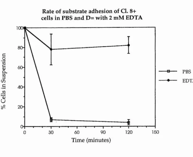
The rate of cell-substrate attachment of imaginai disc cells, either in the presence or absence of divalent cations was also tested. For this assay, 3 day old Cl. 8+ cells were isolated, as for the previous reaggregaton assays, by pipetting and centrifugation. Aliquots of 1 x 10^ cells were measured out and centrifuged down in 1 ml of either PBS or D= plus 2 mM EDTA. These cells were then plated out into individual wells of a 24 well plate (Nunc) as a single cell suspension in 1 ml of each test solution, and were incubated at 25°C. Four replicate samples were taken for cell counting at 0, 30 and 120 minutes for each test solution, by completely removing the medium in each well and leaving any adherent cells.
3.2.2 Cloning
immediately adjacent to them, on the 5 cm dish surface, whilst preventing any possible cross-contamination between these populations.
L127D6 cells for cloning were selected from populations of cells that displayed low reaggregative capacities. To isolate these cells, 4 tubes, each containing 1 x 10^ cells which had been harvested after 6 days growth, were rotated on a Spiramix roller as before and left to reaggregate in MM3 for 2 hours at 25°C. After this period, the tubes were centrifuged at low speed (200 rpm for 15 minutes) to separate the multicellular aggregates from any single cells. After removal of any aggregates, single cells in the supernatant were incubated for a further 2 hours and centrifuged as before. The number of single cells in the supernatant was taken and an aliquot was then removed from each sample and plated at a low density (approxroximately 500 cells/dish) onto the 3 cm Petri dish base in 1 ml of CSM and left to adhere overnight at 25°C. In this way, a very sparse seeding of single L127D6 cells was created, each cell being physically unable to establish contact with the next.
Each feeder layer of L127D6 cells had been grown for 2 days, at a seeding density of 3 x 10^ cells/dish, in a 5 cm Petri dish prior to use. The 3 cm dish bases from each of the four samples were then inverted onto the 5 cm dishes, in which the medium was presumably sufficiently conditioned, and incubated at 25°C. After 1 weeks growth the feeder layer was replaced, and this was repeated for 4 weeks.
plastics used were supplied by Nunc). These cells were then routinely passaged for the next 8 weeks until they had reached a sufficient state of proliferation. Thus 4 new cell lines representing each original sample were created, and were named: L4, L13, L14 and L15 (named after well noumbers in the 24 well plates they were taken from), and were then frozen down as established stocks.
3.3 Results
3.3.1 Reaggregation studies
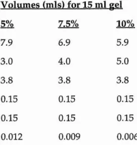
Figure 3. A illustrates the typical reaggregation response of an imaginai disc cell line when incubated in PBS and D= with 2 mM EDTA. The subject for this particular assay was the cloned wing cell line, Cl. 8R. This graph illustrates well a marked difference in response of the cell line to incubation in either of the test solutions. Cells that were suspended in PBS, and were thus exposed to the metal ions Ca^+ and Mg2+, had reaggregated to a great extent, with only around 20% of the cells failing to aggregate after 120 minutes of suspension. Most of this aggregation was achieved within a short space of time (30 minutes of incubation). Cl. 8R cells which had been incubated in D= with EDTA had not reaggregated to the same extent. In this environment cells seemed to adhere more slowly and less efficiently. After 120 minutes of assay, only around 60% of the cells had failed to reaggregate.
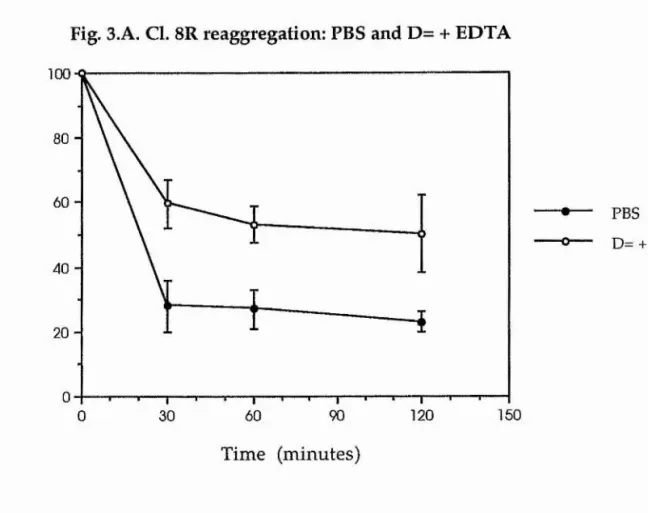
Figure 3.C represents the rate and extent of reaggregation of another cloned wing cell, Cl. C9. This time, however, the rate of reaggregation has been recorded by counting the number of cells in each aggregate within a given field of the haemocytometer, after 120 minutes of incubation, and calculating the average number of cells in an aggregate for that treatment. Those cells which have reaggregated in PBS can again be seen to have formed aggregates more quickly and to a greater extent than those cells which have been incubated in D= with EDTA. What can also be seen from this graph is that there is a large variance in the number of cells found in aggregates which had been formed in PBS; with cell numbers ranging from a few cells to around fifty in different clumps.
In Figure 3.D, the average number of cells at various stages of reaggregation has been calculated for trypsin-treated cells in both PBS and D= with EDTA. Again, trypsin has been used at a concentration of 0.01%. Cells in each treatment here tend to form aggregates of varying sizes; the larger aggregates being found, perhaps predictably, in the PBS treated samples. However, as is also seen when monitoring the decrease in single cells (Figure 3.B), this initial period of adhesivity apparently only existed during the first 60 minutes of culture, which was then followed by a decline in aggregate-cell number over the remaining 60 minutes. The largest cell aggregates, thus, were found in the PBS treated samples midway through the assay, with the average aggregate size dropping by the end of 120 minutes.
Therefore, one can conclude that trypsin seems to have little effect on the early phase of reaggregation in both test solutions. However, by the end of the assay, the presence of trypsin does seem to have a deleterious effect on average aggregate size, dropping to numbers similar to those found at 30 minutes of culture.
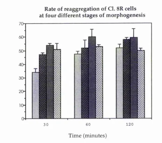
In imaginai cells at different stages of morphogenesis, one might expect there to be a variance in cell-cell adhesivity, depending on the type of CAM expressed at that particular stage. Figure 3.E illustrates the rate of reaggregation of Cl. 8R cells at different stages of growth in culture. These results suggest that Cl. 8R cells are at their most adhesive at around 7 days of growth, i.e. during aggregate formation, although the adhesivity of each sample appears to be fairly similar to the next. These results also indicate that the cells are at their least cell-cell adhesive when either very early on in morphogenesis, as the cells form a monolayer on day 1 with little or no cell cell adhesion, or when very late on in morphogenesis, on day 11 by which time the cells will have been in aggregates for a number of days.
These results provide some evidence that imaginai disc cells may change their modes of adhesion during morphogenesis depending on whether or not they have established contact with other cells. The normal sequence of in vitro morphogenetic events may be characterised by interactions and adhesion with the cell substrate during the early phase of growth, as the cells plate out as a monolayer and migrate or extend cell processes (day 0) followed by a period of cell-cell adhesion, during the intermediate stages of aggregation (days 4-7). Later stages (day 11) may, again, favour a lack of cell-cell adhesivity due to the cells already being found in aggregates thus suppressing the need for such adhesion.
presence or absence of the divalent cations Ca^+ and Mg2+, over a period of 2 hours. In the PBS sample, cell-substrate adhesion appears to be very rapid, with around 90% of all cells adhering to the plastic by 30 minutes of culture. The converse is true of cell-substrate adhesion in the D= plus EDTA samples, where, by the end of the assay only around 20% of all cells have adhered to the substrate.
3.3.2 Cloning
Within a few weeks of culture, some of the single cells plated to the underside of the cloning-plate had begun to proliferate and form isolated patches similar to those seen in Figures 3.G and 3.H, with fibroblast-shaped cells gradually growing outwards from a few aggregates at the centre. The same clonal group photographed in Figure 3.G is also pictured in Figure 3.1, by which time 14 days have elapsed and the feeder layer of cells has been removed: these cells have noticeably proliferated in the intervening period.
Figure 3.N illustrates the rates of reaggregation, in PBS, of four cell lines cloned from the parent line, L127D6. These results indicate that there does not seem to be a common trend of adhesion and that each cloned line has a different potential to reaggregate compared to the next. This reflects the clonal derivation of these lines and suggests that individual cells within an uncloned cell line, such as L127D6, may have different adhesive specificities.
From this assay, L. 14 was selected as a candidate line which exhibits a reduced capacity for short term cell-cell adhesion in roller culture. In Figure 3.N, 20% of all L. 14 cells have reaggregated by 60 minutes of culture, but this number has dropped by 120 minutes of culture to around 5%. Any aggregates that have formed are presumably very weakly cohesive, subsequently dissociating to single cells again.
plus EDTA. This graph clearly illustrates that the divalent cations, Ca^+ and Mg2+, are important, but not essential, for cell-cell adhesion in roller culture. The indication here is that there may be two different
populations of cell adhesion molecules present: one that is cation dependent and one that is cation independent.
8
Î
.B O) wi
T3 100 80 60 --Ô 40-20
-160 120 90 60 30 0 PBS
-o— D= + EDTA
Time (minutes)
Fig. 3.B. Cl. 8R reaggregation (trypsin): PBS and D= + EDTA
U
f
in 100 80 60 40-2 0
-150
120
60 90
0 30
PBS
D= + EDTA
determined by aggregate size. Again, over a period of 120 minutes, the presence of divalent cations is seen to enhance cell-cell adhesion, this time reflected in aggregate size. Here the average aggregate size by the end of the assay is larger in the PBS samples than in the EDTA
containing ones. There is also seen to be a large variation in aggregate size in the PBS samples.
60
50
40
30
-20
-10
-2 Z ^
0 30 60 120
m
PBS□ D= + EDTA
Time (minutes)
Fig. 3.D. Cl. C9 reaggregation (trypsin): PBS and D= + EDTA
bO
I
CL cu U 6050
40
30
-20
-10
-120
60 30
0
m
PBS□ D= + EDTA
O)
u
"OSd
I
rS
cS
El 0 days B3 4 days # 7 days S 11 days
Time (minutes)
Figure 3.E. The rate of reaggregation of Cl. 8R cells at four different stages of one passage in vitro (at 0,4, 7 and 11 days after plating onto tissue culture plastic). These results suggest that Cl. 8R cells have a relatively low capacity for cell-cell adhesion very early on in growth. As the cells progress in culture and begin to aggregate, cell-cell