printed in Great Britain © The Company of Biologists Limited 1989
SXE THE HINDWING CHORDOTONAL ORGANS ELEMENTS
OF THE LOCUST FLIGHT PATTERN GENERATOR?
BY K. G. PEARSON, B. HEDWIG* AND H. WOLFt
Department of Physiology, University of Alberta, Edmonton, Canada T6G 2H7
Accepted 24 February 1989
Summary
1. Anatomical and electrophysiological techniques were used to examine the structure, central nervous connections and discharge patterns of afferents arising from the hindwing chordotonal organs in the locust, Locusta migratoria.
2. The hindwing chordotonal organ afferents were found to be sensitive to low-frequency sounds (approx. 3 kHz) and to vibrations of the supporting surface. During flight in tethered animals these afferents were strongly activated. This flight-related activity was weakly modulated and the maximum occurred between depressor bursts.
3. Low-frequency sound pulses were used to activate selectively the hindwing chordotonal organ afferents during rhythmic flight activity in deefferented preparations (all motor nerves cut). Phasic stimulation of the chordotonal organs, time-locked to every cycle, had no effect on the frequency of the centrally generated rhythm as recorded from proximal nerve stumps.
4. Staining of single afferents from the hindwing chordotonal organ showed that they bifurcate in the metathoracic ganglion, with one branch terminating in the ring tract and the other branch projecting to the mesothoracic ganglion via the ipsilateral connective. None of the terminal processes of chordotonal afferents was observed to be located in dorsal neuropile regions containing the processes of flight neurones. Consistent with this finding, the chordotonal afferents were not observed to make synaptic connections to flight interneurones or motoneurones. 5. The main conclusion of this investigation is that the hindwing chordotonal organs do not contribute to the patterning of motor activity for flight. The hindwing chordotonal organs probably function as detectors of vibration and perhaps low-frequency sound.
Introduction
Associated with the wings of the locust are at least six groups of sensory receptors: wing-hinge stretch receptors, tegulae, campaniform sensillae,
chordo-* Present address: Zoologisches Institiit der Universitat, 3400 Gottigen, FRG.
t Present address: Fakultat fur Biologie, Universitat Konstanz, D-7750 Konstanz, FRG.
has been directed towards determining the role of some of these receptors in patterning motor activity for flight. This has led to a number of specific proposals regarding the function of the stretch receptors (Burrows, 1975, 1976; Mohl, 1985a,fc; Wendler, 1982; Wolf & Pearson, 1988), the tegulae (Neumann, 1985; Pearson & Wolf, 1988; Wolf & Pearson, 1988), the campaniform sensilla (Horsmann & Wendler, 1985; Elson, 1987a,&) and the hair bristles on the wings (Weis Fogh, 1956). Currently we have no knowledge about the function(s) of the chordotonal organs or thoracic hair fields.
The chordotonal organs of the hindwings consist of about 21 scolopidia in close association with each wing-hinge stretch receptor (Gettrup, 1962). The only reported property of the afferents arising from the hindwing chordotonal organ is that they are activated by wing depression (Gettrup, 1962). The effect of this activity on the flight motor pattern has not been established. Indeed, there are at present no indications that the chordotonal organs are involved in patterning flight activity, and at least one finding suggests that the chordotonal organs may be without direct effect on the flight system. This is that stimulation of the hindwing stretch receptor and chordotonal organ afferents produces the same effect on the central oscillator as stimulation of the hindwing stretch receptor alone (Reye & Pearson, 1988).
The aim of this investigation was to establish whether the hindwing chordotonal organs can influence the pattern-generating network for flight. Several approaches were used in this analysis. The first was to determine the timing of activity in the chordotonal organ afferents during tethered flight in intact animals; the second was to stimulate selectively the chordotonal organ afferents and examine the effect on the centrally generated rhythm in deefferented preparations; and the third was to determine the central projections and connections of the chordotonal organ afferents to neurones in the meso- and metathoracic ganglia. The results of this study indicate that activity in hindwing chordotonal organ afferents has no influence on the flight system and that these afferents make no connections to flight interneurones.
Materials and methods Animals
All experiments were performed on adult male and female Locusta migratoria obtained from a colony at the University of Alberta.
Preparations and recording set-up
chordotonal organ afferents in response to sound pulses and to passive and active wing movements.
Dorsal preparation
Following the removal of the wings and legs the animals were mounted dorsal side up on a cork board. The thoracic ganglia were exposed by making a midline incision along the dorsum of the thorax and removing the underlying gut, fat, salivary glands and small muscles. The meso- and metathoracic ganglia were covered with saline and a rigid stainless-steel plate was placed under them.
To determine the conduction properties and connections of afferents in nerve 1D2, monopolar hook electrodes (75/xm silver wire) were placed both distally on nerve 1D2 and proximally on nerve root 1 on one side of the animal. Nerve 1D2 was exposed by removing the hindwing dorsal longitudinal muscle. Both elec-trodes were covered with silicone grease to insulate them from the saline. The distal electrode was used to stimulate the afferents in nerve 1D2 and the proximal electrode on nerve 1 was used to monitor the afferent input to the ganglion. The distance between the two electrodes was about 6 mm.
The central connections of the afferents in nerve 1D2 were determined by recording intracellularly in neurones in the meso- and metathoracic ganglia and observing the response either to electrical stimulation of N1D2 or to low-frequency sound pulses (see below). Recording electrodes were filled with Lucifer Yellow (5% in distilled water). This dye was injected into individual neurones following recording (—5 nA for about 5 min). The ganglia were removed from the animal, fixed in 4% paraformaldehyde, dehydrated, and cleared in methyl salicylate. Filled neurones were viewed as wholemounts under epifluorescent illumination. The structure and central projections of individual afferents in nerve 1D2 were determined by penetrating the afferent close to nerve root 1. Lucifer Yellow was injected and the metathoracic ganglion was fixed and cleared in the described manner. Any ganglion found to contain a well-stained afferent was embedded in Spurr's resin and serially sectioned to determine the location of the processes within the ganglion.
hindwing chordotonal organs (see below).
Ventral preparation
The ventral surface of the thorax was fastened by wax to a rigid support and the animal was mounted ventral side up in a manner that allowed the wings to move naturally (see Wolf & Pearson, 1987, for details). A small piece of cuticle was removed from above the metathoracic ganglion and the nerve roots 1 were exposed to the point where they divide into branches 1C and ID. Metathoracic nerve 1C on one side of the animal was cut to eliminate input from the tegula and wing on that side. An extracellular hook electrode was placed on nerve 1 on the same side to monitor activity in the afferents in nerve 1D2 during flight. Flight activity was initiated by directing a wind stream towards the head.
Stimulation of afferents in nerve 1D2
The metathoracic nerves 1D2 contain afferents arising from the stretch receptor, chordotonal organ and a number of thoracic hair fields (Altman et al. 1978). Electrical stimulation (0-2 ms pulses) of nerve 1D2 was used to activate these afferents in some experiments. The stretch receptor could easily be stimulated selectively (see Fig. 1) because it is significantly larger than all other axons in nerve 1D2 (Altman et al. 1978). The afferents arising from the chordotonal organ and the hair fields do not form two distinct populations and thus it was not possible precisely to set the stimulation strength so that only one of these two groups of afferents was stimulated (see Fig. 1A). Nevertheless, the largest afferents from the chordotonal organ are significantly larger than the largest afferents from the hair fields (Gettrup, 1962). By monitoring the evoked activity in nerve root 1 it was therefore possible to adjust the stimulus strength to excite selectively the largest chordotonal organ afferents in addition to exciting the stretch receptor axon. Thus, to distinguish the central effects of the three groups of receptors three basic stimulus conditions were used: (i) activation of the stretch receptor alone, (ii) activation of the stretch receptor plus the low-threshold afferents from the chordotonal organ, and (iii) activation of all afferents by high stimulus strengths.
Results
Afferents in nerve 1D2
Anatomical studies have shown that the hindwing nerve 1D2 contains approxi-mately 400 axons (Altman et al. 1978). The largest of these (about 6/an in diameter) arises from the wing-hinge stretch receptor. The chordotonal organ gives rise to about 21 axons with diameters in the range 2-5-4/an (Gettrup, 1962; Altman et al. 1978). The origin of the smaller axons (diameters less than 2-5 /an) has not yet been fully established but many arise from hair fields on the thorax (Altman et al. 1978). The distinctly different diameters of the afferents originating from the different receptors was confirmed by recording from the afferents and examining their responses to electrical and natural stimuli. Fig. 1A shows typical potentials recorded extracellularly from nerve 1 in response to electrical stimu-lation of nerve 1D2. With stimulus strengths of about 0-12 V (0-2 ms duration) the axon of the stretch receptor was selectively activated. The origin of this unitary potential was confirmed by elevating the wing stump and noting that the potentials from the stretch receptor axon were identical to those evoked by just-threshold stimuli to nerve 1D2. The stimulus strength required for activating the stretch receptor axon was taken as the reference, T, for establishing the electrical thresholds for activating other afferents in nerve 1D2. The voltage required to just activate the next largest afferents in nerve 1D2 was about 1-3 times the stretch receptor threshold voltage, i.e. 1-3T. The conduction time of these afferents over the .6 mm distance separating the electrodes was longer than the conduction time of the stretch receptor axon (4 ms vs 2 ms). This resulted in two distinct components in the evoked potential recorded from nerve 1, the first being from the stretch receptor axon. The amplitude of the longer-latency component progress-ively increased as the stimulus strength was increased (Fig. 1A) and usually remained triphasic. Thus, the voltage at which the afferents other than those arising from the chordotonal organ were excited could not be distinguished by a noticeable change in the second component. However, intracellular recordings from interneurones not receiving input from the large chordotonal afferents indicated the existence of a group of smaller afferents with a threshold voltage of about 1-6T (see Fig. 9). We concluded, therefore, that within the range 1-3T-1-6T electrical stimulation of nerve 1D2 could be used to excite selectively the larger chordotonal organ afferents.
1-4T
1-6T
2-2T
2 ms 100 ms
Fig. 1. Physiological characterization of afferents in metathoracic nerve 1D2. (A) Re-cordings from nerve root 1 show the afferent volley evoked by electrical stimulation of nerve 1D2. The stimulus voltage required to just activate the stretch receptor axon, T, was taken as reference for establishing the voltages required to activate other afferents (top trace). Selective activation of the stretch receptor axon occurred over the range 1-0T-1-3T. Above 1-3T additional, slower-conducting axons were progressively recruited, yielding a second component in the recordings (traces labelled 1-4T, 1-6T and 2-2T). The afferents contributing to this second component arise from the chordotonal organ and thoracic hair fields. (B-D) Responses recorded from nerve 1D2 to (B) deflecting a single thoracic hair; (C) low-frequency sounds with the thoracic hair fields exposed; and (D) low-frequency sounds with the thoracic hair fields waxed over. The responses are indicated by the solid bars above the records. Note that the amplitude of the spikes originating from the hair afferent is smaller than that of the spikes from the spontaneously active units in nerve 1D2 (these units are from the chordotonal organ) and that waxing the hair fields does not abolish the activation of the sound-sensitive units.
waxing the hair fields presumably prevented any activation of the hair sensilla by sound we conclude that the chordotonal organ can be excited by low-frequency sound and that most of the chordotonal organ afferents are larger than the hair afferents. We also noted that the larger afferents were extremely sensitive to vibrations produced by lightly tapping the supporting platform.
Response of chordotonal organ afferents to sound
4 kHz 12 kHz
H
Sound
50
40 n. E
30
E 20
10
•
5 10 Frequency (kHz)
[image:7.473.106.374.54.336.2]15
Fig. 2. Sensitivity of afferents in metathoracic nerve 1D2 to sound stimuli. (A) Rec-ords from nerve 1 (Nl) show that low- (4 kHz) but not high-frequency (12 kHz) sound pulses excite numerous afferents in nerve 1D2 (nerve 1C was cut to eliminate input from the tegula and wing). (B) Frequency-response curve for the sound-sensitive afferents in nerve 1D2. The data for this curve were derived from four animals using sound pulses of 100 ms duration and 70 db re SPL. About 400 stimuli were given at each frequency and the average number of impulses per stimulus was calculated. The activity at frequencies above about 10 kHz is due to spontaneous background activity and not to auditory stimulation.
nerve 1C cut to eliminate input from the wings and tegulae. We assume that removal of the small piece of ventral cuticle in this preparation minimized any change in the mechanical properties of the thorax that might influence the auditory responsiveness of the chordotonal organ afferents. Low-frequency (2-5 kHz) sound pulses evoked vigorous activity in the chordotonal organ afferents (Fig. 2A, left), whereas high-frequency (10-20kHz) pulses with the same intensity were almost without effect (Fig. 2A, right). The dependence of the chordotonal organ response on frequency is shown in Fig. 2B.
Discharge pattern of chordotonal organ afferents in response to passive and active wing movements
Elevated
Rest
Depressed
[image:8.469.98.379.81.236.2]100 ms
Fig. 3. Maintained elevation or depression of the hindwing does not influence the activity in afferents in metathoracic nerve 1D2. Recordings are from nerve root 1 with nerve 1C cut. (A) Wing held in elevated position. This record was obtained following adaptation of activity in the stretch receptor. (B) Wing folded in normal resting position. (C) Wing held in depressed position.
tf—fM
50 ms
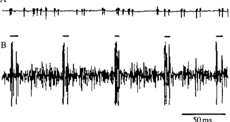
Fig. 4. Activity of afferents in metathoracic nerve 1D2 at rest (A) and during flight in a tethered animal (B). Records are from nerve root 1 with nerve 1C cut. The large spikes in B (indicated by bars) arise from the axons of the dorsal longitudinal motoneurones and the hindwing stretch receptor. Note the intense afferent activity between depressor bursts (compare with A) and the weak modulation of this activity.
was moved and held stationary. An unexpected result was that the spontaneous activity in the chordotonal organ afferents following adaptation was not related to wing position (Fig. 3). In contrast, it has been reported that chordotonal units in Schistocerca gregaria are excited during passive wing depression and the adapted level of activity is related to final wing position (Gettrup, 1962).
[image:8.469.122.348.330.450.2]attributed to the chordotonal organ (some may be from hair afferents) there seems little doubt that most of the activity arose from this receptor. This follows from the large amplitude of the spikes (compare Fig. 4A and 4B), similar to those activated by low-frequency sounds. The afferent activity was modulated during flight but in most preparations there was no time when afferent activity was absent. The peak activity always occurred approximately midway between depressor bursts and the minimal level of activity occurred immediately following each depressor burst (Fig. 4B).
Chordotonal organ activity does not influence the flight rhythm
The sound sensitivity of the chordotonal organs allowed us to examine the effects of input from these receptors on the flight rhythm. In a preparation that was deefferented (i.e. all motor nerves cut to prevent movement) and had both tympanal nerves cut (metathoracic nerves 6), the hindwing chordotonal organs were stimulated by 40 ms sound pulses triggered at a fixed delay on each cycle by activity in the dorsal longitudinal motor axons (Fig. 5A). Fig. 5B shows that the frequency of the centrally generated rhythm was not influenced when the hindwing chordotonal organs were activated midway through a short flight sequence. The record in Fig. 5B is typical of results obtained in all five animals. This negative result was obtained regardless of the delay between depressor activity and the onset of the sound pulse and regardless of sound intensity and duration.
Our observation that chordotonal organ activity had no effect on the flight rhythm was unexpected. Consequently, we sought additional evidence to demon-strate that the chordotonal organs do not participate in motor pattern generation. This evidence came from an examination of the structure of the central projections of the chordotonal organ afferents and the connections these afferents make with thoracic interneurones.
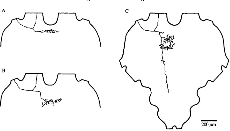
Anatomy of the central projections of the chordotonal organ afferents Backfilling the axons of nerve 1D2 with either Lucifer Yellow or cobalt chloride showed that they divide into three major projections in the metathoracic ganglion (Fig. 6A). One projection joins the dorsal medial tract (DMT) anteriorly in the ganglion and runs caudally into the fused abdominal ganglia. This projection shows only minor arborizations off the main tract. A second projection becomes closely associated with the median ventral tract (MVT) and projects to all three abdominal neuromeres. In the metathoracic neuromere, profuse arborizations from this projection engulf the ventral medial tract (VMT) and many additional processes course into the ring tract (RT). Although all other projections are restricted to the ipsilateral half of the ganglion these latter arborizations also supply the contralateral portions of these areas. Minor arborizations occur in the abdominal neuromeres. The third projection enters the meso-metathoracic connective. In the mesothoracic ganglion this branch runs in the MVT and arborizes in the anterior half of the ganglion where it engulfs the VMT and MVT
N l
-S o u n d •
'DL
20 -i
[image:10.470.115.362.43.373.2]
10-4 Time (s)
Fig. 5. Phasic stimulation of the hindwing chordotonal organ afferents does not influence the central flight rhythm. Rhythmic flight activity was initiated in a deefferented preparation and low-frequency sound pulses (4 kHz, 95 db, 40 ms) were triggered on each cycle commencing midway through the rhythmic sequence. (A) Rec-ords showing that sound pulses (sound) gave an intense burst of activity in the chordotonal organ afferents (CO) as recorded in nerve 1 (Nl). The large spikes produced by the dorsal longitudinal motoneurones (DL) were used to trigger the sound pulses at a pre-set delay, t. In this example the delay was 20 ms. (B) Plotting the instantaneous frequency of the central rhythm versus time shows that phasic stimu-lation of the chordotonal organ afferents midway through the sequence did not influence the frequency. The monitor of the sound pulses is shown below the plot. Each step in the plot was calculated by taking the inverse of the cycle period.
200 f*m
Fig. 6. Central projections of afferents in the metathoracic (A) and the mesothoracic (B) ganglia, as revealed by backfilling axons in nerve 1D2. The locations of the projections relative to the main fibre tracts are drawn into schematic diagrams of the cross-sections of the metathoracic ganglion in B and C, and of the mesothoracic ganglion in E and F. Tracts labelled according to Tyrer & Gregory (1982). MDT, median dorsal tract; LDT, lateral dorsal tract; DMT, dorsal medial tract; VLT, ventral lateral tract; MVT, median ventral tract; VMT, ventral medial tract; RT, ring tract; DIT, dorsal intermedial tract; VIT, ventral intermediate tract.
ganglion, then bifurcated to send a branch into the ipsilateral meso-metathoracic connective. The other branch either entered directly into the frontal auditory neuropile (anterior ring tract) (Figs 7B, 8A) or turned posteriorly before sending secondary processes medially to this neuropile region (Fig. 8B,C). Some of the latter group also projected posteriorly into the fused abdominal ganglia (Fig. 8C). This posterior process was associated with the MVT. We did not attempt to correlate the terminal structure with the auditory characteristics of the afferent.
Afferent «**S»»i
Sound'
n
100 ms
[image:12.470.49.419.39.311.2]200 jan
Fig. 7. Recording and staining of a sound-sensitive afferent in the metathoracic nerve 1D2. The afferent was penetrated close to its point of entry into the metathoracic ganglion. (A) A 4 kHz sound pulse evoked a brief burst of action potentials in the afferent. (B,C) Intracellular staining with Lucifer Yellow revealed that the axon bifurcated to send one branch into the meso-metathoracic connective and the other directly into the frontal auditory neuropile (ring tract, RT). Abbreviations as in Fig. 6.
ipsilateral connective or to the frontal auditory neuropile. Instead, all of them initially ran medially then turned to project posteriorly in the DMT. Because of their small size (as judged by their high threshold for activation) and absence of auditory response we presume these afferents arose from the thoracic hair fields.
Central connections of chordotonal organ afferents
200 fan
Fig. 8. Variability in structure of the central processes of sound-sensitive afferents in metathoracic nerve 1D2.
the largest afferents (Fig. 9C). Similar results were obtained in all the recordings we made in flight motoneurones and interneurones (N > 100).
Although flight neurones were not influenced by activity in the chordotonal organ afferents, many did receive an excitatory synaptic input from smaller afferents (presumably arising from the hair fields). Fig. 9D shows this input for intemeurone 511.
200 ms
Fig. 9. Flight interneurone 511 does not receive input from the hindwing chordotonal organs. (A) Drawing of interneurone 511 in the metathoracic ganglion. (B) Rhythmic activity recorded intracellularly from interneurone 511 (top trace). The depolarizations occur in antiphase to activity in depressors [bottom trace, electromyogram (EMG) from dorsal longitudinal muscles]. (C) Intracellular recording from neurone 511 (top trace) does not show any signs of synaptic input when nerve 1D2 was stimulated to activate the large afferents of the chordotonal organ. The bottom trace monitors the afferent input via nerve 1. The first potential was produced by the stretch receptor axon and the second potential (marked by the arrowhead) represents the afferent volley in the large chordotonal organ afferents. (D) Increasing the stimulus strength to activate all afferents in nerve 1D2 results in an EPSP in interneurone 511. (E) Low-frequency sound pulses (indicated in the bottom trace by intense bursts of activity recorded from nerve 1) do not produce a noticeable depolarization in interneurone 511 (top trace).
2mV
10 mV
c
—f*
T *
200 urn
601
Nl
Nl
5 m V
5 ms
5mV
[image:15.471.67.408.52.434.2]100 ms
Fig. 10. Excitatory synaptic input from hindwing -chordotonal organ afferents to 'auditory' intemeurones 601. (A) Drawing of interneurone 601 in the metathoracic ganglion. (B) Depolarization of interneurone 601 in response to low-frequency sound (tympanal nerves cut). Top trace, intracellular recording from 601; bottom trace, extracellular recording from nerve 1. The auditory signals evoked bursts of activity in afferents in nerve 1 and a corresponding depolarization in 601. (C-E) EPSPs evoked in 601 by electrical stimulation of nerve 1D2. Top traces, intracellular recordings; bottom traces, extracellular recordings from nerve 1. In C the stimulus strength was adjusted to excite both the stretch receptor axon (arrow) and the largest afferents of the chordotonal organ (arrowhead). Note the small EPSP evoked by the just-threshold stimulation of the large afferents of the chordotonal organ. As the stimulus strength was increased (indicated by the increase in amplitude of the afferent volley, arrowheads) the amplitude of the EPSP increased (D,E).
607
Nl
D 607
5 ms
200/mi
200 ms
llOmV
10 mV
Fig. 11. Interneurone 607 in the mesothoracic ganglion (A) and interneurone 608 in the metathoracic ganglion (B) receive excitatory synaptic input from the hindwing chordotonal organs. ( C - D ) EPSPs evoked in 607 by electrical stimulation of nerve 1D2. Top traces, intracellular recordings; bottom traces, extracellular recordings from nerve root 1. In this preparation no potential was recorded from the hindwing stretch receptor and thus only a single evoked potential is apparent in nerve 1. Note that as the stimulus strength was increased from C to D (indicated by the increase in amplitude of the afferent volley) the amplitude of the EPSP increased. (E) Strong depolarizations of interneurone 607 in response to low-frequency sounds (tympanal nerves cut). Top trace, intracellular recording from 607; bottom trace, extracellular recording from nerve 1. The auditory signals evoked bursts of activity in nerve 1 afferents and corresponding depolarizations in 607.
The chordotonal organ afferents were also found to make weaker, more variable and longer-latency excitatory connections with a number of other interneurones. Most of these were auditory interneurones that have been described previously. We did not examine the characteristics of these connections in detail.
Discussion
Do the hindwing chordotonal organs contribute to the patterning of flight motor activity?
The main finding of this investigation was that phasic activation of the afferents from the hindwing chordotonal organs had no influence on the flight rhythm produced in deefferented locusts (Fig. 5). Two other findings consistent with this result were: (1) that the chordotonal organ afferents did not terminate in neuropile regions known to contain processes of neurones involved in patterning flight activity, and (2) that the chordotonal organ afferents did not make connections with any interneurones or motoneurones in the flight system. These findings suggest that the hindwing chordotonal organs are not elements in the pattern-generating system for flight. However, this conclusion depends critically on two important assumptions. The first is that afferents from the chordotonal organ all respond to sound and none project to the dorsal neuropile, and the second is that examining the effect of activity in the chordotonal organ afferents on the centrally generated flight rhythm is an appropriate method for assessing the contribution of the chordotonal organs in patterning flight activity. We consider each of these assumptions separately in the following discussion.
Sound-sensitivity of the chordotonal organ afferents
Each hindwing chordotonal organ gives rise to about 21 afferent fibres (Gettrup, 1962). Anatomical studies have shown that these afferents are fairly uniform in diameter (2-5-4 //m) and that most of them are larger than all other afferents in nerve 1D2 with the exception of the single axon from the stretch receptor (Gettrup, 1962; Altman et al. 1978). The only other afferents known to exist in nerve 1D2 arise from thoracic hair fields. Since the spikes from sound-sensitive afferents in nerve 1D2 are larger than the spikes from the hair afferents (Fig. 1) and the chordotonal organ afferents are larger than hair afferents, it follows that the sound-sensitive afferents arise from the chordotonal organ. Supporting this conclusion is the finding that waxing the thoracic hair fields does not prevent sound from activating the larger afferents in nerve 1D2 (Fig. IE).
1D2 reveals that the minimal number of afferents that can be excited by sound is greater than 10 (K. G. Pearson, B. Hedwig & H. Wolf, unpublished observations). Assuming that only the chordotonal organ afferents are sound-sensitive, then it appears safe to conclude that the majority, but not necessarily all, of the chordotonal organ afferents can be excited by sound. If a sound-insensitive group exists it is unlikely to include the largest afferents from the chordotonal organ. This follows from the failure to find any neurone that could be depolarized by just-threshold electrical stimulation of the large chordotonal organ afferents in nerve 1D2 and not be depolarized by sound stimuli. However, electrical stimulation of nerve 1D2 at high voltages did produce excitatory input to some neurones that were not depolarized by low-frequency sounds (e.g. interneurone 511, Fig. 9). Thus the possibility remains that some of the smaller afferents from the chordotonal organ are not sensitive to sound and that these could influence the flight neurones.
Our anatomical studies have shown that the sensitive and the insensitive afferents in nerve 1D2 have distinctly different structures. All sound-sensitive afferents send projections into the frontal auditory neuropile (ring tract) of the metathoracic ganglion and an axonal branch to the mesothoracic ganglion. By contrast, sound-insensitive afferents are all smaller in diameter (as judged by the larger stimulus voltage required to activate them) and none have central projections to the frontal auditory neuropile region of the metathoracic ganglion (K. G. Pearson, B. Hedwig & H. Wolf, unpublished observations). Assuming that all chordotonal organ afferents are sound-sensitive, then it follows that none of these afferents send projections into the dorsal neuropile regions that contain the processes of neurones in the flight system (Ramirez & Pearson, 1988).
Sensitivity of the method
clear effect that the tegulae and stretch receptors can have on the central rhythm is the finding that these receptors send afferent projections into the flight neuropile. By contrast, the chordotonal organ afferents do not influence the central rhythm and do not project to the flight neuropile.
Although we have been unable to observe an influence of chordotonal input on the frequency of the deefferented rhythm, we have not excluded the possibility that chordotonal input could influence more subtle aspects of the motor pattern, such as the relative timing of activity in different motoneurones, without affecting the frequency. To test this possibility will require a detailed analysis of the flight motor pattern with and without chordotonal input.
Conclusion
Although we cannot provide a definite answer to the question posed in the heading of this section, there is considerable evidence that the chordotonal organs are not involved in the generation of the flight rhythm. The main evidence is that phasic input from the chordotonal organs does not influence the centrally generated rhythm, and that chordotonal afferents do not project to the flight neuropile or to flight interneurones or motoneurones. The main uncertainty that prevents full acceptance of this negative conclusion is the possibility that some of the smaller afferents from the chordotonal organ may not be sensitive to sound. If this subgroup of afferents exists then our technique of using sound pulses to activate chordotonal organ afferents would not have activated all the afferents. If this is the case then it is conceivable that the sound-insensitive subgroup could influence the flight system.
Possible function of the hindwing chordotonal organ
The striking feature of the afferents arising from the hindwing chordotonal organs is their sensitivity to low-frequency sound and to vibrations of the supporting platform. The latter property is consistent with the vibrational sensitivity of the chordotonal organ in the forewing base of crickets (Moss, 1971). It suggests that the wing chordotonal organs may function primarily as detectors of vibratory stimuli. This idea is further supported by the connections of the chordotonal organ to the thoracic low-frequency neurone 601. In locusts (Cokl etal. 1977), bushcrickets (Kiihne, 1982) and crickets (Kuhne etal. 1984) neurones of the auditory pathway receive additional vibratory input at least from the subgenual organs, which are vibration receptors in the legs. Thus, the hindwing chordotonal organ may be an additional sensory organ feeding information about vibration into the auditory centres of the nervous system. The simultaneous processing of auditory and vibratory stimuli enhances the responses of central auditory/vibratory neurones and is probably an important factor assisting orien-tation behaviour (Kalmring, 1983).
Heritage Foundation for Medical Research to HW and BH.
References
ALTMAN, J. S., ANSELMENT, E. & KUTSCH, W. (1978). Postembryonic development of an insect sensory system: ingrowth of axons from the hindwing sense organs in Locusta migratoria. Proc. R. Soc. B 202, 497-516.
BURROWS, M. (1975). Monosynaptic connexions between wing stretch receptors and flight motoneurones of the locust. J. exp. Biol. 62, 189-219.
BURROWS, M. (1976). The influence of sensory inflow on the flight system of the locust. Persp. exp. Biol. 1, 399-409.
COKL, A., KALMRING, K. & Wrrnc, H. (1977). The response of auditory ventral-cord neurones of Locusta migratoria to vibration stimuli. /. comp. Physiol. 120, 161-172.
ELSON, R. C. (1987a). Flight motor neurone reflexes driven by strain-sensitive wing mechanoreceptors in the locust. /. comp. Physiol. 161, 747-760.
ELSON, R. C. (1987/J). Interneuronal processing of inputs from the campaniform sensilla to the locust hindwing. J. comp. Physiol. 161, 761-776.
GETTRUP, E. (1962). Thoracic proprioceptors in the flight system of locusts. Nature, Lond. 193, 498-499.
HORSMANN, U. & WENDLER, G. (1985). The role of fast wing reflex in locust flight. In Insect Locomotion (ed. M. Gewecke & G. Wendler), pp. 157-166. Berlin: Paul Parey.
KALMRING, K. (1983). Convergence of auditory and vibratory senses at the neuronal level of the ventral cord in grasshoppers; its probable importance for behaviour in the habitat. Fortschr. Zool. 28, 129-141.
KOHNE, R. (1982). Neurophysiology of the vibration sense in locusts and bushcrickets: the response of ventral cord neurones. J. Insect Physiol. 28, 616-623.
KOHNE, R., SILVER, S. & LEWIS, B. (1984). Processing of vibratory and acoustic signals by ventral cord neurones in the cricket Gryllus campestris. J. Insect Physiol. 30, 575-585. MOHL, B. (1985a). The role of proprioception in locust flight control, n . Information signalled
by forewing stretch receptors during flight. J. comp. Physiol. 156, 103-116.
MOHL, B. (1985£>). The role of proprioception in locust flight control. III. The influence of afferent stimulation of the stretch receptor nerve. J. comp. Physiol. 156, 281-292.
MOss, D. (1971). Sinnesorgane im Bereich de Flugels der Feldgrille {Gryllus campestril L.) und ihre Bedeutung fiir die Kontrolle der Singbewegung und die Einstellung de Flugellage. Z. vergl. Physiol. 73, 53-83.
NEUMANN, L. (1985). Experiments on tegula function for flight coordination in the locust. In Insect Locomotion (ed. M. Gewecke & G. Wendler), pp. 149-156. Hamburg: Paul Parey. PEARSON, K. G. & WOLF, H. (1988). Connections of hindwing tegulae with flight neurones in the
locust, Locusta migratoria. J. exp. Biol. 135, 381-409.
RAMIREZ, J. M. & PEARSON, K. G. (1988). Generation of motor patterns for walking and flight in motoneurons supplying bifunctional muscles in the locust. J. Neurobiol. 19, 257-282. REYE, D. N. & PEARSON, K. G. (1988). Entrainment of the locust central flight oscillator by wing
stretch receptor stimulation. /. comp. Physiol. 162, 77-89.
ROBERTSON, R. M. & PEARSON, K. G. (1982). A preparation for the intracellular analysis of neuronal activity during flight in the locust. J. comp. Physiol. 146, 311-320.
ROMER, H. & MARQUART, V. (1984). Morphology and physiology of auditory interneurons in the metathoracic ganglion of the locust. J. comp. Physiol. 155, 249-262.
TYRER, N. M. & GREGORY, G. E. (1982). A guide to the neuroanatomy of the locust suboesophageal and thoracic ganglia. Phil. Trans. R. Soc. Ser. B 297, 91-124.
WEIS-FOGH, T. (1956). Biology and physics of locust flight. IV. Notes on sensory mechanisms in locust flight. Phil. Trans. R. Soc. Ser. B 239, 553-584.
WOLF, H. & PEARSON, K. G. (1987). Comparison of motor patterns in the intact and deafferented flight system of the locust. II. Intracellular recordings from flight motoneurons. J. comp. Physiol. 160, 269-279.