1,2,3,4-Tetrahydrophenazine
5,10-dioxide
Tao Sun,aJianye Li,aHongwei Qiao,bAiyou Haoa* and Yueming Lia
aSchool of Chemistry and Chemical Engineering and Key Laboratory of Colloid and
Interface Chemistry of the Ministry of Education, Shandong University, Shanda Nanlu 27, Jinan 250100, People’s Republic of China, andbShandong Shengquan Chemical
Co. Ltd, Zhangqiu Jinan, 250204, People’s Republic of China Correspondence e-mail: [email protected]
Received 10 June 2010; accepted 29 July 2010
Key indicators: single-crystal X-ray study;T= 296 K; mean(C–C) = 0.002 A˚; disorder in main residue;Rfactor = 0.040;wRfactor = 0.127; data-to-parameter ratio = 10.2.
The complete molecule of the title compound, C12H12N2O2, lies on two crystallographic symmetry elements: a twofold axis and a mirror plane. In the molecular structure, the quinoxaline ring and two methylene substituents lie on the mirror plane while the other two methylene groups are disordered about the plane. The crystal packing is stabilized by weak inter-molecular – stacking interactions with centroid–centroid distances of 3.6803 (7) A˚ .
Related literature
For the synthetic preparation, see: Haddadin & Issidorides (1965); Issidorides & Haddadin (1966). For background to quinoxaline di-N-oxide compounds, see: Edwardset al.(1975) and for their biological activity, see: Urquiolaet al.(2008). For a related structure, see: Wanget al.(2010).
Experimental
Crystal data
C12H12N2O2
Mr= 216.24 Orthorhombic,Cmcm a= 11.7780 (2) A˚
b= 13.1938 (3) A˚
c= 6.5561 (1) A˚
V= 1018.80 (3) A˚3
Z= 4
MoKradiation = 0.10 mm 1
T= 296 K
0.310.290.26 mm
Data collection
Bruker APEXII CCD area-detector diffractometer
Absorption correction: multi-scan (SADABS; Sheldrick, 1996)
Tmin= 0.67,Tmax= 0.74
3311 measured reflections 620 independent reflections 534 reflections withI> 2(I)
Rint= 0.016
Refinement
R[F2> 2(F2)] = 0.040
wR(F2) = 0.127
S= 1.10 620 reflections 61 parameters
H atoms treated by a mixture of independent and constrained refinement
max= 0.23 e A˚ 3 min= 0.29 e A˚ 3
Data collection:APEX2(Bruker, 2007); cell refinement:SAINT
(Bruker, 2007); data reduction:SAINT; program(s) used to solve structure:SIR97(Altomare et al., 1999); program(s) used to refine structure: SHELXTL (Sheldrick, 2008); molecular graphics:
SHELXTL; software used to prepare material for publication:
WinGX(Farrugia, 1999).
This work was supported by the NSFC (grant No. 20625307), the National Basic Research Program of China (973 Program, 2009CB930103) and the Graduate Independent Innovation Foundation of Shandong University (GIIFSDU).
Supplementary data and figures for this paper are available from the IUCr electronic archives (Reference: NK2042).
References
Altomare, A., Burla, M. C., Camalli, M., Cascarano, G. L., Giacovazzo, C., Guagliardi, A., Moliterni, A. G. G., Polidori, G. & Spagna, R. (1999).J. Appl. Cryst.32, 115–119.
Bruker (2007).APEX2andSAINT. Bruker AXS Inc., Madison, Wisconsin, USA.
Edwards, M. L., Bambury, R. E. & Ritter, H. W. (1975).J. Med. Chem.18, 637– 639.
Farrugia, L. J. (1999).J. Appl. Cryst.32, 837–838.
Haddadin, M. J. & Issidorides, C. H. (1965).Tetrahedron Lett.6, 3253–3256. Issidorides, C. H. & Haddadin, M. J. (1966).J. Org. Chem.31, 4067–4068. Sheldrick, G. M. (1996).SADABS. University of Go¨ttingen, Germany. Sheldrick, G. M. (2008).Acta Cryst.A64, 112–122.
Urquiola, C., Cabrera, M., Lavaggi, M. L., Cerecetto, H., Gonzalez, M., Cerain, A. L., Monge, A., Costa-Filho, A. J. & Torre, M. H. (2008).J. Inorg. Biochem.102, 119–126.
Wang, Z., Jia, W., Yao, H., Qiu, H. & Wang, W. (2010).Acta Cryst.E66, o1380. Acta Crystallographica Section E
Structure Reports Online
supporting information
Acta Cryst. (2010). E66, o2425 [https://doi.org/10.1107/S1600536810030242]
1,2,3,4-Tetrahydrophenazine 5,10-dioxide
Tao Sun, Jianye Li, Hongwei Qiao, Aiyou Hao and Yueming Li
S1. Comment
Quinoxaline di-N-oxide compounds are widely used in sterilization and growth-promoting of animals, pharmacological
properties usable as intermediates for producing plant protection agents (Edwards et al.,1975). There has been a growing
interest in the syntheses of quinoxaline di-N-oxide compounds that have both biological and commercial importance
(Urquiola et al., 2008). Now, we report herein the crystal structure of the title benzotriazole derivative.
The complete molecule of the title compound, C12H12N2O2, is generated by a crystallographic symmetry operation along
a twofold axis. In the moleclcular structure of the crystal, the quinoxaline ring and two methylene substituents of the
quinoxaline ring locate at a mirror plane of the Cmcm group. The other two methylenes of the cyclohexane ring are
disordered over two positions with half occupancy. The crystal packing is stabilized by weak intermolecular π-π aromatic
stacking interactions with centroid-centroid distances of 3.6803 (7) Å.
S2. Experimental
The compound was synthesized as described previously by Haddadin & Issidorides (1965) and Issidorides & Haddadin
(1966). Yellow crystals were obtained by slow evaporation of a methanolic solution.
S3. Refinement
H atoms in the benzene were placed in geometrically calculated positions and refined using a riding model. H atoms in
CH2 groups were located in geometrically calculated positions also but their positions were refined independently and
their isotropic displacement parameters were fixed to 0.08 in the refinement. Two CH2 groups were disordered over
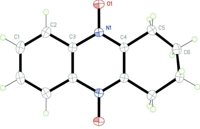
Figure 1
A view of the title compound with displacement ellipsoids are drawn at the 30% probability level. Unlabelled atoms are
related to labelled atoms by a twofold rotation. The second disorder component is omitted.
1,2,3,4-Tetrahydrophenazine 5,10-dioxide
Crystal data
C12H12N2O2
Mr = 216.24
Orthorhombic, Cmcm
Hall symbol: -C 2c 2
a = 11.7780 (2) Å
b = 13.1938 (3) Å
c = 6.5561 (1) Å
V = 1018.80 (3) Å3
Z = 4
F(000) = 456
Dx = 1.410 Mg m−3
Mo Kα radiation, λ = 0.71073 Å Cell parameters from 1577 reflections
θ = 2.3–26.8°
µ = 0.10 mm−1
T = 296 K Prism, yellow
0.31 × 0.29 × 0.26 mm
Data collection
Bruker APEXII CCD area-detector diffractometer
Radiation source: fine-focus sealed tube Graphite monochromator
φ and ω scans
Absorption correction: multi-scan (SADABS; Sheldrick, 1996)
Tmin = 0.67, Tmax = 0.74
3311 measured reflections 620 independent reflections 534 reflections with I > 2σ(I)
Rint = 0.016
θmax = 26.9°, θmin = 2.3°
h = −14→14
k = −16→12
l = −8→8
Refinement
Refinement on F2 Least-squares matrix: full
R[F2 > 2σ(F2)] = 0.040
0 restraints
H atoms treated by a mixture of independent and constrained refinement
w = 1/[σ2(F
o2) + (0.0854P)2 + 0.1004P] where P = (Fo2 + 2Fc2)/3
(Δ/σ)max < 0.001 Δρmax = 0.23 e Å−3 Δρmin = −0.29 e Å−3
Special details
Experimental. 1H NMR (400?MHz, DMSO-d6): δ 8.67 (2H, d, J = 3.5?Hz, Ar—H), 7.89 (2H, d, J = 3.2?Hz, Ar—H),
3.77 (1H, s, CH), 2.66 (3H, s, CH3), 2.51 (2H, m, CH2), 1.45 (6H, s, CH3); Calcd for C13H16N2O2: C, 67.22; H, 6.94; N, 12.06. Found: C, 67.18; H, 6.99; N, 11.95; ESIMS calcd for C13H16N2O2H+ m/z 232.38, found m/z 232.19.
Geometry. All e.s.d.'s (except the e.s.d. in the dihedral angle between two l.s. planes) are estimated using the full
covariance matrix. The cell e.s.d.'s are taken into account individually in the estimation of e.s.d.'s in distances, angles and torsion angles; correlations between e.s.d.'s in cell parameters are only used when they are defined by crystal symmetry. An approximate (isotropic) treatment of cell e.s.d.'s is used for estimating e.s.d.'s involving l.s. planes.
Refinement. Refinement of F2 against ALL reflections. The weighted R-factor wR and goodness of fit S are based on F2,
conventional R-factors R are based on F, with F set to zero for negative F2. The threshold expression of F2 > σ(F2) is used only for calculating R-factors(gt) etc. and is not relevant to the choice of reflections for refinement. R-factors based on F2 are statistically about twice as large as those based on F, and R- factors based on ALL data will be even larger.
Fractional atomic coordinates and isotropic or equivalent isotropic displacement parameters (Å2)
x y z Uiso*/Ueq Occ. (<1)
C1 0.44078 (13) −0.14330 (10) 0.2500 0.0434 (4)
H1 0.4017 −0.2045 0.2500 0.052*
C2 0.38142 (12) −0.05388 (9) 0.2500 0.0399 (4)
H2 0.3025 −0.0544 0.2500 0.048*
C3 0.44062 (11) 0.03818 (9) 0.2500 0.0311 (4)
C4 0.44068 (10) 0.21722 (9) 0.2500 0.0326 (4)
C5 0.37292 (12) 0.31337 (10) 0.2500 0.0470 (4)
H5 0.3247 (10) 0.3113 (9) 0.131 (2) 0.070*
C6 0.44666 (19) 0.40485 (17) 0.1867 (4) 0.0618 (9) 0.50
H6 0.407 (2) 0.4669 (19) 0.199 (5) 0.090* 0.50
H7 0.468 (3) 0.3993 (19) 0.040 (4) 0.090* 0.50
N1 0.38161 (10) 0.12976 (7) 0.2500 0.0336 (4)
O1 0.27161 (9) 0.12938 (6) 0.2500 0.0508 (4)
Atomic displacement parameters (Å2)
U11 U22 U33 U12 U13 U23
C1 0.0608 (9) 0.0288 (7) 0.0407 (7) −0.0082 (5) 0.000 0.000
C2 0.0426 (8) 0.0339 (7) 0.0432 (7) −0.0078 (5) 0.000 0.000
C3 0.0339 (8) 0.0281 (7) 0.0314 (6) −0.0008 (4) 0.000 0.000
C4 0.0318 (7) 0.0277 (7) 0.0384 (7) 0.0008 (4) 0.000 0.000
C5 0.0372 (8) 0.0320 (8) 0.0719 (10) 0.0056 (5) 0.000 0.000
C6 0.0516 (11) 0.0289 (10) 0.105 (3) 0.0038 (7) 0.0002 (10) 0.0110 (10)
N1 0.0282 (6) 0.0313 (6) 0.0412 (6) −0.0005 (3) 0.000 0.000
Geometric parameters (Å, º)
C1—C2 1.3714 (19) C5—C6ii 1.544 (3)
C1—C1i 1.395 (3) C5—C6 1.544 (3)
C1—H1 0.9300 C5—H5 0.965 (13)
C2—C3 1.4005 (17) C6—C6ii 0.831 (5)
C2—H2 0.9300 C6—C6iii 1.256 (5)
C3—N1 1.3939 (15) C6—C6i 1.506 (4)
C3—C3i 1.399 (2) C6—H6 0.95 (2)
C4—N1 1.3474 (15) C6—H7 1.00 (3)
C4—C4i 1.397 (2) N1—O1 1.2956 (16)
C4—C5 1.4987 (16)
C2—C1—C1i 120.65 (9) C6ii—C6—C6iii 90.000 (2)
C2—C1—H1 119.7 C6ii—C6—C6i 56.5 (2)
C1i—C1—H1 119.7 C6iii—C6—C6i 33.5 (2)
C1—C2—C3 119.49 (15) C6ii—C6—C5 74.39 (10)
C1—C2—H2 120.3 C6iii—C6—C5 124.23 (10)
C3—C2—H2 120.3 C6i—C6—C5 108.72 (16)
N1—C3—C3i 119.90 (7) C6ii—C6—H6 85.1 (18)
N1—C3—C2 120.24 (14) C6iii—C6—H6 119.6 (16)
C3i—C3—C2 119.86 (8) C6i—C6—H6 111.4 (17)
N1—C4—C4i 121.08 (7) C5—C6—H6 112.1 (16)
N1—C4—C5 116.74 (12) C6ii—C6—H7 164.4 (18)
C4i—C4—C5 122.17 (7) C6iii—C6—H7 75.0 (18)
C4—C5—C6ii 111.23 (14) C6i—C6—H7 108.4 (18)
C4—C5—C6 111.23 (14) C5—C6—H7 110.3 (16)
C6ii—C5—C6 31.2 (2) H6—C6—H7 106 (2)
C4—C5—H5 106.8 (7) O1—N1—C4 121.31 (9)
C6ii—C5—H5 124.8 (8) O1—N1—C3 119.68 (9)
C6—C5—H5 97.8 (7) C4—N1—C3 119.01 (13)
C1i—C1—C2—C3 0.0 C4—C5—C6—C6i −50.6 (2)
C1—C2—C3—N1 180.0 C6ii—C5—C6—C6i 45.6 (2)
C1—C2—C3—C3i 0.0 C4i—C4—N1—O1 180.0
N1—C4—C5—C6ii 163.23 (11) C5—C4—N1—O1 0.0
C4i—C4—C5—C6ii −16.77 (11) C4i—C4—N1—C3 0.0
N1—C4—C5—C6 −163.23 (11) C5—C4—N1—C3 180.0
C4i—C4—C5—C6 16.77 (11) C3i—C3—N1—O1 180.0
C4—C5—C6—C6ii −96.23 (6) C2—C3—N1—O1 0.0
C4—C5—C6—C6iii −17.19 (11) C3i—C3—N1—C4 0.0
C6ii—C5—C6—C6iii 79.04 (8) C2—C3—N1—C4 180.0