5-Ethyl-4-methyl-1
H
-pyrazol-3(2
H
)-one
Tara Shahani,aHoong-Kun Fun,a*‡ R. Venkat Ragavan,b V. Vijayakumarband S. Sarveswarib
a
X-ray Crystallography Unit, School of Physics, Universiti Sains Malaysia, 11800 USM, Penang, Malaysia, andbOrganic Chemistry Division, School of Advanced Sciences, VIT University, Vellore 632 014, India
Correspondence e-mail: [email protected]
Received 5 May 2010; accepted 10 May 2010
Key indicators: single-crystal X-ray study;T= 100 K; mean(C–C) = 0.001 A˚; Rfactor = 0.039;wRfactor = 0.123; data-to-parameter ratio = 22.5.
In the title compound, C6H10N2O, the 2,3-dihydro-1H
-pyrazole ring is approximately planar, with a maximum deviation of 0.013 (1) A˚ . Pairs of intermolecular N—H O hydrogen bonds link neighboring molecules into dimers, generatingR2
2
(8) ring motifs. These dimers are further linked into two-dimensional arrays parallel to the bc plane by intermolecular N—H O hydrogen bonds. The crystal structure is further stabilized by C—H interactions.
Related literature
For the background to and the biological activity of 3-ethyl-4-methyl-1H-pyrazol-5-ol, see: Brogden (1986); Coersmeier et al. (1986); Gursoy et al.(2000); Ragavan et al.(2009, 2010); Watanabeet al. (1984); Kawaiet al. (1997); Wuet al.(2002). For related structures, see: Shahani et al. (2009, 2010). For hydrogen-bond motifs, see: Bernsteinet al.(1995). For refer-ence bond-length data, see: Allenet al.(1987). For the stability of the temperature controller used for the data collection, see: Cosier & Glazer (1986).
Experimental
Crystal data
C6H10N2O
Mr= 126.16 Monoclinic,P21=c a= 8.374 (2) A˚
b= 7.2881 (16) A˚ c= 11.300 (3) A˚ = 109.955 (5) V= 648.3 (3) A˚3
Z= 4
MoKradiation = 0.09 mm1
T= 100 K
0.520.160.09 mm
Data collection
Bruker APEXII DUO CCD area-detector diffractometer Absorption correction: multi-scan
(SADABS; Bruker, 2009) Tmin= 0.954,Tmax= 0.992
10018 measured reflections 2745 independent reflections 2325 reflections withI> 2(I) Rint= 0.029
Refinement
R[F2> 2(F2)] = 0.039
wR(F2) = 0.123
S= 1.14 2745 reflections
122 parameters
All H-atom parameters refined
max= 0.52 e A˚3 min=0.35 e A˚
[image:1.610.311.566.296.346.2]3
Table 1
Hydrogen-bond geometry (A˚ ,).
Cg1 is the centroid of the 1H-pyrazole ring (C1–C3/N1/N2).
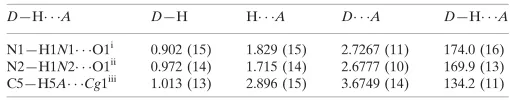
D—H A D—H H A D A D—H A
N1—H1N1 O1i 0.902 (15) 1.829 (15) 2.7267 (11) 174.0 (16) N2—H1N2 O1ii
0.972 (14) 1.715 (14) 2.6777 (10) 169.9 (13) C5—H5A Cg1iii
1.013 (13) 2.896 (15) 3.6749 (14) 134.2 (11)
Symmetry codes: (i)xþ1;yþ1;z; (ii)x;yþ3 2;z
1
2; (iii)x;yþ 1 2;z
1 2.
Data collection:APEX2(Bruker, 2009); cell refinement:SAINT (Bruker, 2009); data reduction:SAINT; program(s) used to solve structure: SHELXTL (Sheldrick, 2008); program(s) used to refine structure:SHELXTL; molecular graphics:SHELXTL; software used to prepare material for publication:SHELXTLandPLATON(Spek, 2009).
TSH and HKF thank Universiti Sains Malaysia (USM) for the Research University Golden Goose Grant (1001/PFIZIK/ 811012). VV is grateful to the DST-India for funding through the Young Scientist Scheme (Fast Track Proposal).
Supplementary data and figures for this paper are available from the IUCr electronic archives (Reference: WN2385).
References
Allen, F. H., Kennard, O., Watson, D. G., Brammer, L., Orpen, A. G. & Taylor, R. (1987).J. Chem. Soc. Perkin Trans. 2, pp. S1–19.
Bernstein, J., Davis, R. E., Shimoni, L. & Chang, N.-L. (1995).Angew. Chem. Int. Ed. Engl.34, 1555–1573.
Brogden, N. R. (1986).Drugs,32, 60–70.
Bruker (2009).APEX2, SAINTandSADABS.Bruker AXS Inc., Madison, Wiscosin, USA.
Coersmeier, C., Wittenberg, H. R., Aehringhaus, U., Dreyling, K. W., Peskar, B. M., Brune, K. & Pesker, B. A. (1986).Agents Actions Suppl.19, 137–153. Cosier, J. & Glazer, A. M. (1986).J. Appl. Cryst.19, 105–107.
Gursoy, A., Demirayak, S., Capan, G., Erol, K. & Vural, K. (2000).Eur. J. Med. Chem.35, 359–364.
Kawai, H., Nakai, H., Suga, M., Yuki, S., Watanabe, T. & Saito, K. I. (1997).J. Pharmacol. Exp. Ther.281, 921–927.
Ragavan, R. V., Vijayakumar, V. & Kumari, N. S. (2009).Eur. J. Med. Chem. 44, 3852–3857.
Ragavan, R. V., Vijayakumar, V. & Kumari, N. S. (2010).Eur. J. Med. Chem. 45, 1173–1180.
organic compounds
Acta Cryst.(2010). E66, o1357–o1358 doi:10.1107/S160053681001696X Shahaniet al.
o1357
Acta Crystallographica Section E
Structure Reports
Online
ISSN 1600-5368
(2009).Acta Cryst.E65, o3249–o3250.
Shahani, T., Fun, H.-K., Ragavan, R. V., Vijayakumar, V. & Sarveswari, S. (2010).Acta Cryst.E66, o142–o143.
Sheldrick, G. M. (2008).Acta Cryst.A64, 112–122.
Watanabe, T., Yuki, S., Egawa, M. & Nishi, H. (1984).J. Pharmacol. Exp. Ther. 268, 1597–1604.
supporting information
sup-1
Acta Cryst. (2010). E66, o1357–o1358
supporting information
Acta Cryst. (2010). E66, o1357–o1358 [https://doi.org/10.1107/S160053681001696X]
5-Ethyl-4-methyl-1
H
-pyrazol-3(2
H
)-one
Tara Shahani, Hoong-Kun Fun, R. Venkat Ragavan, V. Vijayakumar and S. Sarveswari
S1. Comment
Pyrazolone derivatives have a broad spectrum of biological activities as analgesic, antipyretic and anti-inflammatory
therapeutical drugs (Brogden, 1986; Gursoy et al., 2000). A class of new pyrazolone compounds have been synthesized
and reported to exhibit antibacterial and antifungal activities (Ragavan et al., 2010; Ragavan et al., 2009). A new
pyrazolone derivative, edaravone (5-ethyl-4-methyl-1H-pyrazol-3(2H)-one), is being used as a drug in clinical practice
for brain ischemia (Watanabe et al., 1984; Kawai et al., 1997) and it has also been found to be effective against
myocardial ischemia (Wu et al., 2002).
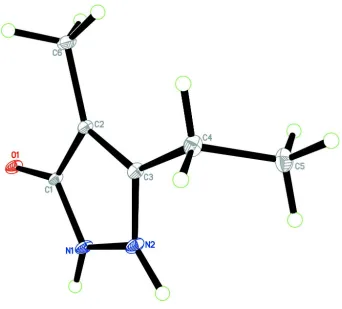
In the crystal structure (Fig. 1), the 2,3-dihydro-1H-pyrazole ring (C1–C3/N1/N2) is approximately planar with a
maximum deviation of 0.013 (1) Å for atoms N1 and N2 (but they are on opposite sides of the plane). The bond lengths
(Allen et al., 1987) and angles are within normal ranges and comparable to those in closely related structures reported
recently (Shahani et al., 2009; 2010).
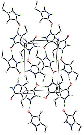
In the crystal packing (Fig. 2), pairs of intermolecular N1—H1N1···O1 hydrogen bonds (Table 1) link neighboring
molecules into dimers, generating R2
2(8) ring motifs (Bernstein et al., 1995). These dimers are further linked into 2D
arrays parallel to the bc plane by intermolecular N2—H1N2···O1 hydrogen bonds (Table 1). The crystal structure is
further stabilized by a C—H···π interaction (Table 1), involving the C1–C3/N1/N2 ring (centroid Cg1) .
S2. Experimental
The compound 5-ethyl-4-methyl-1H-pyrazol-3(2H)-one has been synthesized using the method reported in the literature
(Ragavan et al., 2009, 2010) and purified by column chromatography (MeOH: EtOAc, 1:99). It was recrystallised as a
colourless solid, using ethanol. Mp: 496.4–507.1 K; MS calculated for C6H10N2O: 126.15. Found: 128.0 (M+).
S3. Refinement
All hydrogen atoms were located in a difference map and were refined freely [N–H = 0.902 (14) – 0.972 (14) Å; C–H =
Figure 1
The molecular structure of the title compound, showing 30% probability displacement ellipsoids and the atom numbering
supporting information
sup-3
[image:5.610.171.443.69.503.2]Acta Cryst. (2010). E66, o1357–o1358 Figure 2
The crystal packing of the title compound, showing a 2D array parallel to the bc plane. Hydrogen bonds are denoted by
dashed lines. H atoms not involved in the hydrogen bond interactions have been omitted for clarity.
5-Ethyl-4-methyl-1H-pyrazol-3(2H)-one
Crystal data
C6H10N2O Mr = 126.16
Monoclinic, P21/c
Hall symbol: -P 2ybc
a = 8.374 (2) Å
b = 7.2881 (16) Å
c = 11.300 (3) Å
β = 109.955 (5)°
V = 648.3 (3) Å3 Z = 4
F(000) = 272
Dx = 1.293 Mg m−3
Mo Kα radiation, λ = 0.71073 Å Cell parameters from 3666 reflections
θ = 2.6–34.5°
Bruker APEXII DUO CCD area-detector diffractometer
Radiation source: fine-focus sealed tube Graphite monochromator
φ and ω scans
Absorption correction: multi-scan (SADABS; Bruker, 2009)
Tmin = 0.954, Tmax = 0.992
10018 measured reflections 2745 independent reflections 2325 reflections with I > 2σ(I)
Rint = 0.029
θmax = 34.6°, θmin = 3.4°
h = −13→13
k = −11→11
l = −18→17
Refinement
Refinement on F2
Least-squares matrix: full
R[F2 > 2σ(F2)] = 0.039 wR(F2) = 0.123 S = 1.14 2745 reflections 122 parameters 0 restraints
Primary atom site location: structure-invariant direct methods
Secondary atom site location: difference Fourier map
Hydrogen site location: inferred from neighbouring sites
All H-atom parameters refined
w = 1/[σ2(F
o2) + (0.0715P)2 + 0.0472P]
where P = (Fo2 + 2Fc2)/3
(Δ/σ)max < 0.001
Δρmax = 0.52 e Å−3
Δρmin = −0.35 e Å−3
Special details
Experimental. The crystal was placed in the cold stream of an Oxford Cyrosystems Cobra open-flow nitrogen cryostat (Cosier & Glazer, 1986) operating at 100.0 (1) K.
Geometry. All esds (except the esd in the dihedral angle between two l.s. planes) are estimated using the full covariance matrix. The cell esds are taken into account individually in the estimation of esds in distances, angles and torsion angles; correlations between esds in cell parameters are only used when they are defined by crystal symmetry. An approximate (isotropic) treatment of cell esds is used for estimating esds involving l.s. planes.
Refinement. Refinement of F2 against ALL reflections. The weighted R-factor wR and goodness of fit S are based on F2,
conventional R-factors R are based on F, with F set to zero for negative F2. The threshold expression of F2 > σ(F2) is used
only for calculating R-factors(gt) etc. and is not relevant to the choice of reflections for refinement. R-factors based on F2
are statistically about twice as large as those based on F, and R- factors based on ALL data will be even larger.
Fractional atomic coordinates and isotropic or equivalent isotropic displacement parameters (Å2)
x y z Uiso*/Ueq
O1 0.42822 (7) 0.62337 (8) 0.11992 (5) 0.01463 (13)
N1 0.42529 (8) 0.69441 (9) −0.08076 (6) 0.01353 (13)
N2 0.35794 (9) 0.82809 (9) −0.16813 (6) 0.01431 (13)
C1 0.38533 (9) 0.73007 (10) 0.02374 (6) 0.01110 (13)
C2 0.29351 (9) 0.89787 (9) 0.00188 (6) 0.01142 (13)
C3 0.28136 (9) 0.95309 (10) −0.11791 (7) 0.01249 (14)
C4 0.19811 (10) 1.11714 (10) −0.19250 (7) 0.01638 (15)
C5 0.05452 (11) 1.06785 (12) −0.31308 (8) 0.02089 (17)
C6 0.22769 (10) 0.99011 (11) 0.09370 (7) 0.01700 (15)
H4A 0.1538 (18) 1.1950 (18) −0.1386 (13) 0.026 (3)*
H4B 0.2822 (16) 1.1904 (17) −0.2159 (11) 0.019 (3)*
H5A −0.0064 (17) 1.1779 (18) −0.3632 (13) 0.025 (3)*
H5B −0.0336 (19) 0.991 (2) −0.2946 (14) 0.038 (4)*
supporting information
sup-5
Acta Cryst. (2010). E66, o1357–o1358
H6A 0.3195 (17) 1.0187 (18) 0.1773 (13) 0.027 (3)*
H6B 0.147 (2) 0.9115 (19) 0.1185 (14) 0.033 (4)*
H6C 0.163 (2) 1.103 (2) 0.0557 (16) 0.044 (4)*
H1N1 0.4808 (19) 0.5936 (19) −0.0921 (14) 0.028 (3)*
H1N2 0.3762 (17) 0.8332 (19) −0.2486 (13) 0.028 (3)*
Atomic displacement parameters (Å2)
U11 U22 U33 U12 U13 U23
O1 0.0221 (3) 0.0147 (2) 0.0089 (2) 0.00498 (18) 0.00754 (19) 0.00296 (17)
N1 0.0211 (3) 0.0125 (3) 0.0092 (2) 0.0048 (2) 0.0080 (2) 0.00233 (19)
N2 0.0221 (3) 0.0130 (3) 0.0098 (3) 0.0033 (2) 0.0080 (2) 0.0027 (2)
C1 0.0144 (3) 0.0120 (3) 0.0078 (3) 0.0006 (2) 0.0050 (2) −0.0002 (2)
C2 0.0142 (3) 0.0109 (3) 0.0096 (3) 0.0009 (2) 0.0047 (2) −0.0003 (2)
C3 0.0159 (3) 0.0106 (3) 0.0110 (3) 0.0000 (2) 0.0047 (2) 0.0000 (2)
C4 0.0213 (3) 0.0118 (3) 0.0143 (3) 0.0012 (2) 0.0039 (3) 0.0027 (2)
C5 0.0212 (3) 0.0193 (3) 0.0178 (3) 0.0024 (3) 0.0010 (3) 0.0032 (3)
C6 0.0208 (3) 0.0187 (3) 0.0132 (3) 0.0048 (3) 0.0079 (3) −0.0015 (3)
Geometric parameters (Å, º)
O1—C1 1.2839 (9) C4—C5 1.5209 (12)
N1—C1 1.3578 (9) C4—H4A 0.993 (14)
N1—N2 1.3645 (9) C4—H4B 0.989 (13)
N1—H1N1 0.902 (14) C5—H5A 1.013 (13)
N2—C3 1.3459 (10) C5—H5B 1.003 (15)
N2—H1N2 0.972 (14) C5—H5C 0.992 (16)
C1—C2 1.4206 (10) C6—H6A 1.015 (13)
C2—C3 1.3823 (10) C6—H6B 0.994 (15)
C2—C6 1.4908 (10) C6—H6C 1.000 (16)
C3—C4 1.4916 (11)
C1—N1—N2 109.19 (6) C5—C4—H4A 109.8 (8)
C1—N1—H1N1 124.9 (9) C3—C4—H4B 110.2 (7)
N2—N1—H1N1 125.8 (9) C5—C4—H4B 107.8 (7)
C3—N2—N1 108.49 (6) H4A—C4—H4B 107.7 (11)
C3—N2—H1N2 128.1 (8) C4—C5—H5A 114.0 (8)
N1—N2—H1N2 123.1 (8) C4—C5—H5B 111.0 (9)
O1—C1—N1 122.64 (7) H5A—C5—H5B 106.9 (12)
O1—C1—C2 130.32 (6) C4—C5—H5C 111.5 (9)
N1—C1—C2 107.04 (6) H5A—C5—H5C 106.6 (12)
C3—C2—C1 105.99 (6) H5B—C5—H5C 106.3 (12)
C3—C2—C6 128.98 (7) C2—C6—H6A 113.4 (8)
C1—C2—C6 125.03 (6) C2—C6—H6B 112.5 (9)
N2—C3—C2 109.23 (6) H6A—C6—H6B 103.1 (11)
N2—C3—C4 120.16 (7) C2—C6—H6C 110.4 (10)
C2—C3—C4 130.59 (7) H6A—C6—H6C 110.9 (12)
C1—N1—N2—C3 2.59 (8) N1—N2—C3—C4 179.19 (6)
N2—N1—C1—O1 177.88 (7) C1—C2—C3—N2 0.73 (8)
N2—N1—C1—C2 −2.09 (8) C6—C2—C3—N2 −179.69 (7)
O1—C1—C2—C3 −179.13 (7) C1—C2—C3—C4 179.34 (7)
N1—C1—C2—C3 0.84 (8) C6—C2—C3—C4 −1.08 (13)
O1—C1—C2—C6 1.27 (12) N2—C3—C4—C5 60.72 (10)
N1—C1—C2—C6 −178.76 (7) C2—C3—C4—C5 −117.76 (9)
N1—N2—C3—C2 −2.03 (8)
Hydrogen-bond geometry (Å, º)
Cg1 is the centroid of the 1H-pyrazole ring (C1–C3/N1/N2).
D—H···A D—H H···A D···A D—H···A
N1—H1N1···O1i 0.902 (15) 1.829 (15) 2.7267 (11) 174.0 (16)
N2—H1N2···O1ii 0.972 (14) 1.715 (14) 2.6777 (10) 169.9 (13)
C5—H5A···Cg1iii 1.013 (13) 2.896 (15) 3.6749 (14) 134.2 (11)