warwick.ac.uk/lib-publications
Original citation:
Auckland, Philip, Clarke, Nicholas I., Royle, Stephen J. and McAinsh, Andrew D.. (2017)
Congressing kinetochores progressively load Ska complexes to prevent force-dependent
detachment. The Journal of Cell Biology, 216 (6). pp. 1623-1639.
Permanent WRAP URL:
http://wrap.warwick.ac.uk/91091
Copyright and reuse:
The Warwick Research Archive Portal (WRAP) makes this work of researchers of the
University of Warwick available open access under the following conditions.
This article is made available under the Creative Commons
Attribution-NonCommercial-ShareAlike 4.0 (CC BY-NC-SA 4.0) license and may be reused according to the conditions of
the license. For more details see:
http://creativecommons.org/licenses/by-nc-sa/4.0/
A note on versions:
The version presented in WRAP is the published version, or, version of record, and may be
cited as it appears here.
JCB: Article
T
H
E J
O
U
R
N
A
L O
F C
E
L
L B
IO
L
O
G
Y
The Rockefeller University Press $30.00
Introduction
Congression is the process by which the initially scattered chro-mosomes become aligned at the spindle equator, forming the metaphase plate (Auckland and McAinsh, 2015; Maiato et al., 2017). The force required for this process is generated by ki-netochores, large protein machines that assemble on the cen-tromeric DNA of each sister chromatid and form attachment sites for spindle microtubules (Westhorpe and Straight, 2013; Cheeseman, 2014). Two distinct congression mechanisms have been identified, which together ensure efficient chromosome alignment. Kinetochores located at the periphery of the spindle after nuclear envelope breakdown (NEB) engage the side wall of spindle microtubules, forming lateral attachments (Kapoor et al., 2006; Barisic et al., 2014; Auckland and McAinsh, 2015). Such kinetochores are enriched in the Kinesin-7 CENP-E, which steps toward the microtubule plus-end, pulling chromo-somes to the equator. However, loss of CENP-E activity still allows the vast majority of chromosomes to congress (McEwen et al., 2001; Barisic et al., 2014; Bancroft et al., 2015) and only one-quarter of PtK1 cells contain laterally attached kinetochores (Kapoor et al., 2006). This is in part explained by the observa-tion that sister kinetochore pairs can biorient in the first minutes after NEB (Magidson et al., 2011). Indeed, biorientation is an absolute requirement for the eventual accurate segregation of sister chromatids and is promoted by (a) the conversion of lateral to end-on attachments (Magidson et al., 2011, 2015; Shrestha and Draviam, 2013; Drpic et al., 2015) and (b) stabilization of the bioriented state via the dephosphorylation of outer- kinetochore
Aurora B substrates (Lampson et al., 2004; Liu et al., 2009; Welburn et al., 2010; Lampson and Cheeseman, 2011).
Sister pairs that instantaneously biorient do not necessar-ily require congression, as they are preferentially located at the spindle equator (Magidson et al., 2011). However, those that biorient in a pole-proximal position must generate a directional force to align. This force is thought to be produced by micro-tubule plus-end depolymerization at the kinetochore, which allows the pulling of chromosomes to the equator via the main-tenance of attachment to the shortening fiber (Cassimeris and Salmon, 1991; Skibbens et al., 1993, 1995; Khodjakov and Rie-der, 1996; McEwen et al., 1997; Kapoor et al., 2006). Originally termed Pac-man (Gorbsky et al., 1987), this force-generating mechanism can be described as depolymerization-coupled pull-ing (DCP; Auckland and McAinsh, 2015). DCP demands that the leading (poleward-moving [P]) kinetochore can maintain its attachment to depolymerizing microtubules, whereas the trail-ing (away-from-the-pole–movtrail-ing [AP]) kinetochore is attached to polymerizing microtubules.
In vitro reconstitution experiments have suggested that the heterotrimeric spindle and kinetochore associated (Ska) complex (Ska1-Ska2-Ska3/RAMA1) could mediate P kineto-chore coupling to depolymerizing microtubules, because it can autonomously track depolymerizing plus-ends, bind curved protofilaments, and transduce the force generated by depolym-erization to a polystyrene bead (Welburn et al., 2009; Schmidt et al., 2012). Moreover, siRNA-mediated depletion of the Ska Kinetochores mediate chromosome congression by either sliding along the lattice of spindle microtubules or forming end-on attachments to their depolymerizing plus-ends. By following the fates of individual kinetochores as they congress in live cells, we reveal that the Ska complex is required for a distinct substep of the depolymerization-coupled pulling mechanism. Ska depletion increases the frequency of naturally occurring, force-dependent P kinetochore detachment events, while being dispensable for the initial biorientation and movement of chromosomes. In unperturbed cells, these release events are followed by reattachment and successful congression, whereas in Ska-depleted cells, detached kine-tochores remain in a futile reattachment/detachment cycle that prevents congression. We further find that Ska is progres-sively loaded onto bioriented kinetochore pairs as they congress. We thus propose a model in which kinetochores mature through Ska complex recruitment and that this is required for improved load-bearing capacity and silencing of the spindle assembly checkpoint.
Congressing kinetochores progressively load Ska
complexes to prevent force-dependent detachment
Philip Auckland, Nicholas I. Clarke, Stephen J. Royle, and Andrew D. McAinsh
Centre for Mechanochemical Cell Biology, Division of Biomedical Sciences, Warwick Medical School, University of Warwick, Coventry CV4 7AL, England, UK
© 2017 Auckland et al. This article is distributed under the terms of an Attribution– Noncommercial–Share Alike–No Mirror Sites license for the first six months after the publication date (see http ://www .rupress .org /terms). After six months it is available under a Creative Commons License (Attribution–Noncommercial–Share Alike 4.0 International license, as described at https ://creativecommons .org /licenses /by -nc -sa /4 .0 /).
complex in human cells has been shown to cause congression defects, consistent with its involvement in DCP (Hanisch et al., 2006; Daum et al., 2009; Gaitanos et al., 2009; Theis et al., 2009; Welburn et al., 2009; Jeyaprakash et al., 2012; Schmidt et al., 2012; Abad et al., 2014). Here, we use live-cell imaging of single kinetochores during congression to reveal how the Ska complex is required for a specific substep of DCP. We further show how bioriented kinetochores are maturing through pro-gressive recruitment of the Ska complex and that this may re-flect a mechanical self-check that is coupled to signaling from the spindle assembly checkpoint (SAC). These findings contrib-ute to explaining how kinetochores ensure that anaphase initi-ates only when all sister-pairs have formed mature bioriented attachments and congressed to the spindle equator.
Results
The Ska complex is required for the maintenance of biorientation during congression
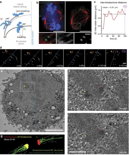
To assay the behavior of congressing chromosomes, we im-aged mid-to-late prometaphase HeLa cells expressing eGFP- CENP-A at 7.5-s intervals for 5 min. Because kinetochores can congress by both lateral sliding and DCP (Fig. 1 a), it was important to identify the latter events in our videos. First, we focused on kinetochores located within the spindle (between pole and equator), because lateral sliding is largely restricted to the peripheral chromosomes (McEwen et al., 2001; Barisic et al., 2014). These kinetochores appeared bioriented based on glutaraldehyde fixation and α-tubulin staining (Fig. 1 b). Sec-ond, we constrained our analysis to sisters whose kinetochore– kinetochore (K–K) axis was <45° relative to local spindle microtubule path (Fig. 1 a), a geometry in which end-on at-tachment to opposite poles is possible. Third, we ensured that kinetochore pairs were undergoing interkinetochore breathing with a mean separation of >0.9 µm, an essential characteristic of biorientation (Cai et al., 2009; Jaqaman et al., 2010; Fig. 1 a). To ensure that kinetochores fulfilling these criteria can be bio-riented, we filmed sister-pairs as they congressed (Fig. 1, c and d) and then fixed/processed the cell for serial block-face scanning EM (SBF-SEM). Kinetochores were clearly visible in the SBF-SEM images, enabling us to correlate the live cell and SEM (Fig. 1, d–f). Importantly, microtubule fibers terminated at both the AP and P kinetochores on the sister-pair that we had tracked (Fig. 1, c–g), confirming that such chromosomes are congressing by DCP.
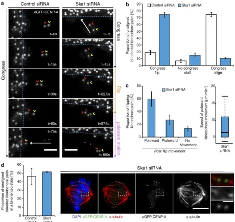
By tracking the behavior of individual bioriented kineto-chore pairs, we documented three phenotypes associated with DCP: (a) successfully congressing to the spindle equator (74.3 ± 3.3%), (b) stalling, where the sister-pair would persist in an im-mobile state (6.8 ± 2.4%), and (c) “flipping” (18.8 ± 3%), where the sister-pair would initiate congression but then rotate through 90° relative to the spindle axis as it approached the metaphase plate (Fig. 2, a and b). (This phenotype is distinct from the pre-viously reported “kinetochore wobbling” by Magidson et al. [2011], where laterally attached kinetochore pairs in early pro-metaphase changed their orientation by >45° in the absence of directed motion.) We then sought to investigate how these phe-notypes differed in cells depleted of Ska1 (Fig. S1). We found a dramatic reduction in successful congression to the spindle equator (to 10.7 ± 1%), which corresponded with a large
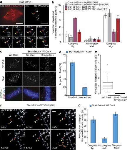
in-crease in kinetochore flipping (to 70 ± 3.7%; Fig. 2, a and b). The majority of these flipped sisters were then transported poleward with a median velocity of ∼7 µm/min (Fig. 2 c). Importantly, the flipping phenotype was not the consequence of a change in attachment state, because the fraction of unaligned bioriented kinetochores in control and Ska1-depleted cells was unchanged (Fig. 2 d). The metaphase plate was identified in these images as the region of highest kinetochore density in a region roughly equidistant from either pole. To control for siRNA off-target ef-fects, we transfected eGFP- CENP-A–expressing cells depleted of Ska1 with an siRNA resistant tagRFP-FKBP-Ska1 trans-gene. This successfully rescued the flipping phenotype, with the majority of unaligned bioriented kinetochore pairs (63 ± 8.5%) now congressing (Fig. 3, a and b). To validate this phenotype in-dependently of siRNA, we targeted the first exon of Ska1 using CRI SPR/Cas9. Quantitative immunofluorescence revealed that ∼20% of cells demonstrated a >95% Ska1 knockdown upon Ska1-GuideA-WTCas9 transfection for 72 h (Fig. 3, c–e; and Fig. S1). In agreement with our siRNA data, transfection with the Ska1-targeted Cas9 augmented kinetochore flipping (to 41 ± 5.1%; Fig. 3, f and g). Together, these data demonstrate that the Ska complex is required for the maintenance of biorienta-tion during congression.
Kinetochore flipping corresponds to lead sister detachment
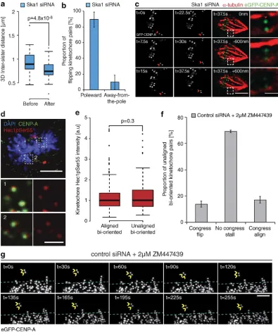
The rotation of a bioriented sister-pair through 90° relative to the spindle axis (Figs. 2 a and 3 f) suggests that a flip event cor-responds to the loss of microtubule attachment at the sister-pair. To validate this, we measured the eGFP-CENP-A–based inter-sister distance of flipping kinetochore pairs at two time points before and after a flip event in cells depleted of Ska1. Consistent with loss of attachment, we observed a significant reduction of the intersister distance after a flip event (from 900 ± 195 nm to 740 ± 190 nm) to a level comparable to that measured for tensionless (nonattached) kinetochores in the presence of the microtubule-depolymerizing drug nocodazole (Fig. 4 a). Given the proposed function of the Ska complex in DCP, where force generation occurs at the P kinetochore, we sought to identify whether there was a P/AP bias in the flipping kinetochore. By determining which kinetochore rotated around its sister, we found that microtubule detachment occurred at the P kineto-chore in ∼90% of cases (Fig. 4 b). To confirm this, we imaged a congressing kinetochore pair depleted of Ska1 and, when a flip initiated, we immediately fixed the cell and stained with α-tubulin antibodies. In Fig. 4 c, the P kinetochore can clearly be seen rotated through 90° and not bound to any microtubules, whereas the AP kinetochore can still be seen bound to a K-fiber.
flipping is restricted to bioriented sister-pairs undergoing con-gression, suggesting that an alternate mechanism underpins this process. We did attempt to directly test whether Aurora B is required for flipping by treating cells with ZM1 (Ditchfield et
[image:4.612.83.516.56.575.2]al., 2003), but the treatment caused unaligned bioriented kineto-chore pairs to stall (Fig. 4, f and g). We have observed flipping only at kinetochore pairs undergoing active congression, so this result is uninformative.
Force-dependent release of the leading kinetochore
An alternative model is that flipping may result from the me-chanical failure of attachment, possibly because of phases of high pulling force on the lead sister. To test this idea, we depleted the microtubule depolymerase mitotic centromere- associated kinesin (MCAK; Hunter et al., 2003), which reduces the speed of kinetochore movement within the metaphase plate as a result of changing the balance of microtubule depolymer-ization at the P kinetochore (Wordeman et al., 2007; Jaqaman et al., 2010; Armond et al., 2015). Quantitative
[image:5.612.83.543.56.490.2]immunofluo-rescence revealed that both MCAK and Ska1 were codepleted efficiently (Fig. 5, a–d). In agreement with studies of aligned kinetochore-pairs (Jaqaman et al., 2010), MCAK deple-tion slowed the speed of congressing kinetochore-pairs from 2.93 ± 1.2 µm/min in control cells to 1.95 ± 0.81 µm/min in MCAK-depleted cells (Fig. 5 e). We then compared the number of flip events in Ska1 and Ska1/MCAK double-depleted cells and found that MCAK codepletion with Ska1 reduced the num-ber of flip events from 63.1 ± 2.7% to 38.3 ± 5.6%, enabling a larger proportion of chromosomes to congress (Fig. 5 f). Inter-pretation of this result is potentially problematic, as MCAK has
been proposed to function in Aurora B–mediated error correc-tion (Bakhoum et al., 2009; Ems-McClung et al., 2013). There-fore, we treated Ska1-depleted cells with 100 nM taxol for 1 h, which reduces microtubule dynamicity and decreases intersister
[image:6.612.82.514.57.565.2]tension (DeLuca et al., 2011). We found that the treatment re-duced the rate of flipping to 41.7 ± 1.55%, allowing more kine-tochore pairs to successfully congress (Fig. 5 g). Thus, we favor a model in which flip events are caused by excess pulling forces
Figure 3. Confirming the role of Ska in congression. (a) Example image sequence of a congressing bioriented kinetochore pair labeled with eGFP-CENP-A in a cell depleted of Ska1 and subsequently rescued with an siRNA-resistant tagRFP-FKBP-Ska1 transgene. Red and yellow arrows, P and AP kinetochores; dotted blue line, metaphase plate periphery. Bars: (top) 5 µm; (bottom) 1 µm. (b) Quantification of unaligned bioriented kinetochore behavior during the Ska1 siRNA rescue experiment. Error bars ± SD; n (control+tagRFP-FKBP) = 126 KT from 39 cells; n (control+tagRFP-FKBP-Ska1) = 132 KT from 27 cells;
being exerted on the kinetochore-depolymerizing microtubule attachment, which can lead to mechanical failure.
Congression is coupled to an increase in microtubule occupancy at kinetochores We suspected that the increase in load on the P kinetochore may be related to the observation that metaphase kinetochores
[image:8.612.98.502.53.537.2]are bound to more microtubules that those in prometaphase (McEwen et al., 1997). However, this experiment did not rule out that differences were caused by cell cycle effects; we needed a comparison of unaligned and congressed kinetochores in the same cell. To do this, we used our SBF-SEM images to quantify the number of microtubules terminating at bioriented kineto-chore pairs. We note that the resolution limit of SEM means that
the observed microtubule fibers may reflect both single micro-tubules and small bundles. Hence, this assay reads out microtu-bule density rather than absolute microtumicrotu-bule number. We found a ∼50% increase in microtubule density at aligned bioriented sister-pairs compared with those that had not yet congressed (Fig. 6, a and b; and Fig. S2). To substantiate this finding, we analyzed prometaphase HeLa cells fixed using glutaraldehyde and stained with an α-tubulin antibody. The tubulin signal
[image:9.612.61.402.53.640.2]in-creased by ∼27% at aligned bioriented sister-pairs (Fig. 6, c and e), a relationship that remained in Ska1-depleted cells (Fig. 6, d and e). We do note that depletion of Ska reduces the number of microtubules bound to the kinetochore, a finding that is con-sistent with previous work (Gaitanos et al., 2009; Raaijmakers et al., 2009; Chan et al., 2012). Together, these data demon-strate that kinetochores recruit additional microtubules in a Ska complex– independent manner during congression.
Figure 6. Congression is coupled to an in-crease in microtubule occupancy at
kineto-chores. (a, I) Single slice from an SBF-SEM
image of a prometaphase HeLa cell; boxes show aligned bioriented (1) and unaligned bioriented (2) kinetochore pairs. Zoom boxes display single slices and their associated segmentation. Kinetochore-attached and non– kinetochore-attached microtubule structures are in light green and dark green, respectively. Dotted blue line, metaphase plate center; white asterisks, spindle pole positions. Bars: (left) 5 µm; (right) 1 µm. (II, top) Box depicting the spindle region used to render microtubule attachment at the sister pairs indicated in 1 and 2. (bottom) Z-projection (slices 56–65) of the kinetochore and microtubule model gener-ated from the cell in I. Kinetochore-attached and non–kinetochore-attached microtubule structures are indicated in light green and dark green, respectively. (b) Quantification of micro-tubule structures terminating at kinetochores in either aligned bioriented or unaligned biori-ented state by SBF-SEM. n = 100 aligned KT and 20 unaligned KT from three cells; P-value calculated using two-sample t test. Image of a prometaphase eGFP-CENP-A cell treated with control (c) or Ska1 (d) siRNA and stained with DAPI and anti–α-tubulin. Zoom boxes depict the staining intensities at aligned bioriented (1) and unaligned bioriented (2) kinetochore pairs, respectively. Bars: (top) 5 µm; (bottom) 1 µm. (e) Quantification of kinetochore- proximal
α-tubulin signal at aligned bioriented and un-aligned bioriented kinetochore pairs in cells treated with either control or Ska1 siRNA. n
The fates of flipped kinetochore pairs
During our initial analysis of congression phenotypes associ-ated with DCP, we found that siRNA control cells have a ∼20% baseline level of flipping (Fig. 2 b), which is also observed in untreated HeLa cells (flip rate of 22.3 ± 3.3%; Fig. 7 a). Ad-ditionally, flipping can be observed in RPE1 cells expressing eGFP-CENP-A (Fig. S3); however, because of the accelerated speed of congression and the instantaneous biorientation of kinetochore pairs in these cells (Magidson et al., 2011), such events are less frequent. In unperturbed cells, this flipping be-havior is identical to that in Ska1-depleted cells, in which the P kinetochore detaches from its associated K-fiber during con-gression, resulting in a reduction in intersister distance (Fig. 7, b and c). Importantly, however, the fates of these flipping kine-tochore pairs are distinct from those in Ska1-depleted cells. In Ska1-depleted cells, kinetochores flip, reattach to spindle mi-crotubules (the K–K axis stabilizes at <45°), and resume con-gression. However, 42 ± 6.4% of these kinetochores undergo a second flip (Fig. 7, d and f), and 91 ± 8.7% of all flipped kinetochores failed to congress during the 5-min video (Fig. 7, d and g). This sequential flipping can be illustrated by track-ing the intersister distance and K–K axis angle of the sister-pair (Fig. 7, h and i). We observed a decrease in sister separation (indicative of detachment), which corresponds with an increase in K–K axis angle after both flip events (Fig. 5 i). In both cases, these flip events were preceded by a period of low K–K axis angle and intersister breathing ∼1 µm, demonstrating that the sister-pair was bioriented before the first flip and reattached to microtubules before the second flip (Fig. 7 i). In contrast, only 15 ± 6.2% of kinetochores in control cells undergo a second flip (Fig. 7 f), and 61 ± 1.7% of all flipped kinetochores successfully congress (Fig. 7, e, g, and j). Given that we have only observed a 5-min snapshot of congression, it is likely that all flipped ki-netochores in control cells reach the metaphase plate. This is supported by 12-h time-lapse videos of HeLa cells expressing Histone-2B-GFP, in which 100% of chromosomes in control cells congressed by 24 min, compared with 50% in Ska1- depleted cells (Fig. S4). The resolution of attachment and sub-sequent congression of flipped kinetochore pairs in control cells highlights two points: (a) flipping does not represent a subset of defective kinetochores, and (b) flipping is a normal feature of chromosome congression because flips can lead to reattachment and successful congression in unperturbed human cells.
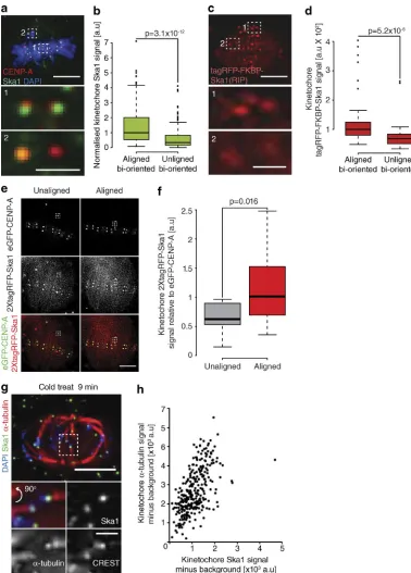
Dynamic maturation of the Ska complex during congression
Our data show that the Ska complex is required to limit the num-ber of lead kinetochore detachment events. Interestingly, we noticed in our immunostaining experiments that Ska1 levels ap-peared to be reduced on unaligned bioriented kinetochore pairs compared with those that had aligned. Indeed, quantification revealed that Ska1 staining intensity was reduced by 51% at un-aligned bioriented kinetochore pairs, a finding we confirmed in live cells using a tagRFP-FKBP–tagged Ska1 transgene (Fig. 8, a–d). This finding suggested that bioriented kinetochores load the Ska complex during congression. To test this, we took a single image of an unaligned kinetochore pair expressing 2xtagRFP-Ska1, allowed it to congress, and imaged the same kinetochore pair once more. We found that the 2xtagRFP-Ska1 signal increased 178% during this alignment (Fig. 8, e and f; and Fig. S5). Thus, bioriented kinetochores progressively load Ska complexes as they congress.
This result raised the possibility that Ska complex matura-tion is coupled to the recruitment of microtubules (Fig. 6, a–f). Ideally, we would have correlated Ska complex and α-tubulin intensities at single kinetochores in different spindle positions. However, our Ska1 antibody was ineffective with glutaralde-hyde fixation, and the noise from non–K-fiber microtubules confounds quantification in cells fixed with paraformaldehyde. To overcome these obstacles, we used cold treatment to remove nonkinetochore microtubules, fixed the cells using paraformal-dehyde, and then quantified the Ska1 and α-tubulin signals at single aligned kinetochores (Fig. 8, g and h). It is important to note that a limitation of this approach is that spindle length decreases in the cold, forming a broad metaphase plate that contains a mix of sister-pairs that were aligned and unaligned before treatment (Fig. 8 g). Nonetheless, our analysis revealed a strong linear correlation (R2 = 0.3444) between the levels of Ska1 and α-tubulin (Fig. 8, g and h). Together, these data demonstrate that bioriented kinetochores can exist in both ma-ture and immama-ture states, and this Ska1 maturation is correlated with both congression and the number of K-fiber microtubules.
Ska maturation correlates with loss of Bub1 from kinetochores
It is well established that unattached kinetochores generate a “wait-anaphase” signal through activation of the SAC. This in-volves the recruitment of Bub and Mad proteins to the kine-tochore (Musacchio, 2015). Our data suggest that immature bioriented kinetochores may also generate a wait-anaphase sig-nal. To investigate this, we stained cells with anti-Bub1 as a readout of checkpoint signaling and anti-Ska1 antibodies as a marker for maturation (Fig. 9 a). First, we measured the levels of both proteins on the same single kinetochores that were aligned, unaligned/bioriented, or unaligned/non-bioriented. These states were defined by the spindle position, orientation, and intersister distance of each sister kinetochore pair (Fig. 9 b). As expected, unaligned/non-bioriented kinetochores had high levels of Bub1 and low levels of Ska1 (Fig. 9, a, c, and d). In contrast, the aligned kinetochores had the opposite phenotype, with high levels of Ska1 and low levels of Bub1 (Fig. 9, a, c, and d). As we previously found, the unaligned but bioriented kinetochores (intersister distances equivalent to the aligned population) had Ska1 levels twofold lower than the aligned kinetochores but significantly higher than the non-bioriented population. How-ever, unlike the aligned kinetochores, Bub1 was still loaded to a level similar to that measured on non-bioriented kinetochores (Fig. 9, a, c, and d). This behavior was also sister kinetochore autonomous, as we observed instances in which one sister was Bub1+/Ska– and the other Bub1–/Ska1+ (Fig. 9 e), thus provid-ing evidence that immature bioriented kinetochores produce an independent SAC signal.
Discussion
plex is not essential for congression per se, because the majority of kinetochores can align in its absence. Presumably, these sisters were not subject to excess load or congressed by lateral sliding (Kapoor et al., 2006), by instantaneous biorientation (Magidson et al., 2011), or through contributions from other redundant factors, with CENP-F being a prime candidate (Volkov et al., 2015).
Here, we have documented a previously overlooked event during mitosis: the detachment and flipping of the P kinetochore during congression (Fig. 10 b). Our data support a model that involves the failure of the kinetochore-microtubule attachment, which would activate the SAC (Fig. 10 b). It could be suggested that we are observing error-correction events mediated by Au-rora B. However, several lines of evidence argue against this idea: (a) our own observations and those from DeLuca et al. (2011) suggest that error correction is antagonized once a bio-riented attachment has formed; (b) we observed flip events at high intersister distance, thus ruling out correction caused by a loss of tension; (c) it is hard to reconcile how a symmetric cue, the loss of tension across the sister-pair, leads to a highly asym-metric response with only the P sister detaching; and (d) the Ska complex has recently been reported to promote Aurora B activ-ity (Redli et al., 2016). Therefore, Ska1-depleted kinetochores display a high rate of microtubule detachment despite having compromised error correction. Nevertheless, the cause of P ki-netochore detachment remains obscure. We favor the idea that kinetochores come under increasing load, perhaps because of the increase in kinetochore-bound microtubules. Moreover, we also found that the poleward-facing sister had more bound mi-crotubules than the sister facing the metaphase plate (unpub-lished data). This may also explain why the P kinetochore needs to recruit microtubules during congression and why it is more sensitive to pulling forces.
We have directly observed that the Ska complex is pro-gressively recruited to bioriented kinetochore pairs as they congress. This shows that once kinetochores biorient, they load Ska complexes in a maturation step dependent on active congression. We propose that this is to handle the escalating pull-ing forces that would occur as microtubules are recruited to the kinetochore (Fig. 10 b). Consistently, the level of kinetochore- bound Ska complex is positively correlated with the number of stably attached microtubules. Importantly, Ska1-depleted cells still display the increase in kinetochore-bound microtu-bules at aligned bioriented sister-pairs, which suggests that the Ska complex is not required for microtubule recruitment to the kinetochore per se, but functions immediately after binding to maintain the attachment during congression. Given that loss of Ska increases the probability of flipping, we suggest that it is the slow maturation of Ska that triggers detachment in normal mitosis. Interestingly, the Ska complex has been reported to recruit PP1, and so it is possible this increase in kinetochore- associated phosphatase activity contributes to maturation (Sivakumar et al., 2016).
Our working model is that these mechanics would allow only bioriented kinetochores that fully mature (in terms
of Ska complexes and microtubules) to persist. Indeed, in wild-type cells, flip events are typically followed by reattach-ment and successful congression, whereas in Ska1-depleted cells, kinetochores are trapped in a futile cycle of detachment- reattachment-detachment. Thus, episodes of lead kinetochore detachment-reattachment in wild-type cells may represent some kind of mechanical self-check on the attachment (Fig. 10 b).
This work provides evidence that unaligned, but bioriented, kinetochores generate a SAC signal that is only extinguished after the kinetochore matures, an event that coincides with congression. This does not, however, mean that the SAC is monitoring congression. In fact, our data show that the maturation state is independent of position; i.e., we can observe kinetochores that are Bub1+/Ska– within the metaphase plate. Our data are consistent with the previous observation that KNL1 and the Bub1 :Bub3 complex (KBB pathway) is required for producing a SAC signal when kinetochores are bioriented but not aligned (Silió et al., 2015). The alternative RZZ pathway can activate the SAC only when the kinetochore is unattached. Our data thus provide further evidence that the KBB pathway is required for immature bioriented kinetochores to generate a SAC signal. The nature of the molecular signals that silence the KBB pathway and drive accumulation of Ska complexes at the metaphase plate remains a crucial open question.
Materials and methods
Cell culture, siRNA transfection, and drug treatmentsHeLa-Kyoto (K) cells were grown in a humidified incubator at 37°C and 5% CO2 in DMEM (Gibco) containing 10% FCS, 100 U/ml peni-cillin, and 100 µg/ml streptomycin supplemented with 0.1 µg/ml puro-mycin (Invitrogen) for maintenance of the eGFP-CENP-A cell line (Jaqaman et al., 2010). The HeLa H2B-GFP cell line was maintained in nonselective medium. The hTERT-RPE1 eGFP-CENP-A cell line was maintained in DMEM/F-12 medium containing 10% FCS, 2.3 g/l sodium bicarbonate, 100 U/ml penicillin, and 100 µg/ml strepto-mycin. siRNA oligonucleotides (53 nM) were transfected using oligo-fectamine (Invitrogen) according to the manufacturer’s guidelines and analyzed at 48 h. The following sequences were used: control, 5′-GGA CCU GGA GGU CUG CUGU-3′; Ska1, 5′-CCG CUU AAC CUA UAA UCAA-3′; and MCAK, 5′-GAU CCA ACG CAG UAA UGGU-3′. For drug treatments, cells were treated with 2 µM ZM447439 (Toc-ris Bioscience) for 30 min or 100 nM taxol (Sigma-Aldrich) for 1 h before live-cell imaging.
Plasmid construction and siRNA rescue experiments
To generate tagRFP-FKBP-Ska1, eGFP was first replaced with tagRFP in pEGFP-C1 (Takara Bio Inc.) using NheI and XhoI, creating pMC387. FKBP12 was then inserted in the XhoI and HindIII restric-tion sites creating pMC390. Next, full-length siRNA-resistant Ska1 was ligated into pMC390 using PstI/MfeI to create tagRFP-FKBP-Ska1 (RIP; pMC393). tagRFP-FKBP-Ska1 was rendered resistant to the tagRFP-FKBP-Ska1 siRNA oligonucleotide (Hanisch et al., 2006) using site-directed mutagenesis
Figure 8. Dynamic maturation of the Ska complex during congression. (a) Image of a prometaphase cell stained with DAPI and antibodies against CENP-A and Ska1. Zoom boxes show aligned (1) and unaligned (2) bioriented kinetochore pairs. Bars: (top) 5 µm; (bottom) 1 µm. (b) Quantification of Ska1 staining intensity at aligned and unaligned bioriented kinetochore pairs. n≥ 157 KT from 37 cells per condition; P-value calculated using two-sample
with the following primer pairs: 5′-CTT CGT ACA TGA AAT CCC GGT TAA CCT ATA ATC AAA TTAA-3′/5′-TTA ATT TGA TTA TAG GTT AAC CGG GAT TTC ATG TAC GAAG-3′, 5′-ATG AAA TCC CGG TTA ACC TAC AAT CAA ATT AAT GAT GTTA-3′/5′-TAA CAT CAT TAA TTT GAT TGT AGG TTA ACC GGG ATT TCAT-3′, and 5′-AAT CCC GGT TAA CC T ACA ACC AAA TTA ATG ATG TTA TTAA-3′/5′-TTA ATA ACA TCA TTA ATT TGG TTG TAG GTT AAC CGG GATT-3′. For siRNA rescue experiments, eGFP-CENP-A cells were transfected with either Ska1 or control siRNA and grown for 12 h in MEM. The medium was then changed to DMEM containing 0.1 µg/ml puromycin, and the cells were transfected with 1 µg of tagRFP-FKBP-Ska1 (pMC393) or tagRFP-FKBP (pMC390) transgene using FuGene6 (Roche) according to the manufacturer’s instructions and incubated for a further 48 h. For Ska1 loading analysis, FKBP was excised from pMC393 and replaced
with PCR-amplified tagRFP fragment using XhoI and HindIII sites, creating pMC463. For expression in cells, 1.5 µg of DNA was trans-fected using FuGene6 (Roche) and incubated for 48 h.
CRI SPR/Cas9
To target Ska1 exon 1, the guide 5′-TAA TTG TTC CAG ATC TGA CG-3′ (Ska1 GuideA top) was cloned into the human codon optimized SpCas9 and chimeric guide expression plasmid (pX330; Addgene) using BbsI. For BbsI compatibility, the sequence CAC CG was added to the 5′ end (creating 5′-CAC CG TAA TTG TTC CAG ATC TGA CG-3′). Because in-sertion requires double-stranded DNA, a complimentary oligo 5′-CGT CAG ATC TGG AAC AAT TA-3′ (Ska1 GuideA bottom) was ordered with 5′-AAAC and 3′-C additions for BbsI compatibility (creating 5′-AAA CCG TCA GAT CTG GAA CAA TTAC-3′). Together, these mod-ifications allow for scarless cloning into the pX330 vector. The
[image:14.612.69.523.55.487.2]gos were phosphorylated and annealed by incubation with T4 PNK (New England Biolabs, Inc.) and T4 ligation buffer (New England Biolabs, Inc.; replacing the supplied PNK buffer). The product was li-gated into the pX330 vector using a single-step digestion-ligation with FastDigest BbsI (Thermo Fisher Scientific) and T4 ligase (New En-gland Biolabs, Inc.). Any residual linearized DNA was digested using the PlasmidSafe exonuclease (EpiBio). Cells were transfected with 1.5 µg plasmid using Fugene6 (Roche) in 1.5 ml DMEM according to the manufacturer’s instructions. After 24 h, 500 µl fresh DMEM was added, and the cells were grown for a further 24 h, at which point the medium was replaced with 2 ml DMEM containing selective antibiot-ics if necessary and grown for 24 h. Cells were imaged, harvested, and fixed after 72 h of transfection. For Surveyor analysis of guide-induced indel mutation, HeLa-K cells were harvested from a single 35-mm dish after guide transfection for 72 h, and genomic DNA was extracted using a QIAamp DNA blood mini kit (QIA GEN). A ∼1-kb fragment containing the target site was amplified using PCR with the primer pair 5′-TTA GAC CCT CCC CTT CTC TCTC-3′ and 5′-CGC TTT TGT
CAG AAC ACA TCTC-3′. 200 ng of PCR product was denatured and reannealed in NEB buffer 2 (New England Biolabs, Inc.; final volume 19 µl) using a thermocycler and the following conditions: 95°C for 5 min; 95°C to 85°C at –2°C/s; and 85°C to 25°C at –0.1°C/s. After cy-cling, the reaction mix was incubated with 1 µl T7 endonuclease (New England Biolabs, Inc.) for 15 min at 37°C.
Immunofluorescence microscopy
[image:15.612.64.375.53.527.2]For anti–CENP-A (mouse; ab13939; Abcam), anti-Ska1 (rabbit; ab46826; Abcam), anti-Hec1PSer55 (rabbit; no longer available; Thermo Fisher Scientific), and anti-Bub1 (mouse; ab54893; Abcam) primary antibodies, cells were fixed for 10 min at RT with 20 mM Pipes, pH 6.8, 10 mM EGTA, 1 mM MgCl2, 0.2% Triton X-100, and 4% formaldehyde. For the cold stable assay, cells were placed on ice for 9 min before fixation. For anti-MCAK antibodies (rabbit; AKIN05; Cytoskeleton, Inc.), cells were fixed for 10 min at −20°C in methanol. In both cases, the cells were then washed three times in PBS for 5 min before blocking in 3% BSA in PBS for 30 min. Cells were incubated for
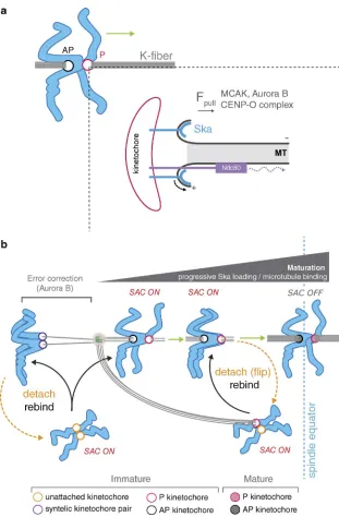
Figure 10. Model for congression via DCP and
in-tegration with checkpoint signaling. (a) Schematic
depicting the contribution of kinetochore factors to movement (green arrow) of bioriented chromosomes by DCP. Inset shows single microtubule-kinetochore attachment site. The Ska complex attaches to, and tracks with, the curving protofilaments as they peel away from the lattice, thus generating a pulling force. This coupling is essential for the maintenance of P ki-netochore attachment when under load. Attachments absolutely require the Ndc80 complex, which may also contribute to force generation by biased diffusion (dotted curved arrow). In addition to these coupling factors, MCAK, Aurora B, and CENP-Q—a subunit of CENP-O complex (Bancroft et al., 2015)—contribute to force generation (Fpull). (b) Model for bioriented
1 h with primary antibodies: anti–CENP-A (1:500), anti-Ska1 (1:400), anti-Hec1pSer55 (1:300), anti-MCAK (1:500), anti-Bub1 (1:500), and CRE ST antisera (1:250) and washed three times in PBS for 5 min. Sec-ondary antibodies were Alexa Fluor–conjugated secSec-ondary antibod-ies (Invitrogen) used at 1:500 for 30 min. For anti–α-tubulin (mouse; T6074-200UL; Sigma-Aldrich) antibodies, cells were preextracted for 30 s in 80 mM Pipes, pH 6.8, 1 mM MgCl2, 4 mM EGTA, and 0.5% Tri-ton X-100 and fixed by adding glutaraldehyde at 0.5% for 10 min. Glu-taraldehyde was quenched using a 7-min treatment with 0.1% NaBH4. Cells were washed three times in PBS for 5 min, followed by blocking in TBS containing 0.1% Triton X-100 and 2% BSA for 10 min. Cells were then incubated with anti–α-tubulin (1:1,000) primary antibody for 30 min, followed by four 5-min washes in TBS containing 0.1% Triton X-100 and incubation with 647 nm Alexa Fluor–conjugated secondary antibodies (Invitrogen) in TBS containing 0.1% Triton X-100 and 2% BSA for 30 min. Cells were mounted and imaged in Vectashield (Vector Laboratories). 3D image stacks were acquired (75 × 0.2-µm z-sections) using a 100× oil NA 1.4 objective on an Olympus Deltavision Elite microscope (Applied Precision Ltd.) equipped with a DAPI, FITC, Rhodamine, or Texas Red and Cy5 filter set, solid-state light source, and a CoolSNAP HQ2 camera (Roper Technologies) at 37°C. Images were acquired, the stacks were deconvolved using SoftWorx, and fluo-rescence intensity measurements were made manually after background subtraction and normalization to the CENP-A or CRE ST signal. Figures were constructed in Illustrator CS5 (Adobe Systems).
Statistical methods
Staining intensity distributions were compared using a two-sample
t test in R. Data distributions were assumed to be normal, but this was not formally tested.
Live-cell imaging
To film kinetochore fates, HeLa-K eGFP-CENP-A–expressing cells were seeded in FluoroDishes (World Precision) and imaged in DMEM supplemented with 10% FCS, 100 U/ml penicillin, 100 µg/ml streptomy-cin, and 0.1 µg/ml puromycin. 3D image stacks (25 × 0.5-µm z-sections) were acquired every 7.5 s using a 100× oil NA 1.4 objective on an Olym-pus Deltavision Elite microscope equipped with a eGFP and mCherry filter set, Quad-mCherry dichroic mirror, solid-state light source, and CoolSNAP HQ2 camera. Environment was tightly controlled at 37°C and 5% CO2 using a stage-top incubator (Tokai Hit) and a weather sta-tion (Precision Control). Image stacks were deconvolved using Soft-Worx (Applied Precision Ltd.), and kinetochore fates were determined manually. Measurements of kinetochore velocity were taken manually from tracks of persistent movement that lasted at least three time frames. Images were deconvolved using SoftWorx, and fluorescence intensity measurements were made manually after background subtraction. To film kinetochore fates in the hTERT-RPE1 eGFP-CENP-A cell line, cells were seeded in FluoroDishes and imaged in Leibovitz L-15 sup-plemented with 10% FCS. 3D image stacks (25 z-sections, 0.5 µm) were acquired every 2 s for 5 min using a 100× 1.4 NA oil objective on a confocal spinning-disk microscope (VOX Ultraview; PerkinElmer) equipped with a Hamamatsu Photonics ORCA-R2 camera, controlled by Volocity 6.0 (PerkinElmer) running on a Windows 7 64-bit (Micro-soft) PC (IBM) at 37°C. Images were deconvolved with Huygens 4.1 (SVI) using a point spread function measured from microbead images (using the Huygens 4.1 PSF distiller), and visualized in Fiji.
EM
For correlative light EM experiments, HeLa-K eGFP-CENP-A– expressing cells were imaged on gridded glass dishes (P35G-1.5-14-CGRD; MatTek Corporation) using a Deltavision microscope as
described earlier. The photo-etched grid coordinate containing the cell of interest was recorded using bright-field illumination at 20×. The same cell was then relocated, and live-cell imaging was acquired at 100×. Kinetochore pairs of interest where tracked as described earlier, and once the congression event had occurred, cells were immediately chemically fixed in 3% glutaraldehyde, 0.5% paraformaldehyde, and 0.1% tannic acid in 0.05 M phosphate buffer, pH 7.4, for 1 h and pro-cessed for SBF-SEM. Samples were propro-cessed and images segmented as previously described (Nixon et al., 2016 Preprint). In brief, cells were postfixed in reduced osmium (2% osmium tetroxide, 1.5% potas-sium ferricyanide) for 1 h, incubated in 0.1% thiocarbohydrazide in dH2O for 20 min, and incubated in 2% osmium tetroxide in dH2O for 30 min before being incubated overnight in 1% uranyl acetate dH2O at 4°C. Cells were incubated in Walton’s lead aspartate at 60°C for 30 min and then dehydrated in graded series ethanol before being infiltrated and embedded in EPON 812 hard premixed resin (TAAB). Once the resin had fully polymerized, the coverslip was re-moved and the cell of interest was located using previously acquired grid coordinates. The block of resin containing the cell of interest was then excised and mounted on to a cryo pin using Silver Dag (Agar Scientific). Excess resin was trimmed away with an Ultrami-crotome (Leica Biosystems), leaving a 200 × 200-µm block face. To reduce charging, the block was first painted with silver conductive paste (TAAB) before 10 nm gold/palladium was evaporated onto it (Quorum Technologies). Imaging was done using Gatan 3View Sys-tem and Digital Micrograph software (Gatan) and FEI Quanta 250 (FEI). To image the whole cell including all chromosomes and visu-alize both microtubules and kinetochores at a resolution of ∼5 nm in x and y and 60 nm in z, the following imaging parameters were used: 4,000 × 4,000 frame size, 12-µs dwell time, 2.3 KeV, 50 Pa, and magnification 9500×. Kinetochores were identified in the images as electron-dense regions on chromatin that span two to three z-slices (120–180 nm).
Online supplemental material
Fig. S1 shows effectiveness of siRNA and CRI SPR/Cas9 targeting Ska1. Fig. S2 shows slice by slice representation of the EM and its associated render for the kinetochore pairs shown in Fig. 6. Fig. S3 shows an example flip event from an untreated RPE1 cell. Fig. S4 shows changes in eGFP-CENP-A signal do not contribute to the ob-served increase in Ska1 at congressed kinetochore pairs. Fig. S5 shows quantification of eGFP-CENP-A and tagRFP intensity.
Acknowledgments
We thank H. Drechsler, E. Vladimirou, J. Armond, E. Roscioli, P. Meraldi, and J. Millar for discussion and comments on the man-uscript. We thank C. Smith for generation of the tagRFP-C1 vector and imaging of kinetochores in RPE1 cells and P. Jallepalli for the Ska1 cDNA. We acknowledge technical assistance from A. Beckett (Liverpool Biomedical EM Unit).
P. Auckland is funded by the Medical Research Council Doc-toral Training Partnership grant MR/J003964/1, N.I. Clarke by a Cancer Research UK studentship (C25425/A16141), and S.J. Royle by a Senior Fellowship from Cancer Research UK (C25425/A15182). A.D. McAinsh was supported by a Wellcome Trust Senior Investigator Award (grant 106151/Z/14/Z) and a Royal Society Wolfson Re-search Merit Award (grant WM150020).
The authors declare no competing financial interests.
ex-cept for the SBF-SEM, which was completed and analyzed by N.I. Clarke and S.J. Royle.
Submitted: 25 July 2016 Revised: 9 December 2016 Accepted: 13 March 2017
References
Abad, M.A., B. Medina, A. Santamaria, J. Zou, C. Plasberg-Hill, A. Madhumalar, U. Jayachandran, P.M. Redli, J. Rappsilber, E.A. Nigg, and A.A. Jeyaprakash. 2014. Structural basis for microtubule recognition by the human kinetochore Ska complex. Nat. Commun. 5:2964. http ://dx .doi .org /10 .1038 /ncomms3964
Armond, J.W., E. Vladimirou, M. Erent, A.D. McAinsh, and N.J. Burroughs. 2015. Probing microtubule polymerisation state at single kinetochores during metaphase chromosome motion. J. Cell Sci. 128:1991–2001. http ://dx .doi .org /10 .1242 /jcs .168682
Auckland, P., and A.D. McAinsh. 2015. Building an integrated model of chromosome congression. J. Cell Sci. 128:3363–3374. http ://dx .doi .org /10 .1242 /jcs .169367
Bakhoum, S.F., S.L. Thompson, A.L. Manning, and D.A. Compton. 2009. Genome stability is ensured by temporal control of kinetochore-microtubule dynamics. Nat. Cell Biol. 11:27–35. http ://dx .doi .org /10 .1038 /ncb1809
Bancroft, J., P. Auckland, C.P. Samora, and A.D. McAinsh. 2015. Chromosome congression is promoted by CENP-Q- and CENP-E-dependent pathways. J. Cell Sci. 128:171–184. http ://dx .doi .org /10 .1242 /jcs .163659 Barisic, M., P. Aguiar, S. Geley, and H. Maiato. 2014. Kinetochore motors drive
congression of peripheral polar chromosomes by overcoming random arm-ejection forces. Nat. Cell Biol. 16:1249–1256. http ://dx .doi .org /10 .1038 /ncb3060
Cai, S., C.B. O’Connell, A. Khodjakov, and C.E. Walczak. 2009. Chromosome congression in the absence of kinetochore fibres. Nat. Cell Biol. 11:832– 838. http ://dx .doi .org /10 .1038 /ncb1890
Cassimeris, L., and E.D. Salmon. 1991. Kinetochore microtubules shorten by loss of subunits at the kinetochores of prometaphase chromosomes. J. Cell Sci. 98:151–158.
Chan, Y.W., A.A. Jeyaprakash, E.A. Nigg, and A. Santamaria. 2012. Aurora B controls kinetochore–microtubule attachments by inhibiting Ska complex–KMN network interaction. J. Cell Biol. 196:563–571. http ://dx .doi .org /10 .1083 /jcb .201109001
Cheeseman, I.M. 2014. The kinetochore. Cold Spring Harb. Perspect. Biol. 6:a015826. http ://dx .doi .org /10 .1101 /cshperspect .a015826
Daum, J.R., J.D. Wren, J.J. Daniel, S. Sivakumar, J.N. McAvoy, T.A. Potapova, and G.J. Gorbsky. 2009. Ska3 is required for spindle checkpoint silencing and the maintenance of chromosome cohesion in mitosis. Curr. Biol. 19:1467–1472. http ://dx .doi .org /10 .1016 /j .cub .2009 .07 .017
DeLuca, K.F., S.M. Lens, and J.G. DeLuca. 2011. Temporal changes in Hec1 phosphorylation control kinetochore-microtubule attachment stability during mitosis. J. Cell Sci. 124:622–634. http ://dx .doi .org /10 .1242 /jcs .072629
Ditchfield, C., V.L. Johnson, A. Tighe, R. Ellston, C. Haworth, T. Johnson, A. Mortlock, N. Keen, and S.S. Taylor. 2003. Aurora B couples chromosome alignment with anaphase by targeting BubR1, Mad2, and Cenp-E to kinetochores. J. Cell Biol. 161:267–280. http ://dx .doi .org /10 .1083 /jcb .200208091
Drpic, D., A.J. Pereira, M. Barisic, T.J. Maresca, and H. Maiato. 2015. Polar ejection forces promote the conversion from lateral to end-on kinetochore-microtubule attachments on mono-oriented chromosomes. Cell Reports. 13:460–469. http ://dx .doi .org /10 .1016 /j .celrep .2015 .08 .008
Ems-McClung, S.C., S.G. Hainline, J. Devare, H. Zong, S. Cai, S.K. Carnes, S.L. Shaw, and C.E. Walczak. 2013. Aurora B inhibits MCAK activity through a phosphoconformational switch that reduces microtubule association. Curr. Biol. 23:2491–2499. http ://dx .doi .org /10 .1016 /j .cub .2013 .10 .054
Gaitanos, T.N., A. Santamaria, A.A. Jeyaprakash, B. Wang, E. Conti, and E.A. Nigg. 2009. Stable kinetochore-microtubule interactions depend on the Ska complex and its new component Ska3/C13Orf3. EMBO J. 28:1442–1452. http ://dx .doi .org /10 .1038 /emboj .2009 .96
Gorbsky, G.J., P.J. Sammak, and G.G. Borisy. 1987. Chromosomes move poleward in anaphase along stationary microtubules that coordinately
disassemble from their kinetochore ends. J. Cell Biol. 104:9–18. http ://dx .doi .org /10 .1083 /jcb .104 .1 .9
Hanisch, A., H.H. Silljé, and E.A. Nigg. 2006. Timely anaphase onset requires a novel spindle and kinetochore complex comprising Ska1 and Ska2. EMBO J. 25:5504–5515. http ://dx .doi .org /10 .1038 /sj .emboj .7601426 Hunter, A.W., M. Caplow, D.L. Coy, W.O. Hancock, S. Diez, L. Wordeman, and
J. Howard. 2003. The kinesin-related protein MCAK is a microtubule depolymerase that forms an ATP-hydrolyzing complex at microtubule ends. Mol. Cell. 11:445–457. http ://dx .doi .org /10 .1016 /S1097 -2765(03)00049 -2
Jaqaman, K., E.M. King, A.C. Amaro, J.R. Winter, J.F. Dorn, H.L. Elliott, N. McHedlishvili, S.E. McClelland, I.M. Porter, M. Posch, et al. 2010. Kinetochore alignment within the metaphase plate is regulated by centromere stiffness and microtubule depolymerases. J. Cell Biol. 188:665–679. http ://dx .doi .org /10 .1083 /jcb .200909005
Jeyaprakash, A.A., A. Santamaria, U. Jayachandran, Y.W. Chan, C. Benda, E.A. Nigg, and E. Conti. 2012. Structural and functional organization of the Ska complex, a key component of the kinetochore-microtubule interface. Mol. Cell. 46:274–286. http ://dx .doi .org /10 .1016 /j .molcel .2012 .03 .005
Kapoor, T.M., M.A. Lampson, P. Hergert, L. Cameron, D. Cimini, E.D. Salmon, B.F. McEwen, and A. Khodjakov. 2006. Chromosomes can congress to the metaphase plate before biorientation. Science. 311:388–391. http ://dx .doi .org /10 .1126 /science .1122142
Khodjakov, A., and C.L. Rieder. 1996. Kinetochores moving away from their associated pole do not exert a significant pushing force on the chromosome. J. Cell Biol. 135:315–327. http ://dx .doi .org /10 .1083 /jcb .135 .2 .315
Lampson, M.A., and I.M. Cheeseman. 2011. Sensing centromere tension: Aurora B and the regulation of kinetochore function. Trends Cell Biol. 21:133–140. http ://dx .doi .org /10 .1016 /j .tcb .2010 .10 .007
Lampson, M.A., K. Renduchitala, A. Khodjakov, and T.M. Kapoor. 2004. Correcting improper chromosome-spindle attachments during cell division. Nat. Cell Biol. 6:232–237. http ://dx .doi .org /10 .1038 /ncb1102 Liu, D., G. Vader, M.J.M. Vromans, M.A. Lampson, and S.M. Lens. 2009.
Sensing chromosome bi-orientation by spatial separation of aurora B kinase from kinetochore substrates. Science. 323:1350–1353. http ://dx .doi .org /10 .1126 /science .1167000
Magidson, V., C.B. O’Connell, J. Lončarek, R. Paul, A. Mogilner, and A. Khodjakov. 2011. The spatial arrangement of chromosomes during prometaphase facilitates spindle assembly. Cell. 146:555–567. http ://dx .doi .org /10 .1016 /j .cell .2011 .07 .012
Magidson, V., C.B. O’Connell, J. Lončarek, R. Paul, A. Mogilner, and A. Khodjakov. 2011. The spatial arrangement of chromosomes during prometaphase facilitates spindle assembly. Cell. 146:555–567. http ://dx .doi .org /10 .1016 /j .cell .2011 .07 .012
Magidson, V., R. Paul, N. Yang, J.G. Ault, C.B. O’Connell, I. Tikhonenko, B.F. McEwen, A. Mogilner, and A. Khodjakov. 2015. Adaptive changes in the kinetochore architecture facilitate proper spindle assembly. Nat. Cell Biol. 17:1134–1144. http ://dx .doi .org /10 .1038 /ncb3223
Maiato, H., A.M. Gomes, F. Sousa, and M. Barisic. 2017. Mechanisms of chro-mosome congression during mitosis. Biology (Basel). 6:6.
McEwen, B.F., A.B. Heagle, G.O. Cassels, K.F. Buttle, and C.L. Rieder. 1997. Kinetochore fiber maturation in PtK1 cells and its implications for the mechanisms of chromosome congression and anaphase onset. J. Cell Biol. 137:1567–1580. http ://dx .doi .org /10 .1083 /jcb .137 .7 .1567
McEwen, B.F., G.K.T. Chan, B. Zubrowski, M.S. Savoian, M.T. Sauer, and T.J. Yen. 2001. CENP-E is essential for reliable bioriented spindle attachment, but chromosome alignment can be achieved via redundant mechanisms in mammalian cells. Mol. Biol. Cell. 12:2776–2789. http :// dx .doi .org /10 .1091 /mbc .12 .9 .2776
Musacchio, A. 2015. The molecular biology of spindle assembly checkpoint signaling dynamics. Curr. Biol. 25:R1002–R1018. http ://dx .doi .org /10 .1016 /j .cub .2015 .08 .051
Nixon, F.M., T.R. Honnor, G.P. Starling, A.J. Beckett, A.M. Johansen, J.A. Brettschneider, I.A. Prior, and S.J. Royle. 2016. Microtubule organization within mitotic spindles revealed by serial block face scanning EM and image analysis. bioRxiv. doi :10 .1101 /087866 (Preprint posted November 15, 2016)
Raaijmakers, J.A., M.E. Tanenbaum, A.F. Maia, and R.H. Medema. 2009. RAMA1 is a novel kinetochore protein involved in kinetochore-microtubule attachment. J. Cell Sci. 122:2436–2445. http ://dx .doi .org /10 .1242 /jcs .051912
biorientation. J. Cell Biol. 215:77–93. http ://dx .doi .org /10 .1083 /jcb .201603019
Schmidt, J.C., H. Arthanari, A. Boeszoermenyi, N.M. Dashkevich, E.M. Wilson-Kubalek, N. Monnier, M. Markus, M. Oberer, R.A. Milligan, M. Bathe, et al. 2012. The kinetochore-bound Ska1 complex tracks depolymerizing microtubules and binds to curved protofilaments. Dev. Cell. 23:968–980. http ://dx .doi .org /10 .1016 /j .devcel .2012 .09 .012
Shrestha, R.L., and V.M. Draviam. 2013. Lateral to end-on conversion of chromosome-microtubule attachment requires kinesins CENP-E and MCAK. Curr. Biol. 23:1514–1526. http ://dx .doi .org /10 .1016 /j .cub .2013 .06 .040
Silió, V., A.D. McAinsh, and J.B. Millar. 2015. KNL1-bubs and RZZ provide two separable pathways for checkpoint activation at human kinetochores. Dev. Cell. 35:600–613. http ://dx .doi .org /10 .1016 /j .devcel .2015 .11 .012 Sivakumar, S., P.L. Janczyk, Q. Qu, C.A. Brautigam, P.T. Stukenberg, H. Yu,
and G.J. Gorbsky. 2016. The human SKA complex drives the metaphase-anaphase cell cycle transition by recruiting protein phosphatase 1 to kinetochores. eLife. 5:pii :e12902. http ://dx .doi .org /10 .7554 /eLife .12902 Skibbens, R.V., V.P. Skeen, and E.D. Salmon. 1993. Directional instability of
kinetochore motility during chromosome congression and segregation in mitotic newt lung cells: A push-pull mechanism. J. Cell Biol. 122:859– 875. http ://dx .doi .org /10 .1083 /jcb .122 .4 .859
Skibbens, R.V., C.L. Rieder, and E.D. Salmon. 1995. Kinetochore motility after severing between sister centromeres using laser microsurgery: Evidence that kinetochore directional instability and position is regulated by ten-sion. J. Cell Sci. 108:2537–2548.
Theis, M., M. Slabicki, M. Junqueira, M. Paszkowski-Rogacz, J. Sontheimer, R. Kittler, A.K. Heninger, T. Glatter, K. Kruusmaa, I. Poser, et al. 2009. Comparative profiling identifies C13orf3 as a component of the Ska complex required for mammalian cell division. EMBO J. 28:1453–1465. http ://dx .doi .org /10 .1038 /emboj .2009 .114
Volkov, V.A., P.M. Grissom, V.K. Arzhanik, A.V. Zaytsev, K. Renganathan, T. McClure-Begley, W.M. Old, N. Ahn, and J.R. McIntosh. 2015. Centromere protein F includes two sites that couple efficiently to depolymerizing microtubules. J. Cell Biol. 209:813–828. http ://dx .doi .org /10 .1083 /jcb .201408083
Welburn, J.P., E.L. Grishchuk, C.B. Backer, E.M. Wilson-Kubalek, J.R. Yates III, and I.M. Cheeseman. 2009. The human kinetochore Ska1 complex facilitates microtubule depolymerization-coupled motility. Dev. Cell. 16:374–385. http ://dx .doi .org /10 .1016 /j .devcel .2009 .01 .011
Welburn, J.P., M. Vleugel, D. Liu, J.R. Yates III, M.A. Lampson, T. Fukagawa, and I.M. Cheeseman. 2010. Aurora B phosphorylates spatially distinct targets to differentially regulate the kinetochore-microtubule interface. Mol. Cell. 38:383–392. http ://dx .doi .org /10 .1016 /j .molcel .2010 .02 .034
Westhorpe, F.G., and A.F. Straight. 2013. Functions of the centromere and kinetochore in chromosome segregation. Curr. Opin. Cell Biol. 25:334– 340. http ://dx .doi .org /10 .1016 /j .ceb .2013 .02 .001