University of Warwick institutional repository: http://go.warwick.ac.uk/wrap
This paper is made available online in accordance with
publisher policies. Please scroll down to view the document
itself. Please refer to the repository record for this item and our
policy information available from the repository home page for
further information.
To see the final version of this paper please visit the publisher’s website.
Access to the published version may require a subscription.
Author(s): A.J. Thompson, M. Lochner and S.C.R. Lummis
Article Title: Loop B Is a Major Structural Component of the 5-HT
3Receptor
Year of publication: 2008
Link to published version:
Loop B Is a Major Structural Component of the 5-HT
3
Receptor
A. J. Thompson,* M. Lochner,
yand S. C. R. Lummis*
*Department of Biochemistry, University of Cambridge, Cambridge, CB2 1QW, United Kingdom; andyDepartment of Chemistry, University of Warwick, Coventry CV4 7AL, United Kingdom
ABSTRACT
The 5-HT
3receptor belongs to a family of therapeutically important neurotransmitter-gated receptors whose
ligand binding sites are formed by the convergence of six peptide loops (A-F). Here we have mutated 15 amino acid residues in
and around loop B of the 5-HT
3receptor (Ser-177 to Asn-191) to Ala or a residue with similar chemical properties. Changes in
[
3H]granisetron binding affinity (
K
d
) and 5-HT
EC
50were determined using receptors expressed in human embryonic kidney 293
cells. Substitutions at all but one residue (Thr-181) altered or eliminated binding for one or both mutants. Receptors were
nonfunctional or
EC
50values were altered for all but two mutants (S182T, I190L). Homology modeling indicates that loop B
contributes two residues to a hydrophobic core that faces into the
b
-sandwich of the subunit, and the experimental data indicate
that they are important for both the structure and the function of the receptor. The models also show that close to the apex of the
loop (Ser-182 to Ile-190), loop B residues form an extensive network of hydrogen bonds, both with other loop B residues and
with adjacent regions of the protein. Overall, the data suggest that loop B has a major role in maintaining the structure of the
region by a series of noncovalent interactions that are easily disrupted by amino acid substitutions.
INTRODUCTION
The 5-HT
3receptor is a Cys-loop receptor, which plays a role
in synaptic transmission in both the central and peripheral
nervous systems (1). These proteins consist of five
symmetri-cally arranged subunits; each subunit has a large extracellular
N-terminal domain that is responsible for ligand binding, four
transmembrane domains (M1–M4) that surround a central ion
conducting pore, and a large intracellular loop that influences
channel conductance and mediates the actions of intracellular
messengers. The ligand binding site is at the interface of two
adjacent subunits and is formed by the convergence of three
amino acid loops (A–C) from the ‘‘principal’’ subunit and three
b
-strands (D–F) from the ‘‘complementary’’ subunit (2).
Structural insight of this region has been gained from high
resolution structures of homologous acetylcholine binding
proteins (AChBPs), and nACh and bacterial receptors (2–7),
and specific interactions between amino acid residues and a
range of 5-HT
3receptor ligands have been identified within
loops A–F (8–11). Using structures of AChBP derived from
crystals, and from cryo-electron microscopy data of the nACh
receptor, a number of Cys-loop homology models have been
published; the recent high resolution structure of a nACh
receptor subunit has confirmed the accuracy of such models
for the nACh receptor (6). For other receptors, however,
sequence identity is only in the region of 10–20% and
ex-perimental evidence is required to support the structural
ac-curacy of these models at the molecular level. Here we
explore the roles of each residue of loop B, as the specific
functions of the majority of these residues are currently
un-known. The data from extensive mutagenesis, binding and
functional studies reveal the importance of these residues and
also provide a structural explanation for their critical roles.
EXPERIMENTAL PROCEDURES
Materials
All cell culture reagents were obtained from Gibco BRL (Paisley, UK), except fetal calf serum which was from Labtech International (Ringmer, UK). [3H]granisetron (63.5 Ci/mmol) was from PerkinElmer (Boston, MA). All other reagents were of the highest obtainable grade.
Cell culture
Human embryonic kidney (HEK) 293 cells were maintained on 90 mm tissue culture plates at 37°C and 7% CO2in a humidified atmosphere. They were cultured in DMEM:F12 (Dulbecco’s modified Eagle’s medium/Nutrient Mix F12 (1:1)) with GlutaMAX I media containing 10% fetal calf serum. For radioligand binding studies cells in 90 mm dishes were transfected using calcium phosphate precipitation at 80–90% confluency and incubated for 3–4 days before use; optimization of the original method (12) by Jordon (13) resulted in increased levels of binding, which has allowed us to obtain binding parameters for mutant receptors that in the past have been expressed at levels too low to measure (e.g., W183Y and H185A). For functional studies, cells were transfected by electroporation using the AMAXA system (Amaxa GmbH, Cologne, Germany), plated on 96 well plates and incubated 1–2 days before assay.
Site-directed mutagenesis
Mutagenesis reactions were performed using the method described by Kunkel (14) using 5-HT3Areceptor subunit cDNA (Accession: AY605711)
Submitted April 18, 2008, and accepted for publication July 30, 2008.
A. J. Thompson and M. Lochner contributed equally to this work. Address reprint requests to Sarah C. R. Lummis, Dept. of Biochemistry, University of Cambridge, Cambridge, CB2 1QW, UK. Tel: 44-1223-765950; Fax: 44-1223-333345; E-mail: [email protected].
This is an Open Access article distributed under the terms of the Creative Commons-Attribution Noncommercial License (http://creativecommons. org/licenses/by-nc/2.0/), which permits unrestricted noncommercial use, distribution, and reproduction in any medium, provided the original work is properly cited.
Editor: David S. Weiss.
in pcDNA3.1 (Invitrogen, Paisley, UK) as described previously (15). A silent restriction site was incorporated into each primer to assist rapid identification.
Radioligand binding
This was undertaken as previously described with minor modifications (15). Briefly, transfected HEK293 cell membranes were incubated in 0.5 ml HEPES buffer containing the 5-HT3 receptor antagonist [3H]granisetron (0.1–40 nM). Nonspecific binding was determined using 1mM quipazine. Incubations were terminated by filtration, thus limiting our determination of
Kdvalues to#10 nM, as separation of bound from free ligand would be too slow to determineKd values greater than this (16). Data were analyzed by iterative curve fitting (Prism v3.0, GraphPad Software, San Diego, CA) according to the equation:B¼(Bmax3[L])/(K1[L]), whereBis bound radioligand,Bmaxis maximum binding at equilibrium,Kis the equilibrium dissociation constant, and [L] is the free concentration of radioligand. Values are presented as mean6SE. Statistical analysis was performed using ANOVA in conjunction with a Dunnett’s post test.
Immunofluorescence
This was as described previously (17). Briefly, transfected cells were fixed (4 % paraformaldehyde) and incubated overnight at 4°C in pAb120 at 1:1000 in Tris-buffered saline (0.1 M Tris pH7.4, 0.9 % NaCl). Biotinylated anti-rabbit IgG (Vector Laboratories, Burlingame, CA) and fluorescein isothio-cyanate avidin D (Vector Laboratories) were used to detect bound antibody as instructed by the manufacturer. Coverslips were mounted in Vectashield HardSet mounting medium (Vector Laboratories). Immuno-fluorescence was observed using an UltraVIEW LCI Confocal Imaging System (PerkinElmer).
FlexStation analysis
This technique uses fluorescent voltage-sensitive dyes to detect changes in the membrane potential and has been used to examine a range of ion channels including 5-HT3receptors (19,20). The methods were as previously described (19). Briefly, fluorescent membrane potential dye (Molecular Devices, Wokingham, UK) was diluted in Flex buffer (10 mM HEPES, 115 mM NaCl, 1 mM KCl, 1 mM CaCl2, 1 mM MgCl2, 10 mM glucose, pH 7.4) and added to transfected cells grown on a 96-well plate. The cells were in-cubated at room temperature for 45 min and then fluorescence was measured in a FlexStation (Molecular Devices) every 2 s for 200 s. Control (Flex buffer) or 5-HT (0.001mM–1.0 mM) was added to each well after 20 s. The percent change in fluorescence was calculated asF(peak fluorescence) mi-nusFmin(baseline fluorescence at 20 s) divided byFmax(peak fluorescence at 30mM 5-HT). Concentration-response data were fitted to the four-parameter logistic equation,F¼Fmin1ðFmaxFminÞ=ð1110ðlogðEC50½AÞ3nHÞ;where
Fmaxis the maximum response,Fminis the baseline fluorescence, [A] is the log concentration of agonist, andnHis the Hill coefficient, using Prism software (GraphPad).EC50values determined using this technique are often slightly lower than values determined using electrophysiological studies as different phenomena are being measured: For electrophysiological studies,
EC50values represent the agonist concentration required to open 50% of channels, whereas in fluorescent studies they represent the agonist concen-tration required to depolarize the membrane potential to 50% of its original value. However, relative values for different mutant 5-HT3receptors are the same, and as cellular events are triggered by changes in membrane potential, this may be a more accurate indication of the ligand potency in vivo (19).
Modeling
This was performed as described previously (9,21). Three-dimensional models of the extracellular region of the 5-HT3receptor were built using MODELER 6v2 (22) based on the crystal structure of AChBP in different states; unbound (Protein Data Bank (PDB) ID 2byn), agonist bound
(car-bomylcholine, PDB ID 1uv6 and epibatidine, PDB ID 2byq) and antagonist bound (2-methyllcaconitine, PDB ID 2byr anda-cobratoxin, PDB ID 1yi5). Models were energy minimized in SYBYL v6.8 using the AMBER force field (23). Models and predicted hydrogen bonds were viewed using PyMOL v 0.98.
RESULTS
Effects of mutations
All 15 amino acids within a contiguous sequence of the 5-HT
3Areceptor subunit (Fig. 1) were mutated to either Ala or an amino
acid with properties similar to the wild-type amino acid
(subsequently referred to as a conserved amino acid change).
Mutant receptors were characterized using [
3H]granisetron
binding to explore changes in the ligand binding site (Table 1,
Fig. 2), and 5-HT induced changes in membrane potential
were measured using a voltage-sensitive fluorescent dye
(Table 2, Fig. 2). Wild-type receptors had a [
3H]granisetron
binding affinity (K
d) of 0.5 nM (n
¼
11) and 5-HT
EC
50of
0.24
m
M (n
¼
12), values that are similar to those previously
published using the same techniques (15,24,25).
Alanine mutations
Amino acid substitutions to Ala revealed significant changes
in [
3H]granisetron binding affinities for 9 of the 15 residues
(Table 1). At Phe-180, Trp-183, Asp-189, Ile-190 and
Asn-191, substitution with Ala eliminated any measurable
bind-ing, indicating a particularly important role for these residues.
Data from the functional assay indicated a more significant
effect on function as only 5 of the Ala substitutions were
functional (T181A, W183A, L184A, T186A and I187A),
and each of these displayed a significant increase in the
EC
50when compared to wild-type responses.
Conserved mutations
Amino acid substitutions with conserved residues showed
significant changes in [
3H]granisetron binding affinities for
10 of the 15 residues (Table 1). Five of these were identical to
residues that had displayed sensitivity to Ala mutation
(Leu-178, Phe-180, Trp-183, Asp-189, Asn-191). For the
re-maining five conserved mutations that displayed altered
K
dvalues (Ser-177, Ser-182, His-185, Thr-186, Gln-188),
there was no corresponding change in affinity for the Ala
mutants, indicating that it was not the chemical
character-istics of the residue that was critical at these positions.
Mutations at Leu-178, Gln-188 and Asp-189 caused
com-plete ablation of [
3H]granisetron binding. In the functional
assay five conserved mutations were functional (S182T,
W183Y, L184I, I187L, I190L). Three of these displayed
smaller increases in
EC
50than their Ala counterparts
(W183Y, L184I, I187L), whereas S182T and I190L had values
that were not significantly different to wild-type receptors.
The 5-HT3Receptor B-Loop 5729
Immunocytochemistry
For mutants that were classified as having no specific
radio-ligand binding, cell surface expression was studied using
immunofluorescence and confocal microscopy. Fig. 3 shows
that of the eight mutants, four were not expressed at the cell
surface (F180A, Q188N, D189A and I190A). The remaining
mutants (L178I, W183A, D189E and N191A) all showed a
distinct halo of fluorescence at the cell surface, indicating that
the receptors were correctly assembled and trafficked to the
plasma membrane.
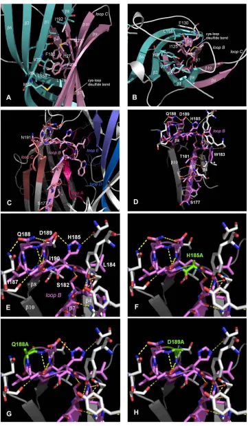
Loop B modeling
[image:4.603.106.401.59.317.2]To gain further insight into the possible orientations and
in-teractions of loop B residues, a range of homology models
were constructed based on AChBP crystal structures that
FIGURE 1 Location of the amino acid residues examined in the current study. (A) Two adjacent subunits (principaland
complementary), showing the positions of the six binding loops A-F. (B) The amino acid sequence for the extracellular domain of the murine 5-HT3Areceptor (accession No. Q6J1J7), aligned with AChBP isolated fromLymnaea stagnalis
were either unbound or bound by different ligands. The
lowest energy states for the models are shown in Fig. 4,
A–E,
and the carbon backbones for each of these are overlaid in
Fig. 4
F. Loop B is similar in each of these models,
sug-gesting that regardless of the bound ligand, the position of the
loop and the orientations of the side-chains remains relatively
unaltered.
The models indicate that there is a hydrophobic core
be-tween the inner and outer
b
sheets (Fig. 5,
A
and
B). They
also predict an extensive network of hydrogen bonds, both
between loop B and adjacent regions, and within the B loop
itself (Fig. 5,
C
and
D). In the first group, the
b
-strand of
loop B is stabilized by hydrogen bonds between the
back-bones of residues from Leu-178 to His-185, and the adjacent
anti-parallel strands of
b
4 and
b
10, and the backbones of
Asp-189 and Asn-191 form hydrogen bonds with the
back-bones of Thr-64 and Val-66 in the adjacent
b
1-strand. The
second group includes the side-chains of residues His-185,
Gln-188 and Asp-189 which stabilize a tight
b
7-
b
8 turn at
the top of loop B. Our homology models predict that amino
acid substitutions in this region would disrupt this hydrogen
bonding network (Fig. 5,
E–H).
DISCUSSION
This study shows that the 5-HT
3receptor loop B is extremely
sensitive to amino acid changes; our data show that
substi-tutions of most of the residues from Ser-177 to Asn-191 have
a significant effect on [
3H]granisetron binding and 5-HT
evoked currents. Loop B in all Cys-loop receptors has long
been known to play an important role in receptor function,
but it was surprising that so many of the residues in this
re-gion were sensitive to amino acid substitution. A possible
explanation for these data is provided by our homology
models, which show that many of these residues could
sta-bilize the structure of the region through hydrophobic
inter-actions and hydrogen bonds.
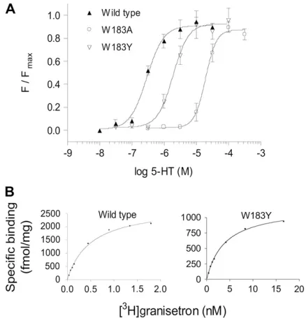
FIGURE 2 Example data for receptor function and radioligand binding at wild-type and mutant receptors. (A) Channel function was measured using a voltage-sensitive fluorometric dye on a FlexStation. Values from a series of experiments were normalized, averaged and fitted with a four-parameter logistic equation. The calculatedEC50values are shown in Table 2. (B)Kd values were estimated using the 5-HT3 antagonist [
3
[image:5.603.53.294.78.269.2]H]granisetron. The examples show binding for single experiments, fitted with a one site binding equation.Kdvalues for a series of experiments were averaged for each mutant, and are presented in Table 1.
TABLE 2 Functional effects of Ala and conserved substitutions at the 5-HT3receptor
Alanine mutant
pEC50 Mean6SE
EC50 (mM) n
Conserved mutant
pEC50 Mean6SE
EC50 (mM) n
Wild-type 6.6260.06 0.24 12 Wild-type 6.6260.06 0.24 12
S177A NR* — 4 S177T NR* — 4
L178A NR* — 4 L178I NR* — 4
T179A NR* — 4 T179S NR* — 4
F180A NR* — 4 F180Y NR* — 4
T181A 5.8660.08* 1.38 4 T181S NR* — 6
S182A NR* — 4 S182T 6.2160.07 0.62 4
W183A 4.4060.11* 39.8 6 W183Y 5.2860.26* 5.25 6 L184A 4.4760.07* 33.9 4 L184I 5.4760.17* 3.39 4
H185A NR* 4 H185N NR* — 4
T186A 4.2460.03* 57.5 6 T186S NR* — 4 I187A 4.6560.07* 22.4 4 I187L 5.4860.10* 3.31 4
Q188A NR* 4 Q188N NR* — 4
D189A NR* 4 D189E NR* — 4
I190A NR* 4 I190L 6.3360.01 0.47 4
N191A NR* 4 N191Q NR* — 4
*Significantly different to wild-type (ANOVA with Dunnett’s post test:p,
0.05). NR, No response.
TABLE 1 Effects of Ala and conserved substitutions on [3H]granisetron binding affinities at the 5-HT
3receptor
Alanine mutant
pKd(nM) Mean6SE
Kd (nM) n
Conserved mutant
pKd(nM) Mean6SE
Kd (nM) n
Wild-type 9.3060.12 0.5 11 Wild-type 9.3060.12 0.5 11 S177A 9.2360.26 0.7 5 S177T 7.8660.09y 14* 3 L178A 8.0260.09y 10 4 L178I NBy — 4 T179A 8.5560.09y 3.2 8 T179S 9.3360.22 0.5 7 F180A NBy — 5 F180Y 8.1960.08y 6.5 3 T181A 9.3860.18 0.4 7 T181S 9.1560.11 0.7 4 S182A 9.0660.10 0.9 6 S182T 8.7160.03y 1.9 8 W183A NBy — 6 W183Y 8.3860.10y 4.2 6 L184A 8.4260.10y 4.1 4 L184I 9.1260.03 0.8 6 H185A 8.8660.11 1.4 5 H185N 8.4560.07y 3.5 5 T186A 9.2660.06 0.6 9 T186S 8.1760.06y 6.8 4 I187A 7.9060.04y 13* 4 I187L 8.9060.14 1.3 8
Q188A 9.2260.15 0.6 7 Q188N NBy — 4
D189A NBy — 4 D189E NBy — 7
I190A NBy — 5 I190L 8.9360.02 1.2 4
N191A NBy — 5 N191Q 8.2060.11y 6.3 4
ySignificantly different when compared to wild-type (ANOVA with
Dunnett’s post test:p,0.05). NB, No binding orKdvalues$10 nM, which is the resolution of the technique (16); thus values obtained that are .10 nM (*) may be inaccurate.
The 5-HT3Receptor B-Loop 5731
[image:5.603.310.552.79.269.2] [image:5.603.63.282.387.615.2]Loop B is a stable, static structure
Comparing a range of homology models based on AChBP
bound to different agonists (which are presumed to represent
the open state of the receptor), and antagonists (the closed
state), shows that loop B is similarly positioned in each of the
models. This indicates that there is little or no movement of
this loop between the open and closed states, suggesting a
stable and static structure that does not change conformation
when ligands bind. Our homology models indicate that loop
B residues could play an important role in stabilizing the
structure of the whole region, as many loop B residues
con-tribute to a global network of noncovalent interactions. In
agreement, a similar homology-based study has found that
distances between Trp-183 residues of adjacent 5-HT
3sub-units were comparable regardless of the AChBP template
used (25). Data from previous studies also indicate that this
region of the protein has an important structural role in
ho-mologous receptors. For example, in the nACh receptor the
loop B residue Lys-145 (equivalent to Thr-179 in the 5-HT
3receptor) has been shown to form a salt-bridge with Asp-200
(Glu-236 in 5-HT
3), and is proposed as one of a number of
strong interactions that stabilize the binding region in this
receptor (26). We must, of course, be cautious when using
bound AChBP structures as templates for open and closed
states of the receptor, but comparisons with nACh receptor
structures and molecular dynamic simulations indicate they
are reasonably accurate (3,5,6,27). This observation is further
supported by the more recent structure determination of a
homologous prokaryotic ion channel (7).
Loop B contributes to a hydrophobic core
Our modeling data indicate that in each subunit there is a
central hydrophobic core formed by the converging side
chains of residues distributed throughout the 5-HT
3receptor
subunit sequence. The residues are mostly located in the
in-ner and outer
b
-sheets comprising the
b
-sandwich of the
extracellular part of the subunit, and their side chains all point
into the interior of the
b
-sandwich (see Figs. 1 and 5). Two
of these residues are contributed by loop B, and these are
surrounded by other hydrophobic residues; Leu-178 is
sur-rounded by Ile-127, Phe-130, Phe-180 and Phe-239, whereas
Phe-180 is surrounded by Val-70, Tyr-91, Ile-125, Ile-127,
Leu-178 and Phe-239. In total there are 16 hydrophobic
residues within 5 A
˚ of Leu-178 and Phe-180 (Fig. 5). This
region may be critical for receptor folding, as hydrophobic
regions are usually the first to form, and indeed our data
suggest that substituting Phe-180 for Ala prevents correct
assembly and targeting of the receptor. Tyr-91 and Phe-130
have also been proposed to play a role in assembly and/or
targeting (15,28). We suggest, therefore, that this hydrophobic
region is important for the correct folding and assembly of the
5-HT
3receptor, and as 15 of the 16 hydrophobic residues
align with hydrophobic residues across the entire Cys-loop
family, this region probably has a similar role in all Cys-loop
receptors.
[image:6.603.64.281.56.599.2]Our data also suggest that this region has a role in receptor
function. The mutations L178A and F180Y both yield
re-ceptors that bind antagonist, indicating that these rere-ceptors
are correctly assembled and targeted, and their binding sites
FIGURE 4 Homology models showing the amino acid backbone of the loop B region of the 5-HT3receptor. (A) Unbound structure (modeled from AChBP with no ligand, PDB ID 2byn). (B) Agonist bound, (modeled from AChBP with carbomylcholine, PDB ID 1uv6). (C) Agonist bound, (modeled from AChBP with epibatidine, PDB ID 2byq). (D) Large antagonist bound (modeled from AChBP witha-cobratoxin, PDB ID 1yi5). (E) Small antagonist bound, (modeled from AChBP with 2-methyllcaconitine, PDB ID 2byr) (F) Overlays of the modeled loop B backbones fromA–Ecompiled with Swiss-PdbViewer ‘‘magic fit’’, using residues Ser-177 to Asn-191 as a reference point.
The 5-HT3Receptor B-Loop 5733
are intact. However they do not function. We propose that
specific hydrophobic interactions in this part of loop B are a
critical component of a hydrophobic core, where weak and
easily interchangeable interactions are necessary for the
lat-eral movement of the outer and inner
b
-sheets when a ligand
binds (a proposed step in the transduction pathway that
ul-timately leads to pore opening (29)).
Loop B contributes to receptor gating
In addition to the gating role played by residues in the
hy-drophobic core, we propose that other amino acids in loop B
also have a significant role in channel function. One of these
residues is Leu-184 which displays an
;
10-fold increase in
EC
50with a conservative Ile substitution, but shows very
little change in antagonist binding affinity, as previously
re-ported (9).
EC
50is constituted from both agonist affinity and
functional efficacy. The agonist affinity can be modified by
mutations that cause a structural change in the binding site,
or when an agonist-specific binding residue is altered. The
antagonist binding data show there is no structural change in
the binding site with L184I mutant receptors, and the
mod-eling data face Leu-184 away from the binding pocket; thus it
is unlikely to be an agonist-specific ligand binding residue.
The data therefore support a role for this residue in the
con-formational changes that follow, or are concomitant with,
ligand binding, and that ultimately result in channel opening.
In support of this hypothesis the adjacent residue Trp-183,
which has been shown to be involved in agonist binding (30–
33), is located facing into the binding pocket in the model,
and the mutation W183Y results in a similar fold change in
antagonist affinity and
EC
50, suggesting it is primarily the
affinity change that causes the change in
EC
50.
Our models suggest a mechanism by which Leu-184 could
be involved in channel opening: via hydrogen bonds with the
adjacent
b
-sheet of loop A. There is evidence that loop A
plays a dynamic role during receptor gating and it has been
proposed that it acts as a ‘‘latch’ that controls this process in
the nACh receptor (20). In addition, reorientations in this
region have been shown with molecular dynamic studies
(27). Our data suggest that a small residue at position 184 is
much less favorable than a larger one, and we propose that to
allow movement, Leu-184 may function as a ‘spacer’
resi-due, holding loop B at an optimal distance from loop A and
thereby allowing movement of the latter. Similarly, ‘spacer’
residues may be important to allow movement of loop C, as a
bulky hydrophobic side chain was preferred at position 187
(loop B), and our homology models suggest van der Waals
contacts between the side chain of Ile-187 and residues
within the
b
9 and
b
10 strands (loop C) of the same subunit.
Thus Ile-187 may sterically oppose the
b
9 and
b
10 strands
and thereby assure the mobility of loop C. We propose that
loop B forms a rigid frame around which loops A and C move.
Other residues that may play a role in channel function are
Thr-179, Thr-181, Ser-182, His-185 and Thr-186 as mutation
of all of these significantly affected agonist-mediated effects,
but neither Ala nor conserved mutations caused major
changes in the antagonist binding affinity; the latter results
indicating the receptors are correctly assembled. However,
loss of the backbone hydrogen bonding between these
resi-dues and the adjacent
b
-sheets, as indicated in the homology
models, may prevent efficient opening of the channel. It
should also be noted that in previous studies H185A has been
reported as nonbinding, a difference that is likely to reflect the
very low levels of expression (
;
20-fold less than wild-type)
that we observed for this mutant (11). Binding affinities for
Thr-179, Ser-182 and Thr-186 mutants are almost identical to
those shown elsewhere (9).
Our homology models also suggest that there are hydrogen
bonds contributed by residues His-185 to Asn-191 which
may be important for stabilizing the
b
7 -
b
8 turn; substitution
of residues that significantly disrupt this series of bonds
(Q188, D189 and I190; Fig. 5,
G
and
H) results in receptors
that are not folded, assembled and/or targeted correctly.
Substitutions that permit the expression of cell-surface
re-ceptors, possibly because they disrupt fewer hydrogen bonds
(such as in H185A), may result in low levels of expression
or non functional receptors. We propose that a rigid
b
7-
b
8
turn is critical for efficient receptor expression and function.
Asp-189 may also have a role in subunit-subunit interactions,
similar to the equivalent residue (Glu-149) in AChBP (3,11).
In our 5-HT
3homology models Asp-189 is in close
prox-imity to the basic side chains of Lys-112 on the
comple-mentary subunit and a highly conserved Arg-55 on the same
subunit, and there is the potential to form a salt bridge here,
an interaction that would explain why substitution of this
residue was especially sensitive; it was the only residue where
both Ala and conserved amino acid substitutions yielded non
binding receptors.
CONCLUSIONS
We have demonstrated that residues from the 5-HT
3receptor
loop B region have a major influence on binding and function.
These data can be explained by our model, which indicates
that noncovalent interactions within and between loop B and
neighboring residues, both in the binding region and within the
b
-sandwich structure of the subunit, are vital for the integrity
of the binding site and for the dynamic mobility of structural
elements involved in the gating process.
We thank the Wellcome Trust for funding (S.C.R.L. and A.J.T.) and the Swiss National Science Foundation for a postdoctoral fellowship to M.L. (PA00A-105073). S.C.R.L. is a Wellcome Trust Senior Research Fellow in Basic Biomedical Studies.
REFERENCES
1. Thompson, A. J., and S. C. R. Lummis. 2007. The 5-HT3Receptor as a therapeutic target.Expert Opin. Ther. Targets.11:527–540.
The 5-HT3Receptor B-Loop 5735
2. Brejc, K., W. J. van Dijk, R. V. Klaassen, M. Schuurmans, J. van Der Oost, A. B. Smit, and T. K. Sixma. 2001. Crystal structure of an ACh-binding protein reveals the ligand-binding domain of nicotinic receptors.Nature.411:269–276.
3. Celie, P. H., S. E. van Rossum-Fikkert, W. J. van Dijk, K. Brejc, A. B. Smit, and T. K. Sixma. 2004. Nicotine and carbamylcholine binding to nicotinic acetylcholine receptors as studied in AChBP crystal struc-tures.Neuron.41:907–914.
4. Celie, P. H., R. V. Klaassen, S. E. van Rossum-Fikkert, R. van Elk, P. van Nierop, A. B. Smit, and T. K. Sixma. 2005. Crystal structure of acetylcholine-binding protein from Bulinus truncatusreveals the conserved structural scaffold and sites of variation in nicotinic acetyl-choline receptors.J. Biol. Chem.280:26457–26466.
5. Unwin, N. 2005. Refined structure of the nicotinic acetylcholine receptor at 4A˚ resolution.J. Mol. Biol.346:967–989.
6. Dellisanti, C. D., Y. Yao, J. C. Stroud, Z. Z. Wang, and L. Chen. 2007. Crystal structure of the extracellular domain of nAChRa1 bound to
a-bungarotoxin at 1.94 A˚ resolution.Nat. Neurosci.10:953–962. 7. Hilf, R. J., and R. Dutzler. 2008. X-ray structure of a prokaryotic
pentameric ligand-gated ion channel.Nature.452:375–379. 8. Thompson, A. J., and S. C. Lummis. 2006. 5-HT3 receptors.Curr.
Pharm. Des.12:3615–3630.
9. Thompson, A. J., K. L. Price, D. C. Reeves, S. L. Chan, P. L. Chau, and S. C. Lummis. 2005. Locating an antagonist in the 5-HT3receptor binding site using modeling and radioligand binding.J. Biol. Chem.
280:20476–20482.
10. Yan, D., and M. M. White. 2005. Spatial orientation of the antagonist granisetron in the ligand-binding site of the 5-HT3 receptor. Mol.
Pharmacol.68:365–371.
11. Joshi, P. R., A. Suryanarayanan, E. Hazai, M. K. Schulte, G. Maksay, and Z. Bikadi. 2006. Interactions of granisetron with an agonist-free 5-HT3Areceptor model.Biochemistry.45:1099–1105.
12. Chen, C. A., and H. Okayama. 1988. Calcium phosphate-mediated gene transfer: a highly efficient transfection system for stably trans-forming cells with plasmid DNA.Biotechniques.6:632–638. 13. Jordan, M., A. Schallhorn, and F. M. Wurm. 1996. Transfecting
mammalian cells: optimization of critical parameters affecting calcium-phosphate precipitate formation.Nucleic Acids Res.24:596–601. 14. Kunkel, T. A. 1985. Rapid and efficient site-specific mutagenesis
without phenotypic selection.Proc. Natl. Acad. Sci. USA.82:488–492. 15. Price, K. L., and S. C. Lummis. 2004. The role of tyrosine residues in the extracellular domain of the 5-hydroxytryptamine3receptor.J. Biol.
Chem.279:23294–23301.
16. Bylund, D. B., and M. L. Toews. 1993. Radioligand binding methods: practical guide and tips.Am. J. Physiol.265:421–429.
17. Spier, A. D., G. Wotherspoon, S. V. Nayak, R. A. Nichols, J. V. Priestley, and S. C. R. Lummis. 1999. Antibodies against the extra-cellular domain of the 5-HT3receptor label both native and recombi-nant receptors.Brain Res. Mol. Brain Res.67:221–230.
18. Fitch, R.W., Y. Xiao, Y., K.J. Kellar, and J. W. Daly. 2003 Membrane potential fluorescence: a rapid and highly sensitive assay for
nico-tinic receptor channel function.Proc. Natl. Acad. Sci. USA.100:4909– 4914.
19. Price, K. L., and S. C. Lummis. 2005. FlexStation examination of 5-HT(3) receptor function using Ca21- and membrane potential-sensitive dyes: advantages and potential problems. J. Neurosci. Methods.149: 172–177.
20. Chakrapani, S., T. D. Bailey, and A. Auerbach. 2003. The role of loop 5 in acetylcholine receptor channel gating.J. Gen. Physiol.122:521–539. 21. Reeves, D. C., M. F. Sayed, P. L. Chau, K. L. Price, and S. C. Lummis. 2003. Prediction of 5-HT3 receptor agonist-binding residues using homology modeling.Biophys. J.84:2338–2344.
22. Sali, A., and T. L. Blundell. 1993. Comparative protein modelling by satisfaction of spatial restraints.J. Mol. Biol.234:779–815.
23. Weiner, S. J., P. A. Kollman, D. A. Case, U. C. Singh, C. Ghio, G. Alagona, S. Profeta, and P. Weiner. 1984. A new force-field for molecular mechanical simulation of nucleic-acids and proteins.J. Am. Chem. Soc.106:765–784.
24. Spier, A. D., and S. C. Lummis. 2000. The role of tryptophan residues in the 5-Hydroxytryptamine3receptor ligand binding domain.J. Biol.
Chem.275:5620–5625.
25. Thompson, A. J., C. L. Padgett, and S. C. Lummis. 2006. Mutagenesis and molecular modeling reveal the importance of the 5-HT3receptor F-loop.J. Biol. Chem.281:16576–16582.
26. Mukhtasimova, N., C. Free, and S. M. Sine. 2005. Initial coupling of binding to gating mediated by conserved residues in the muscle nicotinic receptor.J. Gen. Physiol.126:23–39.
27. Cashin, A. L., E. J. Petersson, H. A. Lester, and D. A. Dougherty. 2005. Using physical chemistry to differentiate nicotinic from cholin-ergic agonists at the nicotinic acetylcholine receptor.J. Am. Chem. Soc.
127:350–356.
28. Sullivan, N. L., A. J. Thompson, K. Price, and S. C. R. Lummis. 2006. Defining the roles of Asn-128, Glu-129 and Phe-130 in loop A of the 5-HT3receptor.Mol. Membr. Biol.23:1–10.
29. Unwin, N., A. Miyazawa, J. Li, and Y. Fujiyoshi. 2002. Activation of the nicotinic acetylcholine receptor involves a switch in conformation of the alpha subunits.J. Mol. Biol.319:1165–1176.
30. Beene, D. L., G. S. Brandt, W. Zhong, N. M. Zacharias, H. A. Lester, and D. A. Dougherty. 2002. Cation-pinteractions in ligand recognition by serotonergic (5-HT3A) and nicotinic acetylcholine receptors: the anomalous binding properties of nicotine.Biochemistry.41:10262–10269. 31. Lummis, S. C., D. L. Beene, N. J. Harrison, H. A. Lester, and D. A. Dougherty. 2005. A cation-pbinding interaction with a tyrosine in the binding site of the GABACreceptor.Chem. Biol.12:993–997. 32. Zhong, W. G., J. P. Gallivan, Y. O. Zhang, L. T. Li, H. A. Lester, and
D. A. Dougherty. 1998. From ab initio quantum mechanics to molec-ular neurobiology: A cation-pbinding site in the nicotinic receptor.
Proc. Natl. Acad. Sci. USA.95:12088–12093.