Natural Antioxidants: Function and Sources
Yevgenia Shebis1, David Iluz1,2, Yael Kinel-Tahan1*, Zvy Dubinsky1, Yaron Yehoshua1
1
The Mina & Everard Goodman Faculty of Life Sciences, Bar-Ilan University, Ramat-Gan, Israel; 2Department of Environmental Science and Agriculture, Beit Berl College, Kfar Saba, Israel.
Email: *[email protected]
Received April 10th, 2013; revised May 12th, 2013; accepted May 20th, 2013
Copyright © 2013 Yevgenia Shebis et al. This is an open access article distributed under the Creative Commons Attribution License, which permits unrestricted use, distribution, and reproduction in any medium, provided the original work is properly cited.
ABSTRACT
The definition of antioxidants, given in 1995 by Halliwell and Gutteridge, stated that an antioxidant is “any substance that, when present at low concentrations compared with that of an oxidizable substrate, significantly delays or inhibits oxidation of that substrate” [1]. In 2007, Halliwell gave a more specific definition, stating that an antioxidant is “any substance that delays, prevents or removes oxidative damage to a target molecule” [2]. Oxidation reactions produce free radicals that can start multiple chain reactions that eventually cause damage or death to the cell. Antioxidants remove these free-radical intermediates by being oxidized themselves, and inhibit other oxidation reactions, thus stopping the harmful chain reactions. Such oxidative processes are dangerous for all living cells, especially those in proximity to sites where active oxygen is released by photosynthesis. Spontaneous oxidation causes food rancidity and spoilage of medicines. Furthermore, oxidative stress is an important part of many human diseases that can occur, inter alia, due to a lack of appropriate nutrition and exercise, air pollution, smoking, and more, leading to lethal diseases, such as cancer. Therefore, it is imperative to include antioxidants in our diets. Due to the fact that synthetically produced antioxidants are currently used in the food and pharmaceutical industries in order to prolong product shelf life, there is currently a strong trend to search for large, available, and efficient natural sources of antioxidants to replace the synthetic ones, thus minimizing damage to our cells.
Keywords: Algae; Antioxidant; Disease
1. Introduction
1.1. Antioxidant Cell Functions
There are two major groups of antioxidants in living cells: enzymatic antioxidants and non-enzymatic antioxidants. These groups are divided into several subgroups. The en- zymatic antioxidants are divided into primary and secon- dary enzymatic defenses [3]. The primary defense is com- posed of three important enzymes that prevent the forma- tion of and neutralize free radicals: glutathione peroxi-dase, which donates two electrons to reduce peroxides by forming selenols and also eliminates peroxides as poten- tial substrates for the Fenton reaction; catalase, which turns hydrogen peroxide into water and molecular oxy- gen—one of the most important and efficient antioxidants known today, when just one molecule of catalase con- verts 6 billion molecules of hydrogen peroxide [4]; and lastly, superoxide dismutase, which converts superoxide anions into hydrogen peroxide as a substrate for subse-
quent catalase action. The secondary enzymatic defense includes glutathione reductase and glucose-6-phosphate dehydrogenase. Glutathione reductase reduces glutathi- one (antioxidant) from its oxidized to its reduced form, and by this recycling, to continue neutralizing more free radicals [5]. Glucose-6-phosphate regenerates NADPH, which creates a reducing environment. These two enzy- mes support the primary enzymatic defense antioxidants and do not neutralize free radicals directly. The group of non-enzymatic antioxidants contains several subgroups, the main ones being: vitamins (A, E, C), enzyme cofac- tors (Q10), minerals (zinc and selenium), peptides (gluta- thione), phenolic acids, and nitrogen compounds (uric acid) [3].
There is great importance in maintaining the fragile balance between these antioxidants and the ROS mole- cules. For instance, in humans, disturbing this balance can cause serious health problems, such as cancer, car- diovascular and neurodegenerative diseases, and prema- ture aging [6].
*
Synthetic Antioxidants in Pharmaceutical and Food Industries
Nowadays, most food & pharmaceutical products contain synthetic antioxidants. These compounds are added to food in order to prolong product shelf life, mainly by pre- venting the oxidation of unsaturated double bonds of fat- ty acids. In pharmaceutical products to antioxidants are added to enhance the stability of therapeutic agents that are susceptible to chemical degradation by oxidation. The two most common synthetic antioxidants used today are butylated hydroxyanisole (BHA) and butylated hydroxyl- toluene (BHT). Propyl gallate and tert-butylhy-droqui- none (TBHQ) are other widely used synthetic antioxi- dants in the processed-food industry. For example, TBHQ is usually added to food products such as beef and chicken. Though no harmful effect of these synthetic an- tioxidants has been shown in man, in 2012, the Euro- pean Food Safety Authority (EFSA) evaluated informa- tion regarding several of these antioxidants and estab- lished revised acceptable daily intakes of antioxidants for human consumption, setting a proper scale for their use by food companies [7]. Ascorbic acid derivatives, such as ascorbic acid and erythorbic acid; thiol derivatives, such as thioglycerol, cysteine, dithiothreitol, and gluta- thione; sulfurous-acid salts, such as sodium sulfite, so- dium formaldehyde sulfoxylate, and tocopherols, are wi- dely used in the pharmaceutical industry.
Unfortunately, new data indicating that the synthetic antioxidants used in the industry could have carcinogenic effects on human cells resurface every year, thus fueling an intense search for new, natural and efficient antioxi- dants.
2. Antioxidants in Plants: Relation to
Photosynthesis
Photosynthesis is an important source of cellular oxidants, and the importance of antioxidants in maintaining high rates of photosynthesis has been shown in many studies [8]. Studies showed that photosynthesis is the source of reactive oxygen species (ROS) and that the photosyn- thetic electron transport chain operates as a regulatory system for minimizing ROS production in an aerobic en- vironment. In addition, there is a need for a strong and efficient antioxidant network to process ROS effectively and to maintain intracellular ROS pools at low levels [9]. Originally, ROS were recognized as toxic by-products of aerobic metabolism, molecules that have the potential to cause irreversible damage to photosynthetic components and that are removed by antioxidants and antioxidative enzymes. Now, it has become clear that these molecules play an important signaling role in plants, controlling processes such as growth, development, and even pro- grammed cell death [10]. Due to the recent findings, it is of great importance to maintain the fragile balance of the
ROS molecules and antioxidants. Plants usually contain a wide variety of free-radical scavenging molecules, such as phenolic compounds, nitrogen compounds, vitamins, and more [11-14]. Studies have proven that many of these antioxidant compounds exhibit anti-inflammatory, anti- carcinogenic, antibacterial, antitumor, or antimutagenic effects in cells [15-19]. Nowadays, the intake of natural antioxidants is associated with reduced risks of cancer, cardiovascular disease, and other diseases.
It has been widely proven that green tea leaves contain a high concentration of polyphenols that act as antioxi- dants both in vitro and in vivo (in animal and human cells), thus reducing and controlling ROS molecules [20, 21]. Studies conducted on Chinese medical and other me- dical herbs demonstrated that some herbs, such as rose- mary, sage, thyme, and bay, have much stronger antioxi- dant activity and contain significantly more phenolic acids than common vegetables and fruits, which are con- sidered good natural sources of dietary antioxidants [22, 23].
3. Antioxidants in Algae
Due to the widespread use of synthetic antioxidants in food & pharmaceuticals, scientists are also attempting to develop new and efficient antioxidants from another al- ternative source found in nature in large amounts—the algae. Algae are widely available aquatic plants contain- ing natural antioxidative compounds, having biological activities that affect the pathogenesis of several diseases, with a relatively low-cost isolation/extraction process [24]. Seaweeds and microalgae make up a big group of pho- tosynthetic marine and freshwater organisms that have adapted to survive in highly complex and competitive environments, including fast temperature changes, dif- ferent light intensities, nutrient deficiency, pH changes, and more. The microalgae are the ocean’s primary pro- ducers, being consumed by other marine organisms for their metabolic energy-requiring processes along the food chain. Recent studies have shown that some species of algae contain large amounts of antioxidants and phe- nolic compounds. Natural antioxidants found in algae play an important role against various diseases and age- ing processes by protecting the cells from oxidative da- mage [25,26]. It has been shown that the red, brown, and green algae, as well as cyanobacteria, display high radi- cal scavenging activities [27-29]. The main antioxidants found in algae are vitamins C and E [30], carotenoids (β & α-carotene, zeaxanthin, neoxanthin, etc.) [31-34] po- lyphenols [35], and chlorophylls [36]. In addition, red and brown algae contain high levels of folic acid and folate derivatives [37].
biochemicals, such as antioxidants, polysaccharides, pig- ments, fatty acids, vitamins, and more. For example, Du- naliella salina is the most suitable organism for the mass production of β-carotene as it can produce up to 14% of its dry weight and can be easily and rapidly cultivated compared to plants [38,39]. As it is well known, β-caro- tene is one of the most common food colorants in the world and has been applied to a wide range of food and beverage products, improving their appeal to consumers, besides its antioxidant function. Studies show that algae play an important supporting role in the prevention of or recovery from a variety of diseases, including cancer [40, 41]. Moreover, studies show the great benefits of the cy- anobacteria in the prevention of cardiovascular disease, nonalcoholic fatty liver disease, and more. One of the most talked about cyanobacteria that can be used as a dietary supplement is Spirulina. We use two species of these cyanobacteria in order to produce the supplements Spirulina platensis (SP) and Spirulina maxima (SM). Supplementation with these species of Spirulina resulted in lipid-lowering, antioxidant, and anti-inflammatory ef- fects [42-46]. Moreover, its therapeutic benefits were seen in a variety of diseased conditions, such as hyper- cholesterolemia, cardiovascular diseases, viral infections, and cancer [47]. Furthermore, due to its high nutritional
value, Spirulina was recommended by the European
Space Agency (ESA) and National Aeronautics and Space Administration (NASA) as one of the primary and im- portant foods during long-term space missions [48]. In addition, in 2011, the Dietary Supplements Information Expert Committee (DSI-EC) of the United States Phar- macopeial Convention awarded Spirulina (SM and SP) the highest possible safety rating and concluded that it is generally safe for consumption. It is noteworthy that the Spirulina species were used in antiquity as a proteina- ceous food around Lakes Tchad in Africa and Texcoco in Mexico [49]. With the increasing need to scout for new natural sources of antioxidants, more and more species of algae are now being checked. It was found that industri-
ally cultivated samples of Botryococcus braunii, Neo-
chloris oleoabundans, Isochrysis sp., Chlorella vulgaris, Phaeodactylum tricornutum, Turbinaria ornate, Gayra- lia oxysperma, Chaetomorpha antennina, Sargassum vul- gare, Undaria pinnatifida, Himanthalia elongate, Chon- drus crispus, and much more, possess a high antioxidant capacity and could be potential new sources of natural antioxidants [40,50,51].
Screening Methods in Algae
There are two common methods in use today in the search for antioxidative reactions in algae. The first assay is lipoxygenase-activity determination. There are several methods for assessing this activity. The most commonly used of these methods is based on the fact that hydrop-
eroxides generated from the action of the lipoxygenase on linoleic acid with an uptake of oxygen contain a con- jugated diene that strongly absorbs UV light at 234 nm. Reaction rates can be determined by measuring product formation through the changes in absorbance. This me- thod can be combined or used separately from the rate of
O2 consumption which is followed by an oxygen elec-
trode [52-54]. Another assay is the ferric-reducing ability of plasma (FRAP), which is the most commonly used as- say for determination of antioxidant capacity. This is bas- ed on the reduction of the ferric to ferrous ion at low pH, which causes a colored ferrous-tripyridyltriazine com- plex to form. Comparing the absorbance changes at 590 - 593 nm of the reaction mixtures to that of known con-centrations of mixtures (usually ascorbate or α-tocophe- rol) containing ferrous ions, gives us the FRAP value of our test mixture. This assay gives fast and reliable results whether with a single or a mixture of antioxidants in various solutions. The reaction is linearly related to the
molar concentration of the antioxidant [55].An example
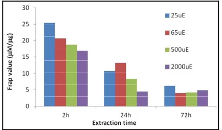
[image:3.595.321.526.360.501.2]of antioxidant activity in microalgae, using FRAP method, are presented in Figures 1 and 2.
Figure 1. Total antioxidant activity presented as total mean FRAP value in M per g crude extract of dry Ankyra sp. The algae were grown under different light intensities, i.e., 65 and 300 mole quanta m−2·s−1 (uE).
[image:3.595.315.532.561.688.2]4. Antioxidants in Food
Regular consumption of vegetables and fruits has been recognized as reducing the risk of chronic diseases [56]. Studies demonstrate that an antioxidant-rich diet has a very positive health impact in the long run [57,58]. It is a well-known fact that citrus fruits (oranges, lemons, etc.) contain a high amount of natural antioxidants, such as vitamin C. Blueberries, strawberries, grapes, plums, prunes, red beans, spinach, kale, broccoli flowers, alfalfa sprouts, and more have been proven to contain a high amount of antioxidants and have been incorporated into many die- tary menus [59,60]. Furthermore, there are some new and unique antioxidants that have been discovered in spinach. NAO is an aqueous spinach-leaf extract that contains de- rivatives of flavonoids and p-coumaric acid. The biolo- gical activity of NAO has been shown to be beneficial in preventing prostate cancer. Moreover, glucosylated fla-vonoids that were found in that extract exhibit anti-in- flammatory activities [61]. Recent studies also suggested that fruit-like jackfruit, araticu-domato, pindo palm, and mandacaru-de-trêsquinas are good sources of vitamins C and A and phenolic compounds [62,63]. In addition, there are studies that research genetic, chemical, or biological modification in order to increase the antioxidant potency of fruits [64].
5. Supplements
The most common antioxidants given as food supple- ments are vitamins C (aka ascorbic acid and ascorbate) and E. Vitamin C is a cofactor in enzymatic reactions, in- cluding several collagen syntheses and an electron do- nor, which makes it a potent water-soluble antioxidant in humans. If these reactions are disrupted or damaged, they can cause severe health problems, such as scurvy [65]. Furthermore, studies showed that some gene expression and protein assimilation functions are dependent on die- tary vitamin C [66]. Vitamin E is a fat-soluble antioxi- dant whose function is to stop the production of ROS formed due to fat oxidation. Among other functions, it is involved in cell signaling, gene expression regulation, immune function, and other metabolic processes [67]. Cur- rent medical supplements of vitamin E usually provide only synthetically produced α-tocopherol, whereas natu- ral supplements provide mixed tocopherols and contain the eight isomeric forms of α-tocopherol. Both vitamins are produced today both synthetically and from natural sources. It is said that the naturally produced vitamins have a much higher percentage of absorbance in the hu- man body. The regular intake of these vitamins is associ- ated with a reduced risk of chronic diseases, such as can- cer, cardiovascular disease, and cataracts, through their antioxidant mechanisms. The antioxidant supplement
market has been increasing rapidly for the past several years. The global vitamin and supplement market is worth close to $68 billion, with the US being the world leader in terms of market share for vitamins and supplements with around 30% of the world market. In 2010, vitamin E imported by the EU reached up to $421 million and vi- tamin C-$361 million. In addition, according to Business Insights, the vitamin and mineral market in the US will see compound annual growth of 4.5%, nearing $30 bil- lion, in 2015 [68].
6. Summary
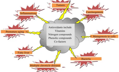
As already said, the main goal of researchers today is to find natural antioxidants that will replace the synthetic ones in the food, pharmaceutical, and cosmetic industries [69]. Though it has not been proven that synthetic anti- oxidants are harmful to human cells, the EFSA has been carefully monitoring their amounts, preventing food com- panies from exaggerating in their dosages. Seaweeds and microalgae are a common, available, and valuable poten- tial source of natural antioxidants. They have a very high concentrated nutritional value, containing a variety of vi- tamins and other non-enzymatic antioxidants, and are re- latively inexpensive to mass-produce and extract. Fur- thermore, several studies have shown the multiple bene- fits of some algal species in preventing chronic and even lethal diseases. It is more imminent than ever that algae might become the main future source of natural antio- xidants (Figure 3).
7. Acknowledgements
[image:4.595.310.536.567.705.2]The authors thank Dr. Alla Alster, Dr. Dovi Kelman, Moran Topf, Barak Vasker, Dr. Miriam Zigman and Said Abu Ghosh for their contribution with cultures of algae and help with the experiments. This research was sup- ported by grants awarded by the Israel Ministry of En- ergy and Water Resources, the Israel Ministry of Science
and Technology, the “Varda Rabin” Foundation, NATO sfp 981,883.
REFERENCES
[1] B. Halliwell and J. M. C. Gutteridge, “The Definition and Measurement of Antioxidants in Biological Systems,” Free Radical Biology and Medicine, Vol. 18, No. 1, 1995, pp. 125-126. doi:10.1016/0891-5849(95)91457-3
[2] B. Halliwell, “Biochemistry of Oxidative Stress,” Bioche- mical Society Transactions, Vol. 35, No. 5, 2007, pp. 1147-1150. doi:10.1042/BST0351147
[3] M. Carocho and I. C. F. R. Ferreira, “A Review on Anti- oxidants, Prooxidants and Related Controversy: Natural and synthetic compounds. Screening and Analysis Metho- dologies and Future Perspectives,” Food and Chemical Toxicology, Vol. 51, 2013, pp. 15-25.
doi:10.1016/j.fct.2012.09.021
[4] K. Rahman, “Studies on Free Radicals, Antioxidants, and Co-Factors,” Clinical Interventions in Aging, Vol. 2, No. 2, 2007, pp. 219-236.
[5] D. V. Ratnam, D. D. Ankola, V. Bhardwaj, D. K. Sahana and M. N. V. R. Kumar, “Role of Antioxidants in Prophy- laxis and Therapy: A Pharmaceutical Perspective,” Jour- nal of Controlled Release, Vol. 113, No. 3, 2006, pp. 189- 207. doi:10.1016/j.jconrel.2006.04.015
[6] M. Valko, D. Leibfritz, J. Moncol, M. T. D. Cronin, M. Mazur and J. Telser, “Free Radicals and Antioxidants in Normal Physiological Functions and Human Disease,” In- ternational Journal of Biochemistry & Cell Biology, Vol. 39, No. 1, 2007, pp. 44-84.
doi:10.1016/j.biocel.2006.07.001
[7] EFSA, “Scientific Opinion on the Reevaluation of Buty- lated Hydroxytoluene BHT (E 321) as a Food Additive. EFSA Panel on Food Additives and Nutrient Sources Ad- ded to Food (ANS),” European Food Safety Authority Journal, Vol. 10, No. 3, 2012, p. 2588.
http://www.efsa.europa.eu/en/efsajournal/doc/2588.pdf
[8] C. H. Foyer and G. Noctor, “Redox Sensing and Signal- ling Associated with Reactive Oxygen in Chloroplasts, Pe- roxisomes and Mitochondria,” Physiologia Plantarum, Vol. 119, No. 3, 2003, pp. 355-364.
doi:10.1034/j.1399-3054.2003.00223.x
[9] C. H. Foyer and S. Shigeoka, “Understanding Oxidative Stress and Antioxidant Functions to Enhance Photosyn- thesis,” Plant Physiology, Vol. 155, No. 1, 2011, pp. 93- 100. doi:10.1104/pp.110.166181
[10] J. Bailey-Serres and R. Mittler, “The Roles of Reactive Oxygen Species in Plant Cells (Editorial),” Plant Physi- ology, Vol. 141, No. 2, 2006, p.
311doi:10.1104/pp.104.900191.
[11] G. Agati, E. Azzarello, S. Pollastri and M. Tattini, “Flavo- noids as Antioxidants in Plants: Location and Functional Significance,” Plant Science, Vol. 196, 2012, pp. 67-76. doi:10.1016/j.plantsci.2012.07.014
[12] C. J. Chiang, H. Kadouh and K. Q. Zhou, “Phenolic Com- pounds and Antioxidant Properties of Gooseberry as Af- fected by in Vitro Digestion,” LWT-Food Science and Te-
chnology, Vol. 51, No. 2, 2013, pp. 417-422. doi:10.1016/j.lwt.2012.11.014
[13] C. C. Wong, H. B. Li, K. W. Cheng and F. Chen, “A Sys- tematic Survey of Antioxidant Activity of 30 Chinese Medicinal Plants Using the Ferric Reducing Antioxidant Power Assay,” Food Chemistry, Vol. 97, No. 4, 2006, pp. 705-711. doi:10.1016/j.foodchem.2005.05.049
[14] Y. Z. Cai, M. Sun and H. Corke, “Antioxidant Activity of Betalains from Plants of the Amaranthaceae,” Journal of Agricultural and Food Chemistry, Vol. 51, No. 8, 2003, pp. 2288-2294. doi:10.1021/jf030045u
[15] B. Halliwell, “Free Radicals, Antioxidants, and Human Di- sease—Curiosity, Cause, or Consequence,” Lancet, Vol. 344, No. 8924, 1994, pp. 721-724.
doi:10.1016/S0140-6736(94)92211-X
[16] R. W. Owen, A. Giacosa, W. E. Hull, R. Haubner, B. Spie- gelhalder and H. Bartsch, “The Antioxidant/Anticancer Po- tential of Phenolic Compounds Isolated from Olive Oil,” European Journal of Cancer, Vol. 36, No. 10, 2000, pp. 1235-1247. doi:10.1016/S0959-8049(00)00103-9
[17] Y. Z. Cai, Q. Luo, M. Sun and H. Corke, “Antioxidant Ac- tivity and Phenolic Compounds of 112 Traditional Chi- nese Medicinal Plants Associated with Anticancer,” Life Sciences, Vol. 74, No. 17, 2004, pp. 2157-2184.
doi:10.1016/j.lfs.2003.09.047
[18] J. H. Xiong, S. C. Li, W. J. Wang, Y. P. Hong, K. J. Tang and Q. S. Luo, “Screening and Identification of the Anti- bacterial Bioactive Compounds from Lonicera japonica Thunb. Leaves,” Food Chemistry, Vol. 138, No. 1, 2013, pp. 327-333. doi:10.1016/j.foodchem.2012.10.127
[19] G. A. El-Chaghaby, A. F. Ahmad and E. S. Ramis, “Eva- luation of the Antioxidant and Antibacterial Properties of Various Solvents Extracts of Annona squamosa L. Leaves,” Arabian Journal of Chemistry, 2011, in press.
doi:10.1016/j.arabjc.2011.06.019
[20] B. Frei and J. V. Higdon, “Antioxidant Activity of Tea Polyphenols in Vivo: Evidence from Animal Studies,” Jour- nal of Nutrition, Vol. 133, No. 1, 2003, pp. 3275s-3284s. [21] J. V. Higdon and B. Frei, “Tea Catechins and Polyphenols:
Health Effects, Metabolism, and Antioxidant Functions,” Critical Reviews in Food Science and Nutrition, Vol. 43, No. 1, 2003, pp. 89-143.
doi:10.1080/10408690390826464
[22] B. Bozin, N. Mimica-Dukic, N. Simin and G. Anackov, “Characterization of the Volatile Composition of Essen- tial Oils of Some Lamiaceae Spices and the Antimicrobial and Antioxidant Activities of the Entire Oils,” Journal of Agricultural and Food Chemistry, Vol. 54, No. 5, 2006, pp. 1822-1828. doi:10.1021/jf051922u
[23] V. Katalinic, M. Milos, T. Kulisic and M. Jukic, “Screen- ing of 70 Medicinal Plant Extracts for Antioxidant Capa- city and Total Phenols,” Food Chemistry, Vol. 94, No. 4, 2006, pp. 550-557. doi:10.1016/j.foodchem.2004.12.004
[24] S. Lordan, R. P. Ross and C. Stanton, “Marine Bioactives as Functional Food Ingredients: Potential to Reduce the Incidence of Chronic Diseases,” Marine Drugs, Vol. 9, No. 6, 2011, pp. 1056-1100. doi:10.3390/md9061056
Y.-J. Lee, S.-K. Kim, et al., “Protective Effect of Enzyma- tic Extracts from Microalgae against DNA Damage In- duced by H2O2,” Marine Biotechnology, Vol. 9, No. 4,
2007, pp. 479-490. doi:10.1007/s10126-007-9007-3
[26] K. N. Kim, S. J. Heo, C. B. Song, J. Lee, M. S. Heo, I. K. Yeo, et al., “Protective Effect of Ecklonia cava Enzyma- tic Extracts on Hydrogen Peroxide-Induced Cell Damage,” Process Biochemistry, Vol. 41, No. 12, 2006, pp. 2393- 2401. doi:10.1016/j.procbio.2006.06.028
[27] Y. X. Li, Y. Li, S. H. Lee, Z. J. Qian and S. K. Kim, “Inhibitors of Oxidation and Matrix Metalloproteinases, Floridoside, and D-Isofloridoside from Marine Red Alga Laurencia undulata,” Journal of Agricultural and Food Chemistry, Vol. 58, No. 1, 2010, pp. 578-586.
doi:10.1021/jf902811j
[28] K. Li, X. M. Li, N. Y. Ji and B. G. Wang, “Natural Bromo- phenols from the Marine Red Alga Polysiphonia Urceo- lata (Rhodomelaceae): Structural Elucidation and DPPH Radical-Scavenging Activity,” Bioorganic and Medicinal Chemistry, Vol. 15, No. 21, 2007, pp. 6627-6631. doi:10.1016/j.bmc.2007.08.023
[29] S. Singh, B. N. Kate and U. C. Banerjee, “Bioactive Com- pounds from Cyanobacteria and Microalgae: An Over- view,” Critical Reviews in Biotechnology, Vol. 25, No. 3, 2005, pp. 73-95. doi:10.1080/07388550500248498
[30] P. MacArtain, C. I. R. Gill, M. Brooks, R. Campbell and I. R. Rowland, “Nutritional Value of Edible Seaweeds,” Nu- trition Reviews, Vol. 65, No. 12, 2007, pp. 535-543. doi:10.1111/j.1753-4887.2007.tb00278.x
[31] C. C. Hu, J. T. Lin, F. J. Lu, F. P. Chou and D. J. Yang, “De- termination of Carotenoids in Dunaliella salina Culti- vated in Taiwan and Antioxidant Capacity of the Algal Carotenoid Extract,” Food Chemistry, Vol. 109, No. 2, 2008, pp. 439-446. doi:10.1016/j.foodchem.2007.12.043
[32] J. A. Haugan and S. Liaaenjensen, “Algal Carotenoids. 54. Carotenoids of Brown Algae (Phaeophyceae),” Biochemi- cal Systematics and Ecology, Vol. 22, No. 1, 1994, pp. 31-41. doi:10.1016/0305-1978(94)90112-0
[33] E. Christaki, E. Bonos, I. Giannenas and P. Florou-Paneri, “Functional Properties of Carotenoids Originating from Algae,” Journal of the Science of Food and Agriculture, Vol. 93, No. 1, 2013, pp. 5-11. doi:10.1002/jsfa.5902
[34] K. H. Cha, H. J. Lee, S. Y. Koo, D. G. Song, D. U. Lee and C. H. Pan, “Optimization of Pressurized Liquid Ex- traction of Carotenoids and Chlorophylls from Chlorella vulgaris,” Journal of Agricultural and Food Chemistry, Vol. 58, No. 2, 2010, pp. 793-797. doi:10.1021/jf902628j
[35] A. Bocanegra, S. Bastida, J. Benedí, S. Ródenas and F. J. Sánchez-Muniz, “Characteristics and Nutritional and Car- diovascular-Health Properties of Seaweeds,” Journal of Medicinal Food, Vol. 12, No. 2, 2009, pp. 236-258. doi:10.1089/jmf.2008.0151
[36] K. H. Cha, S. W. Kang, C. Y. Kim, B. H. Um, Y. R. Na and C. H. Pan, “Effect of Pressurized Liquids on Extrac- tion of Antioxidants from Chlorella vulgaris,” Journal of Agricultural and Food Chemistry, Vol. 58, No. 8, 2010, pp. 4756-4761. doi:10.1021/jf100062m
[37] A. R. B. de Quirós, C. C. de Ron, J. López-Hernández and M. A. Lage-Yusty, “Determination of Folates in Sea-
weeds by High-Performance Liquid Chromatography,” Journal of Chromatography A, Vol. 1032, No. 1-2, 2004, pp. 135-139. doi:10.1016/j.chroma.2003.11.027
[38] F. B. Metting, “Biodiversity and Application of Microal- gae,” Journal of Industrial Microbiology & Biotechnol- ogy, Vol. 17, No. 5-6, 1996, pp. 477-489.
doi:10.1007/BF01574779
[39] K. Miyashita, “Function of Marine Carotenoids,” Food Factors for Health Promotion, Vol. 61, 2009, pp. 136- 146. doi:10.1159/000212746
[40] D. Kelman, E. K. Posner, K. J. McDermid, N. K. Taban- dera, P. R. Wright and A. D. Wright, “Antioxidant Activ- ity of Hawaiian Marine Algae,” Marine Drugs, Vol. 10, No. 2, 2012, pp. 403-416. doi:10.3390/md10020403
[41] C. S. Ku, Y. Yang, Y. Park and J. Lee, “Health Benefits of Blue-Green Algae: Prevention of Cardiovascular Dis- ease and Nonalcoholic Fatty Liver Disease,” Journal of Medicinal Food, Vol. 16, No. 2, 2013, pp. 103-111. doi:10.1089/jmf.2012.2468
[42] P. Parikh, U. Mani and U. Iyer, “Role of Spirulina in the Control of Glycemia and Lipidemia in Type 2 Diabetes Mellitus,” Journal of Medicinal Food, Vol. 4, No. 4, 2001, pp. 193-199. doi:10.1089/10966200152744463
[43] K. Iwata, T. Inayama and T. Kato, “Effects of Spirulina platensis on Plasma Lipoprotein Lipase Activity in Fruc- tose-Induced Hyperlipidemic Rats,” Journal of Nutri- tional Science and Vitaminology, Vol. 36, No. 2, 1990, pp. 165-171. doi:10.3177/jnsv.36.165
[44] A. Ramamoorthy and S. Premakumari, “Effect of sup- plementation of Spirulina on Hypercholesterolemic Pa- tients,” Journal of Food Science and Technology-Mysore, Vol. 33, No. 2, 1996, pp. 124-127.
[45] U. V. Mani, S. Desai and U. Iyer, “Studies on the Long- Term Effect of Spirulina Supplementation on Serum Lipid Profile and Glycated Proteins in NIDDM Pa- tients,” Journal of Neutraceuticals Functional and Medi- cal Foods, Vol. 2, No. 3, 2000, pp. 25-32.
doi:10.1300/J133v02n03_03
[46] H. J. Park, Y. J. Lee, H. K. Ryu, M. H. Kim, H. W. Chung and W. Y. Kim, “A Randomized Double-Blind, Placebo-Controlled Study to Establish the Effects of Spirulina in Elderly Koreans,” Annals of Nutrition and Metabolism, Vol. 52, No. 4, 2008, pp. 322-328.
doi:10.1159/000151486
[47] R. T. Deng and T. J. Chow, “Hypolipidemic, Antioxidant, and Antiinflammatory Activities of Microalgae Spiru- lina,” Cardiovascular Therapeutics, Vol. 28, No. 4, 2010, pp. e33-e45. doi:10.1111/j.1755-5922.2010.00200.x
[48] R. J. Marles, M. L. Barrett, J. Barnes, M. L. Chavez, P. Gardiner, R. Ko, et al., “United States Pharmacopeia Safety Evaluation of Spirulina,” Critical Reviews in Food Science and Nutrition, Vol. 51, No. 7, 2011, pp. 593-604. doi:10.1080/10408391003721719.
[49] M. Sánchez, J. Bernal-Castillo, C. Rozo and I. Rodríguez, “Spirulina (Arthrospira): An Edible Microorganism: A Review,” Universitas Scientiarum, Revista de la Facultad de Ciencias, Pontificia Universidad Javeriana, Vol. 8, No. 1, 2003, pp. 7-24.
rulina(arthrospira)genelozelliklerivebesinselonemi.pdf
[50] K. Goiris, K. Muylaert, I. Fraeye, I. Foubert, J. De Bra- banter and L. De Cooman, “Antioxidant Potential of Mi- croalgae in Relation to Their Phenolic and Carotenoid Content,” Journal of Applied Phycology, Vol. 24, No. 6, 2012, pp. 1477-1486. doi:10.1007/s10811-012-9804-6
[51] M. Plaza, A. Cifuentes and E. Ibánez, “In the Search of New Functional Food Ingredients from Algae,” Trends in Food Science and Technology, Vol. 19, No. 1, 2008, pp. 31-39. doi:10.1016/j.tifs.2007.07.012
[52] B. Axelrod, T. M. Cheesebrough and S. Laakso, “Li- poxygenase from Soybeans,” Methods in Enzymology, Vol. 71, 1981, pp. 441-451.
doi:10.1016/0076-6879(81)71055-3
[53] G. E. Anthon and D. M. Barrett, “Colorimetric Method for the Determination of Lipoxygenase Activity,” Journal of Agricultural and Food Chemistry, Vol. 49, No. 1, 2001, pp. 32-37. doi:10.1021/jf000871s
[54] R. Matsukawa, Z. Dubinsky, K. Masaki, T. Takeuchi and I. Karube, “Enzymatic Screening of Microalgae as a Po- tential Source of Natural Antioxidants,” Applied Bio- chemistry and Biotechnology, Vol. 66, No. 3, 1997, pp. 239-247. doi:10.1007/BF02785590
[55] I. F. F. Benzie and J. J. Strain, “The Ferric Reducing Ability of Plasma (FRAP) as a Measure of ‘Antioxidant Power’: The FRAP Assay,” Analytical Biochemistry, Vol. 239, No. 1, 1996, pp. 70-76. doi:10.1006/abio.1996.0292
[56] A. Dembinska-Kiec, O. Mykkanen, B. Kiec-Wilk and H. Mykkanene, “Antioxidant Phytochemicals against Type 2 Diabetes,” British Journal of Nutrition, Vol. 99, No. ES1, 2008, pp. ES109-ES117.
[57] H. P. Y. Sin, D. T. L. Liu and D. S. C. Lam, “Lifestyle Modification, Nutritional and Vitamins Supplements for Age-Related Macular Degeneration,” Acta Ophthalmolo- gica, Vol. 91, No. 1, 2013, pp. 6-11.
doi:10.1111/j.1755-3768.2011.02357.x
[58] L. M. Willis, B. Shukitt-Hale and J. A. Joseph, “Recent Advances in Berry Supplementation and Age-Related Cognitive Decline,” Current Opinion in Clinical Nutri- tion and Metabolic Care, Vol. 12, No. 1, 2009, pp. 91-94. doi:10.1097/MCO.0b013e32831b9c6e
[59] G. H. Cao, R. M. Russell, N. Lischner and R. L. Prior, “Serum Antioxidant Capacity is Increased by Consump- tion of Strawberries, Spinach, Red Wine or Vitamin C in Elderly Women,” Journal of Nutrition, Vol. 128, No. 12, 1998, pp. 2383-2390.
[60] S. Grossman, R. Reznik, T. Tamari and M. Albeck, “New
Plant Water Soluble Antioxidant (NAO) from Spinach,” In: K. Asada and T. Toshikawa, Eds., Frontiers of Reac- tive Oxygen Species in Biology and Medicine, Elsevier Science, Amsterdam, 1994, pp. 57-73.
[61] S. Grossman, S. Dovrat and M. Bergman, “Natural Anti- oxidants: Just Free Radical Scavengers or Much More?” Trends in Cancer Research, Vol. 7, 2011, pp. 57-73. [62] S. B. Swami, N. J. Thakor, P. M. Haldankar and S. B.
Kalse, “Jackfruit and Its Many Functional Components as Related to Human Health: A Review,” Comprehensive Reviews in Food Science and Food Safety, Vol. 11, No. 6, 2012, pp. 565-576.
doi:10.1111/j.1541-4337.2012.00210.x
[63] M. C. Pereira, R. S. Steffens, A. Jablonski, P. F. Hertz, A. D. Rios, M. Vizzotto, et al., “Characterization, Bioactive Compounds and Antioxidant Potential of Three Brazilian Fruits,” Journal of Food Composition and Analysis, Vol. 29, No. 1, 2013, pp. 19-24.
doi:10.1016/j.jfca.2012.07.013
[64] F. S. Gomes, P. A. Costa, M. B. D. Campos, R. V. Tonon, S. Couri and L. M. C. Cabral, “Watermelon Juice Pre- treatment with Microfiltration Process for Obtaining Ly- copene,” International Journal of Food Science and Te- chnology, Vol. 48, No. 3, 2013, pp. 601-608.
doi:10.1111/ijfs.12005
[65] S. J. Padayatty, A. Katz, Y. H. Wang, P. Eck, O. Kwon, J. H. Lee, et al., “Vitamin C as an Antioxidant: Evaluation of Its Role in Disease Prevention,” Journal of the Ameri- can College of Nutrition, Vol. 22, No. 1, 2003, pp. 18-35. [66] M. Lucock, Z. Yates, L. Boyd, C. Naylor, J. H. Choi, X.
Ng, et al., “Vitamin C-Related Nutrient-Nutrient and Nu- trient-Gene Interactions that Modify Folate Status,” Euro- pean Journal of Nutrition, Vol. 52, No. 2, 2013, pp. 569- 582. doi:10.1007/s00394-012-0359-8
[67] S. Salinthone, A. R. Kerns, V. Tsang and D. W. Carr, “Alpha-Tocopherol (Vitamin E) Stimulates Cyclic AMP Production in Human Peripheral Mononuclear Cells and Alters Immune Function,” Molecular Immunology, Vol. 53, No. 3, 2013, pp. 173-178.
doi:10.1016/j.molimm.2012.08.005
[68] Industry Reports.
http://www.reportlinker.com/ci02037/Vitamin-and-Suppl ement.html