http://www.scirp.org/journal/aim ISSN Online: 2165-3410
ISSN Print: 2165-3402
DOI: 10.4236/aim.2019.91007 Jan. 24, 2019 87 Advances in Microbiology
Evaluation of Surface Roughness and
Streptococcus mutans
Adhesion to
Bulk-Fill Resin Composites Polished with
Different Systems
Wafaa E. Soliman
1, Ashraf I. Ali
2, Walid F. Elkhatib
3,4*1Department of Microbiology and Biotechnology, Faculty of Pharmacy, Delta University for Science and Technology, Cairo, Egypt 2Faculty of Dentistry, Mansoura University, Mansoura, Egypt
3Department of Microbiology and Immunology, Faculty of Pharmacy, Ain Shams University, African Union Organization St.,
Cairo, Egypt
4Department of Microbiology and Immunology, School of Pharmacy & Pharmaceutical Industries, Badr University in Cairo
(BUC), Cairo, Egypt
Abstract
Purpose: Bacterial adhesion represents the initial step in biofilm formation, dental caries and decay. This study aimed to evaluate and compare surface roughness and bacterial adhesion to bulk fill resin composites polished with different systems. Methods: Filtek Z350 XT (Incremental-fill resin compo-site), Filtek Bulk-fill Posterior (Bulk-fill resin compocompo-site), and Tetric N Ce-ram (Bulk-fill resin composite) were used as resin composites. The polishing systems used in this study were Sof-Lex multi-step, PoGo one step, and Mylar strip. Scanning electron microscope (SEM) was used to examine the surface roughness and adhesion of Streptococcus mutans ATCC 25175 standard strain to bulk-fill resin composites. Results: The type of restorative materials did not affect the surface roughness or bacterial adhesion (p > 0.05) but the polishing systems were significant (p < 0.05) influencing factors. Fur-thermore, Pearson correlation revealed a statistically significant (p < 0.001) association (R = 0.943) between surface roughness and bacterial adhesion to the tested surfaces. Conclusion: Regardless of the restorative material, Mylar polishing system revealed the smoothest surface and the lowest adhesion of S. mutans as compared to Pogo one step and Sof-Lex multi-step polishing sys-tems.
Keywords
Bacterial Adhesion, Surface Roughness, Streptococcus mutans, How to cite this paper: Soliman, W.E., Ali,
A.I. and Elkhatib, W.F. (2019) Evaluation of Surface Roughness and Streptococcus mutans Adhesion to Bulk-Fill Resin Com-posites Polished with Different Systems. Advances in Microbiology, 9, 87-101. https://doi.org/10.4236/aim.2019.91007
Received: December 2, 2018 Accepted: January 21, 2019 Published: January 24, 2019
Copyright © 2019 by author(s) and Scientific Research Publishing Inc. This work is licensed under the Creative Commons Attribution International License (CC BY 4.0).
DOI: 10.4236/aim.2019.91007 88 Advances in Microbiology Polishing Systems
1. Introduction
Oral biofilm is formed of miscellaneous microbes found on the tooth surface, and enclosed in a matrix of polymers of bacterial and salivary origin. The main causes for restoration replacement are dental surface biodegradation, secondary caries, and periodontal inflammation associated with oral biofilm formation. The initial and critical step of plaque formation includes adhesion of bacteria known as early colonizers such as oral streptococci [1] [2]. These bacteria bind to various proteins including alpha-amylase, proline-rich proteins, and glyco-protein [3]. Oral biofilm formation was influenced by many factors such as sur-face roughness and sursur-face free energy [4] [5]. The adhesion and initial coloniz-ing of bacteria along cracks and pits in enamel were shown by microscopic ex-amination of early plaque formation, indicating the influence of surface struc-ture on bacterial adhesion [2]. The increasing demands for tooth-colored resto-rations and seek for amalgam replacements have strong association with en-larged requests for direct tooth-colored restorative materials. Within the past few years this esthetic look of tooth-colored restorations is of large significance to each the dentist and patient [6].
One of the predictable disadvantages of dental composites is its polymeriza-tion shrinkage. In the event that occurs while the resin composite materials are inside the cavity bonded to its walls, stresses may develop inside resin composite. Consequently, debolding, postoperative sensitivity, marginal staining, recurrent caries, and 8 cuspal deflections may develop and induce smaller scale breaks as well as cusp cracks [7]. Incremental composites application technique is antic-ipated to decrease the C-factor, enabling a specific amount of flow to reduce the shrinkage stress partially [8]; on the other hand, it has number of impediments, for example, entrapment of voids between the increments, bond failure between the increments and the long time required to cure each increment separately [9] [10].
DOI: 10.4236/aim.2019.91007 89 Advances in Microbiology Surface roughness has effects on recoloring and bacterial attachment to the restoration. Accordingly, many strategies for finishing and polishing of tooth- colored restoratives have been developed. Recently, specialists have implemented different trials to accomplish a high surface quality by applying one-step polish-ing systems [15]. It has been demonstrated that, the outcomes of one-step strat-egy are better or possibly practically identical to multi-step procedures and may be item related [16] [17]. Concurrently in view of the most recent technology, it is hard to acquire all around well-polished restorations even when utilizing ap-propriate restorative materials and the best polishing system may act as a pre-disposing factor for biofilm formation [18].
Salivary pellicles quickly coat every uncovered surface in the mouth. Pellicle arrangement is trailed by the grip of facultative anaerobic pioneer bacteria [19], for example, Streptococcus gordonii, Streptococcus oralis, and Streptococcus sanguine [20]. Early colonizing microbes play a crucial role in the consequent adhesion of cariogenic bacteria, such as Streptococcus mutans. Substratum sur-face roughness (Ra) and sursur-face free energy are supposed to be as the primary variables influencing dental plaque formation [21]. Bacterial plaque formation and secondary caries are caused by buildup of bacteria on the marginal areas of enamel and restorative material [4]. The main reason of replacement is caries formation around dental restorations which require efforts to decrease or avert plaque formation on restorative materials [22]. Multiple in vitro and in vivo models have examined both the adhesion of a variety of microorganisms to den-tal restorations and the mechanisms involved in [23] [24].
The outcome of diverse finishing/polishing systems on surface roughness and bacterial adhesion of composite resins has been reported in the literature [25] [26]. Nevertheless, little data about the bacterial adhesion to bulk-fill resin com-posites are available in the literature. In this context, the current study aimed to compare and evaluate surface roughness and adhesion of Streptococcus mutans to bulk-fill resin composites with different polishing systems.
2. Materials and Methods
2.1. Materials
Filtek Z350 XT (Incremental-fill resin composite), Filtek Bulk-fill Posterior (Bulk-fill resin composite), and Tetric N Ceram (Bulk-fill resin composite) were used as resin composites in this study (Table 1).
The finishing/polishing systems (F⁄P) used in this study were Sof-Lex Pop-on Discs Multi-step, PoGo One step, and Mylar strip. The composition and manu-facturers of different polishing systems are summarized in Table 2. Streptococ-cus mutans ATCC 25175 (S. mutans ATCC 25175) standard strain is used in all bacterial adhesion experiments.
2.2. Methods
2.2.1. Specimen Preparation
DOI: 10.4236/aim.2019.91007 90 Advances in Microbiology
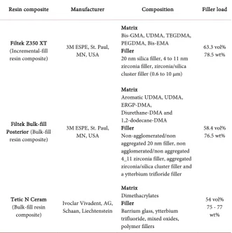
Table 1. Resin composites used in this study.
Resin composite Manufacturer Composition Filler load
Filtek Z350 XT (Incremental-fill resin composite)
3M ESPE, St. Paul, MN, USA
Matrix
Bis-GMA, UDMA, TEGDMA, PEGDMA, Bis-EMA Filler
20 nm silica filler, 4 to 11 nm zirconia filler, zirconia/silica cluster filler (0.6 to 10 μm)
63.3 vol% 78.5 wt%
Filtek Bulk-fill Posterior (Bulk-fill
resin composite)
3M ESPE, St. Paul, MN, USA
Matrix
Aromatic UDMA, UDMA, ERGP-DMA,
Diurethane-DMA and 1,2-dodecane-DMA Filler
Non-agglomerated/non aggregated 20 nm filler, non agglomerated/non aggregated 4_11 zirconia filler, aggregated zirconia/silica cluster filler and a ytterbium trifloride filler
58.4 vol% 76.5 wt%
Tetic N Ceram (Bulk-fill resin composite)
Ivoclar Vivadent, AG, Schaan, Liechtenstein
Matrix Dimethacrylates Filler
Barrium glass, ytterbium trifluoride, mixed oxides, polymer fillers
54 vol% 75 - 77 wt%
Table 2. Polishing systems used in this study.
Polishing
systems Composition Manufacturer
Sof-Lex Pop-On Discs Multi-step
Medium aluminum oxide disc (40 µm) Fine aluminum oxide disc (24 µm) Ultra-fine aluminum oxide disc (8 µm)
3M Dental products, St Paul, MN, USA
PoGo
One step Diamond coated micro-polisher Dentsuply/Caulk, Milford DE, USA
Mylar
Matrix only polyethylene terephthalate matrix SS White, Philadelphia, PA, USA
[image:4.595.209.540.459.587.2]DOI: 10.4236/aim.2019.91007 91 Advances in Microbiology pressed. The light intensity was measured at 800 mW/cm2. Additional 20
seconds curing on both sides of the specimens was done after removing the stripes and glasses. The resulting specimen’s extraneous flanges were removed. All specimens were stored in distilled water at 37˚C for 24 h in the incubator un-til usage [27].
2.2.2. Surface Roughness
To evaluate surface roughness, the tested materials are categorized into the fol-lowing groups; Group 1: These specimens were kept without F/P after removal of Mylar strip to act as control group (30 specimens; ten of each restorative ma-terial). After that, the outermost surface of the remaining 60 specimens were surfaced with the super-fine grit finishing diamond bur (25 μm, No. 837 KREF.314.014, Brasseler) attached to high speed hand piece (W&H, RC-90RM, Austria) for 30 seconds at 200,000 rpm. Group 2: Thirty specimens (ten of each restorative material the specimens) were polished with flat broad surface of the Pogo diamond micro polisher disc for 40 seconds, one-step system according to manufacturer’s instructions. Group 3: Thirty specimens (ten of each restorative material, the specimens) were polished with three step Sof-Lex aluminum oxide disc system according to manufacturer’s instructions. After each polishing step, all the specimens were thoroughly rinsed with water for 10 seconds to remove debris and air-dried for 5 seconds. After completing polishing procedures, spe-cimens were rinsed, cleaned in an ultrasonic cleaner for 3 min, and air dried [28].
2.2.3. Surface Roughness Measurement
All the 90 specimens were assessed for surface roughness by using FEI Quanta 200 FEG ESEM (FEI Co., Hillsboro, OR, USA) combined with image analysis to provide both qualitative and quantitative assessments of surface roughness [28].
2.2.4. Bacterial Adhesion Assay
Samples used for testing surface roughness were used for assessing bacterial ad-hesion with the same grouping. Sterilization of each specimen after packing in dry plastic bag in an autoclave at 121˚C before tested with bacteria. Standard strain of S. mutans ATCC 25175 was used for the in vitro adhesion assay. The standard strain was cultured on blood agar and incubated at 37˚C for 24 h. The colony count was adjusted to 1 × 10⁶ CFU/mL from 0.5 McFarland (1.5 × 10⁸
CFU/mL) equivalence turbidity standard (Thermo Scientific™ Remel, Waltham, MA, USA). In sterile 12-Well Corning microplates (Corning, NY, USA), 2 ml of Muller Hinton (MH) broth culture (1 × 10⁶ CFU/mL) of S. mutans ATCC 25175
DOI: 10.4236/aim.2019.91007 92 Advances in Microbiology NaCl) to remove non-adhering cells. Each disk material was then transferred to a sterile tube containing 1 ml of saline solution and vortexed for 5 min to ensure detachment of bacteria adherent to the discs surfaces. After vortexing, the cell suspensions were tenfold serially diluted in sterile saline and aliquots (10 µl) were surface cultured on blood agar plates, followed by incubation at 37˚C for 24 h for determination of viable cell count as colony forming units per milliliter (CFU/mL) [29].
2.2.5. Scanning Electron Microscopy (SEM)
The samples of disk materials were washed with phosphate buffered saline (PBS) and fixed in solution of 4% v/v paraformaldehyde with 1% glutaraldehyde in PBS for 1 h and rinsed with PBS three times for 2 min each. Finally, samples were washed with deionized water thrice for 2 min each and dehydrated through an ethanol series (50%, 70%, 80%, 95%, and 100%) for 15 min each, desiccated, sputter-coated, and visualized by a SEM (JSM-5310LV JEOL, Tokyo, Japan). Photographs of representative areas of the polished surfaces were captured at 3000× magnifications [30].
2.3. Statistical Analysis
The results were statistically analyzed by IBM SPSS Statistics version 15.0 (SPSS Inc., Chicago, IL, USA). Data were presented as means ± standard deviations (±SD) for each group. Analyses of data variables were performed using ANOVA followed by Tukey’s high significant difference (HSD) test at p-value < 0.05. Pearson correlation coefficient (PCC), at two-tailed, was used to evaluate the potential association between surface roughness and bacterial adhesion to the tested surfaces.
3. Results
3.1. Surface Roughness
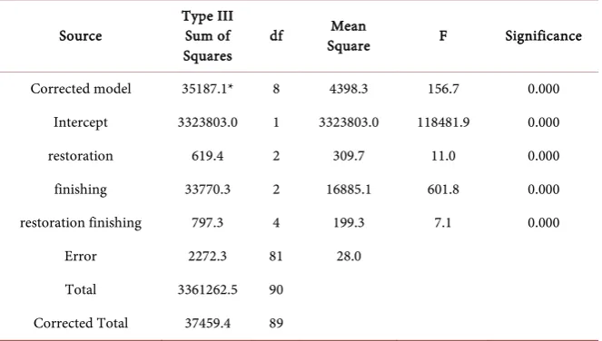
Two-way ANOVA showed that both the restorative materials and the finish-ing/polishing systems have significant effects (p < 0.05) on the surface roughness (Table 3).
The results (Table 4) revealed that the polishing systems significantly (p < 0.05) influenced the surface roughness of different restorative materials. In this context, the Mylar strip showed the smoothest surface followed by PoGo, and the roughest surface was recorded for Sof-lex. On the contrary, different resin composites (FiltekZ350 XT, FiltekZ350 XT, and Tetric N Ceram) had no statis-tically significant (p > 0.05) influence on the surface roughness and this can be attributed to the existence of polishing systems.
DOI: 10.4236/aim.2019.91007 93 Advances in Microbiology
Table 3. Two way ANOVA test results of surface roughness means (nm) among the
tested restorative materials and different finishing/polishing systems.
Source Type III Sum of
Squares df
Mean
Square F Significance
Corrected model 35187.1* 8 4398.3 156.7 0.000
Intercept 3323803.0 1 3323803.0 118481.9 0.000
restoration 619.4 2 309.7 11.0 0.000
finishing 33770.3 2 16885.1 601.8 0.000
restoration finishing 797.3 4 199.3 7.1 0.000
Error 2272.3 81 28.0
Total 3361262.5 90
Corrected Total 37459.4 89
[image:7.595.207.539.361.459.2]*R2 = 0.939 (Adjusted R2 = 0.933).
Table 4. Post hoc Tukey’s test results of surface roughness of the tested restorative
mate-rials with different finishing/polishing systems.
Polishing system Surface roughness of restorative material (nm)
Filtek Z350 XT Filtek Bulk Fill Tetric N Ceram
Mylar strip 161.07 ± 6.82a 162.08 ± 6.82a 164.19 ± 6.82a
Pogo one step 188.33 ± 6.03b 190.41 ± 6.03b 191.42 ± 6.03b
Sof-lex
multi-step 215.87 ± 3.876c 217.89 ± 3.87c 217.88 ± 3.87c
Each value represents mean (±SD) and values with different superscript letters a, b, c indicate statistically significant difference in surface roughness.
3.2. Bacterial Adhesion
The results of two-way ANOVA test revealed that types of polishing systems significantly (p <0.05) influence the bacterial adhesion (Table 5).
The results demonstrated that adhesion S. mutans to Filtek Z350 XT varied significantly (p < 0.05) between different finishing/polishing systems. The high-est bacterial adhesion was observed with Sof-lex multi-step and the lowhigh-est one was observed with Mylarstrip. On the other hand, different types of restorative materials (FiltekZ350 XT, FiltekZ350 XT, and Tetric N Ceram) showed non-significant (p > 0.05) effect on bacterial adhesion (Table 6).
DOI: 10.4236/aim.2019.91007 94 Advances in Microbiology
Figure 1. Two dimensional photomicrograph of resin composites surfaces polished with different polishing systems: (A)
Fil-tekZ350 XT with Mylar strip, (B) Filtek Bulk-fill with Mylar strip, (C) Tetric N Ceram with Mylar strip, (D) FilFil-tekZ350 XT with Pogo one step, (E) Filtek Bulk-fill with Pogo one step, (F) Tetric N Ceram with Pogo one step, (G) FiltekZ350 XT with Sof-lex multi-step, (H) Filtek Bulk-fill with Sof-lex multi-step, (I) Tetric N Ceram with Sof-lex multi-step.
4. Discussion
DOI: 10.4236/aim.2019.91007 95 Advances in Microbiology
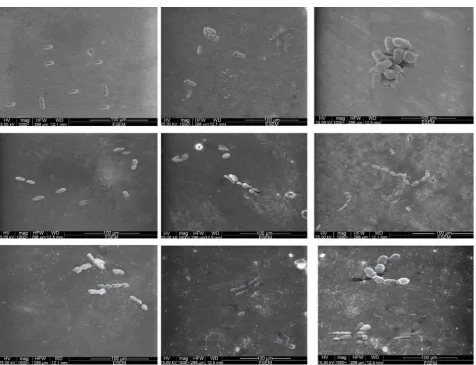
Figure 2. Two dimensional SEM micrographs of Streptococcus mutans ATCC 25,175 adhesion to the surfaces of resin composites
polished with different polishing systems: (A) FiltekZ350 XT with Mylar strip, (B) FiltekZ350 XT with Pogo one step, (C) Fil-tekZ350 XT with Sof-lex multi-step, (D) Filtek Bulk-fill with Mylar strip, (E) Filtek Bulk-fill with Pogo one step, (F) Filtek Bulk-fill with Sof-lex multi-step, (G) Tetric N Ceram with Mylar strip, (H) Tetric N Ceram with Pogo one step, (I) Tetric N Ceram with Sof-lex multi-step.
have effect on the polishing systems with statistical significance.
In the present study, the lowest roughness values were recorded with the My-lar strip samples. The smoothest surface next to MyMy-lar strip was obtained with one-step Pogo polishing system followed by multi-step Sof-lex polishing system. These findings are in agreement with that of Costa et al. [32], who reported that one-step system provides the highest gloss values. Conversely, the outcome of this study contradicts with that of Nasoohi et al. [33], who reported that finish-ing and polishfinish-ing techniques need sequential usage of instrumentation with progressively smaller grained abrasives to finallyatta in the anticipated glossy surface. In this study, Sof-Lex multi-step finishing system revealed rougher sur-faces than PoGo one-step finishing system with all tested composites.
DOI: 10.4236/aim.2019.91007 96 Advances in Microbiology
Table 5. Two way ANOVA test results of bacterial adhesion means among tested
restora-tive materials and different finishing/polishing systems.
Source Type III Sum of Squares df Square Mean F Significance
Corrected model 106012.8* 8 13251.6 255.6 0.000
Intercept 3537870.4 1 3537870.4 68249.8 0.000
restoration 785.0 2 392.5 7.5 0.001
finishing 103731.8 2 51865.9 1000.5 0.000
restoration finishing 1496.0 4 374.0 7.2 0.000
Error 4198.8 81 51.8
Total 3648082.0 90
Corrected total 110211.6 89
*R2 = 0.939 (Adjusted R2 = 0.933).
Table 6. Post hoc Tukey’s test results of Streptococcus mutans ATCC 25,175 adhesion to
the tested restorative materials with different finishing/polishing systems.
Polishing system Bacterial adhesion to restorative material (CFU ×10
3/mL)
Filtek Z350 XT Filtek Bulk Fill Tetric N Ceram
Mylar strip 156.40 ± 6.50a 158.40 ± 6.50a 159.40 ± 6.50a
Pogo one step 199.40 ± 5.31b 201.40 ± 5.31b 202.40 ± 5.31b
Sof-lex multi-step 226.50 ± 4.00c 228.50 ± 4.00c 229.50 ± 4.00c
Each value represents mean (±SD) and values with different superscript letters a, b, c indicate statistically significant difference in bacterial adhesion.
may be due to its highly lustrous feature, which cannot be created with other po-lishing system, and this findings are consistent with some previous studies [34] [35]. Both Filtek Z350XT and Filtek Bulk-fill have nearly the same surface roughness which may be attributed to the similarity of the filler particle size (pure nano-filled) as compared to Tetric N Ceram which has higher na-no-hybrid size as described previously [36] [37].
Cariopathogenic biofilms on tooth surfaces or artificial dental substrata are primarily formed by initial adhesion of specific oral bacteria to such surfaces. Within the complex process of biofilm development, S. mutans is primarily re-sponsible for the initiation of tooth decay as well as for the progression of an es-tablished lesion[38].The selection of S. mutans for adhesion assay in this study was based on the fact that S. mutans is considered as a major etiological agent of dental caries [39] [40]. In this study, the adhered cells were removed for subse-quent quantification after 4 h. This time of exposure was chosen because initial steps of biofilm development in the oral cavity normally occur within 4 h [41] [42].
[image:10.595.207.539.312.391.2]DOI: 10.4236/aim.2019.91007 97 Advances in Microbiology and the surface roughness of these systems which significantly influence the bacterial adhesion to the substrata as describe previously [43] [44] [45]. Bacterial adhesion is governed by non-specific interactions (physico-chemical interac-tions) and specific (ligand-receptor like interacinterac-tions). Non-Specific interactions involve van der Waals, acid-base, and electrostatic interactions. The integration of such interactions plays a fundamental role in the initial bacterial adhesion as well as in biofilm formation [46].
In the current study, Pearson correlation test indicated a strongly positive correlation (PCC = 0.943) between surface roughness and S. mutans adhesion to the tested surfaces. In accordance with our results, some investigators have men-tioned potential correlations between surface roughness and bacterial adhesion
[47] [48]. Similarly, it has been reported that S. aureus adhesion was strongly
correlated to the surface roughness [49]. Furthermore, qualitative and quantita-tive adhesion analyses on different surfaces demonstrated significant aggregation of bacterial cells on untreated surfaces than on electro polished smooth surfaces
[50]. Conversely, Eick et al. [51] disagreed with this relationship and reported
that nocorrelation was observed between surface roughness and the number of colony forming units (CFU) of S. mutans in their study.
5. Conclusion
The current study revealed a strong association between surface roughness and S. mutans adhesion to the tested surfaces. Irrespective of the restorative material, Mylar polishing system presented the smoothest surface and the least bacterial adhesion as compared to Sof-Lex multi-step and Pogo one step polishing sys-tems. Consequently, Mylar polishing system would be more recommended for clinical application.
Conflicts of Interest
The authors declare no conflict of interest.
Funding Statement
This research has not received any specific grants from funding agencies in the public, commercial, or nonprofit sectors.
References
[1] Filoche, S., Wong, L. and Sissons, C. (2010) Oral Biofilms: Emerging Concepts in Microbial Ecology. Journal of Dental Research, 89, 8-18.
https://doi.org/10.1177/0022034509351812
[2] Ono, M., Nikaido, T., Ikeda, M., Imai, S., Hanada, N., Tagami, J., et al. (2007) Sur-face Properties of Resin Composite Materials Relative to Biofilm Formation. Dental Materials Journal,26, 613-622.https://doi.org/10.4012/dmj.26.613
[3] Hojo, K., Nagaoka, S., Ohshima, T. and Maeda, N. (2009) Bacterial Interactions in Dental Biofilm Development. Journal of Dental Research,88, 982-990.
DOI: 10.4236/aim.2019.91007 98 Advances in Microbiology
[4] Teughels, W., Van Assche, N., Sliepen, I. and Quirynen, M. (2006) Effect of Materi-al Characteristics and/or Surface Topography on Biofilm Development. Clinical Oral Implants Research, 17, 68-81.https://doi.org/10.1111/j.1600-0501.2006.01353.x
[5] Subramani, K., Jung, R.E., Molenberg, A. and Hämmerle, C.H. (2009) Biofilm on Dental Implants: A Review of the Literature. International Journal of Oral & Maxil-lofacial Implants, 24, 616-626.
[6] Lutz, F., Krejci, I. and Barbakow, F. (1991) Quality and Durability of Marginal Adaptation in Bonded Composite Restorations. Dental Materials,7, 107-113. https://doi.org/10.1016/0109-5641(91)90055-4
[7] Dauvillier, B.S., Aarnts, M.P. and Feilzer, A.J. (2000) Developments in Shrinkage Control of Adhesive Restoratives. Journal of Esthetic and Restorative Dentistry, 12, 291-299.https://doi.org/10.1111/j.1708-8240.2000.tb00238.x
[8] Versluis, A., Douglas, W., Cross, M. and Sakaguchi, R. (1996) Does an Incremental Filling Technique Reduce Polymerization Shrinkage Stresses? Journal of Dental Re-search,75, 871-878.https://doi.org/10.1177/00220345960750030301
[9] Kwon, Y., Ferracane, J. and Lee, I.-B. (2012) Effect of Layering Methods, Composite Type, and Flowable Liner on the Polymerization Shrinkage Stress of Light Cured Composites. Dental Materials, 2, 801-809.
https://doi.org/10.1016/j.dental.2012.04.028
[10] Tjan, A.H., Bergh, B.H. and Lidner, C. (1992) Effect of Various Incremental Tech-niques on the Marginal Adaptation of Class II Composite Resin Restorations. Jour-nal of Prosthetic Dentistry,67, 62-66.
https://doi.org/10.1016/0022-3913(92)90051-B
[11] Czasch, P. and Ilie, N. (2013) In Vitro Comparison of Mechanical Properties and Degree of Cure of Bulk Fill Composites. Clinical Oral Investigations, 17, 227-235. https://doi.org/10.1007/s00784-012-0702-8
[12] Bollenl, C.M., Lambrechts, P. and Quirynen, M. (1997) Comparison of Surface Roughness of Oral Hard Materials to the Threshold Surface Roughness for Bacterial Plaque Retention: A Review of the Literature. Dental Materials, 13, 258-269. https://doi.org/10.1016/S0109-5641(97)80038-3
[13] Cilli, R., de Mattos, M.C.R., Honorio, H.M., Rios, D., de Araujo, P.A. and Prakki, A. (2009) The Role of Surface Sealants in the Roughness of Composites after a Simu-lated Toothbrushing Test. Journal of Dentistry, 37, 970-977.
https://doi.org/10.1016/j.jdent.2009.08.002
[14] Bürgers, R., Cariaga, T., Müller, R., Rosentritt, M., Reischl, U., Handel, G., et al. (2009) Effects of Aging on Surface Properties and Adhesion of Streptococcus mu-tans on Various Fissure Sealants. Clinical Oral Investigations, 13, 419-426. https://doi.org/10.1007/s00784-009-0256-6
[15] Paravina, R., Roeder, L., Lu, H., Vogel, K. and Powers, J. (2004) Effect of Finishing and Polishing Procedures on Surface Roughness, Gloss and Color of Resin-Based Composites. American Journal of Dentistry, 17, 262-266.
[16] St-Georges, A., Bolla, M., Fortin, D., Muller-Bolla, M., Thompson, J. and Stama-tiades, P. (2005) Surface Finish Produced on Three Resin Composites by New Po-lishing Systems. Operative Dentistry, 30, 593-597.
[17] Yap, A., Ng, J., Yap, S. and Teo, C. (2004) Surface Finish of Resin-Modified and Highly Viscous Glass Ionomer Cements Produced by New One-Step Systems.
Operative Dentistry, 29, 87-91.
DOI: 10.4236/aim.2019.91007 99 Advances in Microbiology
Posterior Resin Composite Restorations: Considerations on Finishing/Polishing. Clinical Procedures. Quintessence International, 35, 359-366.
[19] Li, J., Helmerhorst, E., Leone, C.W., Troxler, R., Yaskell, T., Haffajee, A., et al. (2004) Identification of Early Microbial Colonizers in Human Dental Biofilm.
Journal of Applied Microbiology, 97, 1311-1318. https://doi.org/10.1111/j.1365-2672.2004.02420.x
[20] Stewart, P.S. and Franklin, M.J. (2008) Physiological Heterogeneity in Biofilms.
Nature Reviews Microbiology, 6, 199. https://doi.org/10.1038/nrmicro1838
[21] Pihlstrom, B.L., Michalowicz, B.S. and Johnson, N.W. (2005) Periodontal Diseases.
The Lancet, 366, 1809-1820. https://doi.org/10.1016/S0140-6736(05)67728-8
[22] Bernardo, M., Luis, H., Martin, M.D., Leroux, B.G., Rue, T., Leitão, J., et al. (2007) Survival and Reasons for Failure of Amalgam versus Composite Posterior Restora-tions Placed in a Randomized Clinical Trial. The Journal of the American Dental Association, 138, 775-783. https://doi.org/10.14219/jada.archive.2007.0265
[23] Zijnge, V., van Leeuwen, M.B.M., Degener, J.E., Abbas, F., Thurnheer, T., Gmür, R.,
et al. (2010) Oral Biofilm Architecture on Natural Teeth. PLoS ONE, 5, e9321. https://doi.org/10.1371/journal.pone.0009321
[24] Guggenheim, B., Guggenheim, M., Gmür, R., Giertsen, E. and Thurnheer, T. (2004) Application of the Zürich Biofilm Model to Problems of Cariology. Caries Research, 38, 212-222. https://doi.org/10.1159/000077757
[25] Katsikogianni, M. and Missirlis, Y. (2004) Concise Review of Mechanisms of Bac-terial Adhesion to BiomaBac-terials and of Techniques Used in Estimating Bacte-ria-Material Interactions. European Cells & Materials, 8, 37-57.
https://doi.org/10.22203/eCM.v008a05
[26] Sungurtekin-Ekci, E., Ozdemir-Ozenen, D., Duman, S., Acuner, I.C. and Sandalli, N. (2015) Antibacterial Surface Properties of Various Fluoride-Releasing Restora-tive Materials in Vitro. Journal of Applied Biomaterials & Functional Materials, 13, 169-173. https://doi.org/10.5301/jabfm.5000212
[27] Moshaverinia, A., Roohpour, N., Chee, W.W. and Schricker, S.R. (2011) A Review of Powder Modifications in Conventional Glass-Ionomer Dental Cements. Journal of Materials Chemistry, 21, 1319-1328. https://doi.org/10.1039/C0JM02309D
[28] Hafez, R., Ahmed, D., Yousry, M., El-Badrawy, W. and El-Mowafy, O. (2010) Effect of In-Office Bleaching on Color and Surface Roughness of Composite Restoratives.
European Journal of Dentistry, 4, 118.
[29] Vyavahare, N.G.S., Raghavendra, S.S. and Kazi, M.M. (2014) Effect of Finishing and Polishing Procedures on Biofilm Adhesion to Composite Surfaces: An ex Vivo
Study. JDAS, 3, 70-73.
[30] Kim, D.H. and Kwon, T.-Y. (2017) In Vitro Study of Streptococcus Mutans Adhe-sion on Composite Resin Coated with Three Surface Sealants. Restorative Dentistry & Endodontics, 42, 39-47. https://doi.org/10.5395/rde.2017.42.1.39
[31] Aytac, F., Karaarslan, E.S., Agaccioglu, M., Tastan, E., Buldur, M. and Kuyucu, E. (2016) Effects of Novel Finishing and Polishing Systems on Surface Roughness and Morphology of Nanocomposites. Journal of Esthetic and Restorative Dentistry, 28, 247-261. https://doi.org/10.1111/jerd.12215
DOI: 10.4236/aim.2019.91007 100 Advances in Microbiology
[33] Nasoohi, N., Hoorizad, M. and Tabatabaei, S.F. (2017) Effects of Wet and Dry Fi-nishing and Polishing on Surface Roughness and Microhardness of Composite Re-sins. Journal of Dentistry, 14, 69.
[34] Zimmerli, B., Lussi, A. and Flury, S. (2011) Operator Variability Using Different Polishing Methods and Surface Geometry of a Nanohybrid Composite. Operative Dentistry, 36, 52-59. https://doi.org/10.2341/10-096-LR1
[35] Turssi, C.P., Ferracane, J.L. and Serra, M.C. (2005) Abrasive Wear of Resin Compo-sites as Related to Finishing and Polishing Procedures. Dental Materials, 21, 641-648. https://doi.org/10.1016/j.dental.2004.10.011
[36] Ferracane, J. (1994) Elution of Leachable Components from Composites. Journal of Oral Rehabilitation, 21, 441-452.
https://doi.org/10.1111/j.1365-2842.1994.tb01158.x
[37] Jung, M., Eichelberger, K. and Klimek, J. (2007) Surface Geometry of Four Nanofil-ler and One Hybrid Composite after One-Step and Multiple-Step Polishing. Opera-tive Dentistry, 32, 347-355. https://doi.org/10.2341/06-101
[38] Brambilla, E., Cagetti, M.G., Gagliani, M., Fadini, L., García-Godoy, F. and Stroh-menger, L. (2005) Influence of Different Adhesive Restorative Materials on Mutans Streptococci Colonization. American Journal of Dentistry, 18, 173.
[39] Kolenbrander, P.E., Palmer, R.J., Rickard, A.H., Jakubovics, N.S., Chalmers, N.I. and Diaz, P.I. (2000) Bacterial Interactions and Successions during Plaque Devel-opment. Periodontology, 42, 47-79.
https://doi.org/10.1111/j.1600-0757.2006.00187.x
[40] Ozel, G.S., Guneser, M.B., Inan, O. and Eldeniz, A.U. (2017) Evaluation of C. albi-cans and S. mutans Adherence on Different Provisional Crown Materials. The Journal of Advanced Prosthodontics, 9, 335-340.
https://doi.org/10.4047/jap.2017.9.5.335
[41] Poggio, C., Arciola, C.R., Rosti, F., Scribante, A., Saino, E. and Visai, L. (2009) Ad-hesion of Streptococcus mutans to Different Restorative Materials. The Internation-al JournInternation-al of ArtificiInternation-al Organs, 32, 671-677.
https://doi.org/10.1177/039139880903200917
[42] Montanaro, L., Campoccia, D., Rizzi, S., Donati, M.E., Breschi, L., Prati, C., et al. (2004) Evaluation of Bacterial Adhesion of Streptococcus mutans on Dental Restor-ative Materials. Biomaterials, 25, 4457-4463.
https://doi.org/10.1016/j.biomaterials.2003.11.031
[43] Kawai, K., Urano, M. and Ebisu, S. (2000) Effect of Surface Roughness of Porcelain on Adhesion of Bacteria and Their Synthesizing Glucans. Journal of Prosthetic Dentistry, 83, 664-667. https://doi.org/10.1067/mpr.2000.107442
[44] Saku, S., Kotake, H., Scougall-Vilchis, R.J., Ohashi, S., Hotta, M., Horiuchi, S., et al. (2010) Antibacterial Activity of Composite Resin with Glass-Ionomer Filler Par-ticles. Dental Materials Journal, 29, 193-198. https://doi.org/10.4012/dmj.2009-050
[45] Meier, R., Hauser-Gerspach, I., Lüthy, H. and Meyer, J. (2008) Adhesion of Oral Streptococci to All-Ceramics Dental Restorative Materials in Vitro. Journal of Ma-terials Science: Materials in Medicine, 19, 3249.
https://doi.org/10.1007/s10856-008-3457-7
[46] Hsu, L.C., Fang, J., Borca-Tasciuc, D.A., Worobo, R.W. and Moraru, C.I. (2013) Ef-fect of Micro- and Nanoscale Topography on the Adhesion of Bacterial Cells to Solid Surfaces. Applied and Environmental Microbiology, 79, 2703-2712.
https://doi.org/10.1128/AEM.03436-12
DOI: 10.4236/aim.2019.91007 101 Advances in Microbiology sanguinis Adhesion on Titanium Rough Surfaces: Effect of Shot-Blasting Particles.
Journal of Materials Science: Materials in Medicine, 22, 1913-1922. https://doi.org/10.1007/s10856-011-4366-8
[48] Martínez-Gomis, J., Bizar, J., Anglada, J.M., Samsó, J. and Peraire, M. (2003) Com-parative Evaluation of Four Finishing Systems on One Ceramic Surface. Interna-tional Journal of Prosthodontics, 16, 74-77.
[49] Harris, L.G., Meredith, D.O., Eschbach, L. and Richards, R.G. (2007) Staphylococ-cus aureus Adhesion to Standard Micro-Rough and Electropolished Implant Mate-rials. Journal of Materials Science: Materials in Medicine, 18, 1151-1156.
https://doi.org/10.1007/s10856-007-0143-0
[50] Wu, S., Altenried, S., Zogg, A., Zuber, F., Maniura-Weber, K. and Ren, Q. (2018) Role of the Surface Nanoscale Roughness of Stainless Steel on Bacterial Adhesion and Microcolony Formation. ACS Omega, 3, 6456-6464.
https://doi.org/10.1021/acsomega.8b00769
[51] Eick, S., Glockmann, E., Brandl, B. and Pfister, W. (2004) Adherence of Strepto-coccus mutans to Various Restorative Materials in a Continuous Flow System.