A COMPARISON BETWEEN CLEARING TECHNIQUE AND CONE BEAM COMPUTED TOMOGRAPHY
FOR DETECTION OF ACCESSORY CANALS IN PRIMARY MOLARS: AN
*Dr. Wakpanjar Mayur, M., Dr. Katge Farhin,
Department of Pedodontics and Preventive Dentistry,
Navi Mumbai 400706,
ARTICLE INFO ABSTRACT
Aim:
molars for detection of accessory canals. Materials and Methods:
mandibular molars were collected. The study compared number of accessory canals using Cone Beam Computed Tomography and clearing technique in primary molars. The access
observed in furcation area; coronal one Results:
CBCT technique, the difference was Conclusion:
this method is that it cannot be used in
clinically feasible can be considered over the clearing technique for detection of accessory canals.
Copyright©2017, Dr. Wakpanjar Mayur et al. This
unrestricted use, distribution, and reproduction in any medium, provided the original work is properly cited.
INTRODUCTION
Successful endodontic therapy is dependent on the cleaning, shaping and obturation of the entire root canal system (Ingle and Bakland, 2002; Walton and Torabinejad, 1996). The presence of accessory canals and the ability to cleanse and seal these canals can have an impact on prognosis (Zolty, 2001). Accessory canal is any branch of the main pulp canal or chamber that communicates with the external surface of the root (American Assocation of Endodontists, 2003).
canals extend from the pulp to the periodontium.
canals result due to a localized failure in the formation of Hertwig’s epithelial root sheath during the embryonic stages of tooth formation (Poornima and Subba Reddy, 2008).
to a failure in odontoblastic differentiation and dentine formation and eventually to the formation of the accessory
canal. This was demonstrated by Hess et al as early as 1925 by
injecting India ink into the pulp chamber (Moabita and Defabians, 1992). A variety of techniques such as radiographic
studies, microscopic studies, histological sectioning,
decalcification have been used to study the anatomy of the root canal system (Poornima and Subba Reddy, 2008; Urbas
1996; Koenig, 1964)Bacterial toxins or products of the pulp
*Corresponding author: Dr. Wakpanjar Mayur,
Department of Pedodontics and Preventive Dentistry, Terna Dental College, Sector 22, plot No. 12, Nerul (W) Navi Mumbai 400706, Maharashtra, India.
ISSN: 0975-833X
Article History:
Received 21st April, 2017 Received in revised form 11th May, 2017
Accepted 07th June, 2017
Published online 26th July, 2017
Citation: Dr. Wakpanjar Mayur, M., Dr. Katge Farhin, Dr. Vamsi Krishna, C. and Dr.
technique and cone beam computed tomography for detection of accessory canals in primary molars: An
Research, 9, (07), 54152-24156.
Key words:
Accessory Canal, Cone Beam Computed Tomography, Clearing Technique.
RESEARCH ARTICLE
A COMPARISON BETWEEN CLEARING TECHNIQUE AND CONE BEAM COMPUTED TOMOGRAPHY
FOR DETECTION OF ACCESSORY CANALS IN PRIMARY MOLARS: AN
Dr. Katge Farhin, Dr. Vamsi Krishna, C. and Dr. Shivasharan Pooja
Department of Pedodontics and Preventive Dentistry, Terna Dental College, Sector 22, plot No. 12, Nerul (W)
Navi Mumbai 400706, Maharashtra, India
ABSTRACT
Comparing Cone Beam Computed Tomography and clearing technique in human primary molars for detection of accessory canals.
Materials and Methods: 100 extracted human primary molars constituting of maxillary and mandibular molars were collected. The study compared number of accessory canals using Cone Beam Computed Tomography and clearing technique in primary molars. The access
observed in furcation area; coronal one-third, middle one-third and apical one
Results: Although, the clearing technique showed more number of accessory canals as compared to CBCT technique, the difference was statistical significant insignificant.
Conclusion: The clearing technique is nondestructive and more accurate but the main disadvantage of this method is that it cannot be used in vivo. Hence Cone Beam Computed Tomography which is clinically feasible can be considered over the clearing technique for detection of accessory canals.
This is an open access article distributed under the Creative Commons Att use, distribution, and reproduction in any medium, provided the original work is properly cited.
Successful endodontic therapy is dependent on the cleaning, shaping and obturation of the entire root canal system (Ingle and Bakland, 2002; Walton and Torabinejad, 1996). The canals and the ability to cleanse and seal these canals can have an impact on prognosis (Zolty, 2001). Accessory canal is any branch of the main pulp canal or chamber that communicates with the external surface of the
sts, 2003).Accessory
canals extend from the pulp to the periodontium. Accessory canals result due to a localized failure in the formation of Hertwig’s epithelial root sheath during the embryonic stages of
tooth formation (Poornima and Subba Reddy, 2008).This leads
to a failure in odontoblastic differentiation and dentine formation and eventually to the formation of the accessory as early as 1925 by injecting India ink into the pulp chamber (Moabita and A variety of techniques such as radiographic
studies, microscopic studies, histological sectioning,
decalcification have been used to study the anatomy of the root
canal system (Poornima and Subba Reddy, 2008; Urbas et al.,
erial toxins or products of the pulp
Department of Pedodontics and Preventive Dentistry, Terna Dental College, Navi Mumbai 400706, Maharashtra, India.
tissue decomposition diffuses through accessory canals to
periodontal tissues(Moabita and Defabians, 1992; Woo and
Miller, 1981). However, the documentation of accessory canals in the primary molars is scanty. Hence, an
planned to know the prevalence of acc
molars and to compare the efficiency of digital radiography, decalcification, and histological sectioning in detection.
MATERIALS AND METHODS
100 extracted human primary molars constituting of maxillary and mandibular molars were
divided into 4 groups each consisting of 25 teeth:
Group I – Maxillary first molars (n=25) Group II – Mandibular first molars (n=25) Group III – Maxillary second molars (n=25) Group IV – Mandibular second molars (n=25)
The extracted teeth were collected from different dental colleges and private clinics. As the samples were not extracted for the purpose of this study, the exact data pertaining to age and sex of individual sources was unknown.
conduct the study was sought and obtained from the Institutional Review Board of Ethics. The study was carried out in the Department of Paedodontics and Preventive
International Journal of Current Research Vol. 9, Issue, 07, pp.54152-24156, July, 2017
Dr. Wakpanjar Mayur, M., Dr. Katge Farhin, Dr. Vamsi Krishna, C. and Dr. Shivasharan Pooja, 2017.
technique and cone beam computed tomography for detection of accessory canals in primary molars: An in vitro study
A COMPARISON BETWEEN CLEARING TECHNIQUE AND CONE BEAM COMPUTED TOMOGRAPHY
FOR DETECTION OF ACCESSORY CANALS IN PRIMARY MOLARS: AN
IN VITRO
STUDY
Dr. Shivasharan Pooja
Sector 22, plot No. 12, Nerul (W)
Comparing Cone Beam Computed Tomography and clearing technique in human primary
100 extracted human primary molars constituting of maxillary and mandibular molars were collected. The study compared number of accessory canals using Cone Beam Computed Tomography and clearing technique in primary molars. The accessory canals were
third and apical one-third of root canals. number of accessory canals as compared to statistical significant insignificant.
The clearing technique is nondestructive and more accurate but the main disadvantage of vivo. Hence Cone Beam Computed Tomography which is clinically feasible can be considered over the clearing technique for detection of accessory canals.
Creative Commons Attribution License, which permits
diffuses through accessory canals to (Moabita and Defabians, 1992; Woo and However, the documentation of accessory canals
in the primary molars is scanty. Hence, an in vitro study was
planned to know the prevalence of accessory canals in primary molars and to compare the efficiency of digital radiography, decalcification, and histological sectioning in detection.
MATERIALS AND METHODS
100 extracted human primary molars constituting of maxillary and mandibular molars were collected. The samples were divided into 4 groups each consisting of 25 teeth:
Maxillary first molars (n=25) Mandibular first molars (n=25)
Maxillary second molars (n=25) Mandibular second molars (n=25)
tracted teeth were collected from different dental colleges and private clinics. As the samples were not extracted for the purpose of this study, the exact data pertaining to age and sex of individual sources was unknown. Approval to sought and obtained from the Institutional Review Board of Ethics. The study was carried out in the Department of Paedodontics and Preventive
INTERNATIONAL JOURNAL OF CURRENT RESEARCH
2017. “A comparison between clearing
Dentistry, Terna Dental College, Navi Mumbai. The teeth included in the study were based on these conditions:
Extracted teeth with atleast two intact roots.
At least two-third of the root length needed to be intact.
If there was any evidence of fractured root or the teeth had undergone pulpectomy procedures, then they were excluded from the study. The sample teeth were then washed under tap water in a glass container for 30 minutes followed by immersion in 3% sodium hypochlorite (Mumbai Healthcare Industries, India) for 30 minutes to remove adherent soft tissue. Any remaining adherent soft tissue was physically scraped using a scalpel blade and an ultrasonic scaler was used to remove calculus or stains. Subsequently, the teeth were stored in distilled water with thymol iodide crystals until the
next procedure was performed (Gulabivala et al., 2001). The
aim of the present study was to compare clearing technique and Cone Beam Computed Tomography (CBCT) for detection of accessory canals in primary molars. The accessory canals were observed in furcation area; coronal third, middle one-third and apical one-one-third of root canals.
Cone Beam Computed Tomography (CBCT)
The teeth were mounted in a straight line on modeling wax after determining the various aspects of the tooth, i.e., buccal, lingual, mesial and distal, so as to maintain uniformity in the samples. The mounted teeth were then scanned using Orthophos XG3D Cept CBCT scanner (Sirona, Germany) which provided voxel size of 0.1 mm. The CBCT images were displayed by volume-rendering software (Galileos Viewer). Once the sample data was acquired or data for a sample was
loaded, the software immediately reconstructed the tooth
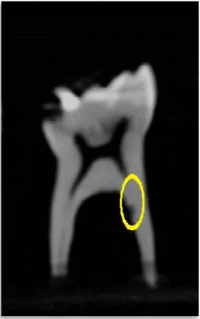
images in sagittal, axial and coronal planes. The lateral canals of each were determined by observing for radiolucent line arising from pulp chamber and main pulp canals (figure 1.a-d).
Clearing Technique
After performing CBCT, the teeth were decalcified by immersing them in 6% hydrochloric acid (Qualigens Fine Chemicals, Mumbai, Maharashtra, India) at room temperature
(Bagherian et al., 2010). The end point of decalcification was
determined by checking the teeth for softening with dental
probe (API® Germany). The hydrochloric acid solution was
changed daily and agitated frequently because
demineralization occurs at the top of the static acid rather than at the bottom. At the end of decalcification, the teeth were again washed under running tap water for 1 hour. The teeth were later kept in varying concentrations of ethanol for dehydration. The sequence of concentrations employed was ascending concentrations of ethanol (Himedia Laboratories, Mumbai, Maharashtra, India) at 60%, 70%, 80%, 90% and 100% (absolute ethanol) consecutively for 5 hours each
(Bagherian et al., 2010). For clearing, the teeth were immersed
into solution of methyl salicylate and absolute ethanol in ratio
of 1:1 for 6 hours (Azar et al., 2012).The teeth were immersed
in methyl salicylate solution (Qualigens Fine Chemicals, Mumbai, Maharashtra, India) to render them transparent. The teeth were then placed on tissue paper for 2 hours so as to
allow them to dry. Drying was important so as to aid the
penetration of ink in the next stage. A plastic disposable endodontic irrigating syringe with a 30 gauge needle was used
to inject the methylene blue dye (Qualigens Fine Chemicals,
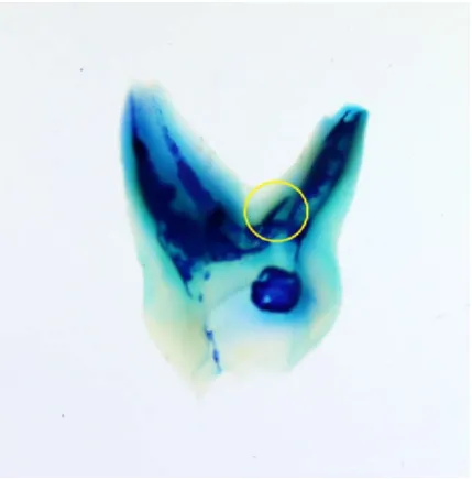
[image:2.595.354.517.276.514.2]Mumbai, Maharashtra, India) into the coronal end of canal orifice. At the same time, a suction tip was placed at the root apex to draw the excess ink away from the root canal. The appearance of ink at the apical foramen indicated the end of the process. The transparent specimens were then examined by the naked eye as well as a magnifying lens (3x magnification) by dipping the teeth in methyl salicylate under special halogen lighting to improve the coefficient of refraction. Photographs of the specimens were taken with a Canon 600-D DSLR camera with lens (18-55mm 0.25m / 0.8ft EFS macro). The accessory canals in both the techniques were recorded by two independent precalibrated examiners for determination of location of canals. The accessory canals were observed in the furcation area (figure 2.a), coronal one-third (figure 2.b), middle one-third (figure 2.c) and apical one-third of root canal (figure 2.d). Statistical analysis was performed using SPSS-21 software. The scores obtained were analyzed using Fisher’s exact probability test and level of significance was p< 0.05.
Figure 1.a furcation area
[image:2.595.362.508.546.778.2]Figure 1.c middle one-third of root canal
[image:3.595.321.546.49.276.2]Figure 1.d apical one-third of root canal
[image:3.595.90.239.56.235.2]Figure 1. Detection of accessory canals by Cone Beam Computed Tomography at
Figure 2.a furcation area
[image:3.595.89.242.259.483.2]Figure 2.b coronal one-third of root canal
Figure 2.c middle one-third of root canal
Figure 2.d apical one-third of root canal
[image:3.595.328.541.314.543.2] [image:3.595.334.536.361.733.2] [image:3.595.57.272.544.762.2]RESULTS
The results of the present study revealed that CBCT technique showed maximum number of accessory canals in maxillary and mandibular second molars (44%). Out of 100 samples, the prevalence of accessory canals in the furcation area of mandibular second molars was 16%. Apical region of maxillary and mandibular second molars had 16% prevalence of accessory canals. In clearing technique, accessory canals showed maximum prevalence in apical one-third (20%) of mandibular second molars (48%). Accessory canals were observed in furcation area; coronal one-third, middle one-third and apical one-third of primary maxillary and mandibular molars. Although, the clearing technique showed more number of accessory canals as compared to CBCT technique, the difference was statistical significant insignificant (Table 1-4).
DISCUSSION
The furcation area of a primary molar tooth, which encompasses the region around the division of the roots, is of special significance in the primary dentition due to its close anatomical relationship with the follicle of the succedaneous premolars. Interradicular periodontal lesions can be initiated and perpetuated by inflamed and/or necrotic pulps. Thus extensive pulp lesions can cause periodontal changes through
the lateral and accessory foramina (Moabita and Defabians,
1992). Various methods to detect accessory canals used were
decalcification (Poornima and Subba Reddy, 2008), canal staining (Poornima and Subba Reddy, 2008), radiographic
techniques, microscopic and histological sectioning (Urbas et
al., 1996; Moabita and Defabians, 1992; Koenig, 1964). In the
present study, the prevalence of accessory canals in furcation area in maxillary molars was 12% and in mandibular molars it was 14%. The overall prevalence in primary molars was 13%.
Similar study conducted by Poornima P et al using
decalcification technique found 25% of accessory canals in
furcation area (Poornima and Subba Reddy, 2008). Vertucci et
al conducted a similar study in permanent first molar using a
dissecting microscope and found that 45% of teeth exhibited
accessory canals in the furcation region (Vertucci et al., 1974).
De Deus studied 1140 transparent teeth and found that 27.4% of teeth demonstrated lateral secondary accessory canals,
which was more when compared to the present study (De Deus 1975).
The other method used in this study was CBCT. The prevalence of accessory canals using CBCT in the present study in maxillary first and second molars was 28% and 44% respectively. According to similar study conducted by
Aggrawal N et al using CBCT, accessory canals were detected
[image:4.595.56.535.276.328.2]in 32% and 24% of maxillary first and second molars respectively. The same study showed accessory canals in 12%
Table 1. Number of accessory canals in primary molars by CBCT and Clearing technique at furcation area of tooth
Groups CBCTtechnique± (n=25) Clearing technique (n=25) p value*
Group I (Maxillary 1st molar)
Group II (Mandibular 1st molar)
Group III (Maxillary 2nd molar)
2 (8) 3 (12) 3 (12)
2 (8) 3 (12) 4 (16)
0.695 0.666 0.50
Group IV (Mandibular 2ND molar) 4 (16) 4 (16) 0.649
*Fisher's exact probability test and level of significance was set at p<0.05; number in parenthesis indicates percentage; ± Cone Beam Computed Tomography
[image:4.595.66.534.377.436.2]technique.
Table 2. Number of accessory canals in primary molars by CBCT and Clearing technique at coronal one-third region of root canal
Groups CBCTtechnique± (n=25) Clearing technique (n=25) p value*
Group I (Maxillary 1st molar)
Group II (Mandibular 1st molar)
Group III (Maxillary 2nd molar)
1 (4) 0 (0) 2 (8)
2 (8) 1 (4) 2 (8)
0.50 0.50 0.695
Group IV (Mandibular 2nd molar) 2 (8) 2 (8) 0.695
*Fisher's exact probability test and level of significance was set at p<0.05; number in parenthesis indicates percentage; ± Cone Beam Computed Tomography
technique.
Table 3. Number of accessory canals in primary molars by CBCT and Clearing technique at middle one-third region of root canal
Groups CBCTtechnique± (n=25) Clearing technique (n=25) p value*
Group I (Maxillary 1st molar)
Group II (Mandibular 1st molar)
Group III (Maxillary 2nd molar)
1 (4) 2 (8) 2 (8)
1 (4) 1 (4) 1 (4)
0.755 0.50 0.50
Group IV (Mandibular 2nd molar) 1 (4) 1 (4) 0.755
*Fisher's exact probability test and level of significance was set at p<0.05; number in parenthesis indicates percentage; ± Cone Beam Computed Tomography
technique.
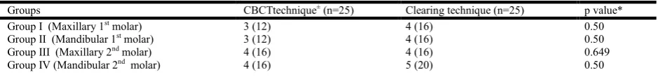
Table 4. Number of accessory canals in primary molars by CBCT and Clearing technique at apical one-third region of root canal
Groups CBCTtechnique± (n=25) Clearing technique (n=25) p value*
Group I (Maxillary 1st molar)
Group II (Mandibular 1st molar)
Group III (Maxillary 2nd molar)
3 (12) 3 (12) 4 (16)
4 (16) 4 (16) 4 (16)
0.50 0.50 0.649
Group IV (Mandibular 2nd molar) 4 (16) 5 (20) 0.50
*Fisher's exact probability test and level of significance was set at p<0.05; number in parenthesis indicates percentage; ± Cone Beam Computed Tomography
[image:4.595.51.543.487.546.2] [image:4.595.65.536.597.657.2]and 28% whereas, the present study showed the same in 32% and 44% of mandibular first and second molars respectively. Although, clearing technique (37%) showed more number of accessory canals overall as compared to CBCT technique (41%), the result was statistically insignificant. The clearing technique is considered the gold standard method for studying root canal anatomy. We used the clearing technique followed by dye injection in this study. Although more accurate, the main disadvantage of this method is that it cannot be used in vivo. A method that has the accuracy of clearing technique followed by dye injection and yet is clinically feasible is
essential in endodontic practice (Weng et al., 2009).
Conclusion
The results obtained while comparing CBCT with clearing technique for the detection of lateral and accessory canals were
statistically insignificant. The clearing technique is
nondestructive and more accurate but the main disadvantage of this method is that it cannot be used in vivo. Hence CBCT which is clinically feasible can be considered over the clearing technique for detection of accessory canals.
REFERENCES
American Association of Endodontists, 2003. Glossary of Endodontic Terms, sixth edn, Chicago IL.
Azar MR, Safi L, Nikaein A. 2012. Comparison of the cleaning capacity of Mtwo and ProTaper rotary systems
and manual instruments in primary teeth. Dent Res J., 9,
146-151.
Bagherian A, Kalhori K.A, Sadeghi M, Mirhosseini F, Parisay I. 2010. An in vitro study of root and canal morphology of
human deciduous molars in an Iranian population. J Oral
Sci., 52, 397-340.
De Deus QD. 1975. Frequency, location and direction of the
lateral, secondary and accessory canals. J Endod., 1,
361-366.
Gulabivala K, Aung TH, Alavi A, Ng YL. 2001. Root and
canal morphology of Burmese mandibular molars. Int
Endod J., 34, 359–370.
Ingle J. and Bakland L. 2002. Endodontics, fifth edn, Hamilton: BC Decker.
Koenig JF, Brilliant JD, Foreman DW. 1964. Preliminary scanning electron microscope investigations of accessory
foramina in the furcation areas of human molar teeth. Oral
Surg Oral Med Oral Pathol., 38, 663-682.
Morabito A. and Defabians P 1992. A SEM investigation of
pulpal periodontal connections in primary teeth. ASDC J
Dent Child 59, 53-56.
Poornima P. and Subba Reddy VV. 2008. Comparison of
digital radiography, decalcification and histologic
sectioning in the detection of accessory canals in furcation
areas of human primary molars. J Indian Soc Pedod
Prevent Dent., 6, 49-52.
Urbas KT, Kielbassa AM, Hellwig E 1996. Microscopic studies of accessory canals in primary molar furcations.
ASDC J Dent Child., 64, 118-122.
Vertucci FJ, Williams RG, Brooklys NY, Middletown NJ 1974. Furcation canals in the human mandibular first
molars. Oral Surg Oral Med Oral Pathol., 38, 306-314.
Walton R. and Torabinejad M 1996. Principles and Practice of Endodontics, second edn, Philadelphia WB: Saunders Co publication.
Weng XL, Yu SB, Zhao SL et al. 2009. Root canal
morphology of permanent maxillary teeth in the Han nationality in Chinese Guanzhong area: a new modified
root canal staining technique. J Endod., 35, 651–656.
Woo RK. and Miller J. 1981. Accessory canals in deciduous
molars. J Int Ass Dent Child 12, 51-56.
Zolty G. 2001. The prevalence and significance of sealing
accessory and lateral canals: a literature review. SADJ, 56,
416–424.
![Mechanical Properties of Concrete [M60] With Electric Arc Furnace Slag as Aggregate and Partially Replacing Cement with Nano Silica](data:image/gif;base64,R0lGODlhAQABAIAAAP///wAAACH5BAEAAAAALAAAAAABAAEAAAICRAEAOw==)