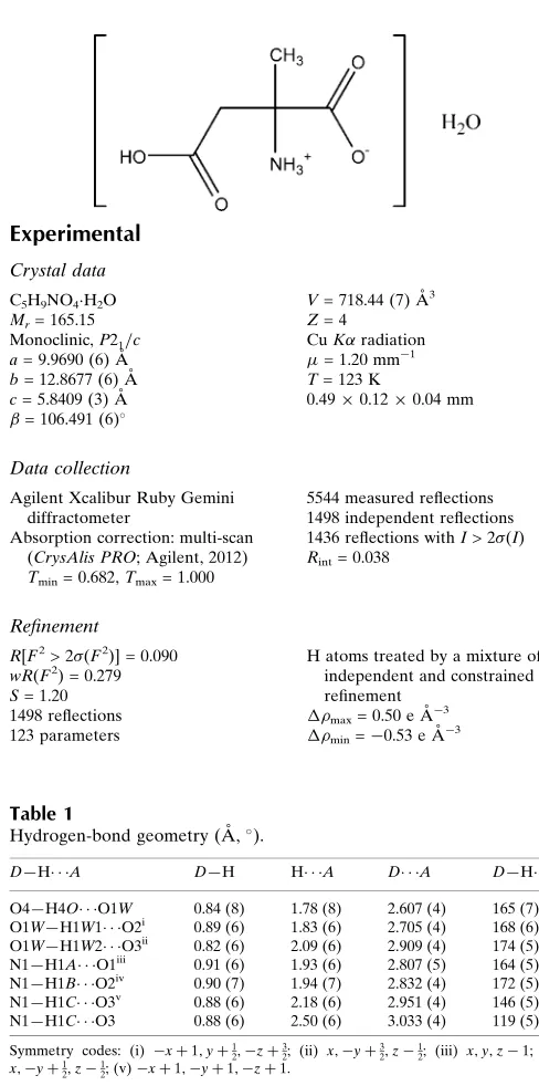
2-Methylaspartic acid monohydrate
Greg Brewer,aAaron S. Burton,bJason P. Dworkincand Ray J. Butcherd*
a
Department of Chemistry, Catholic University of America, Washington, DC 20064, USA,bNASA Goddard Space Flight Center, Greenbelt, MD 20771, USA,cSolar System Exploration Division, NASA Goddard Space Flight Center, Greenbelt, MD 20771, USA, anddDepartment of Chemistry, Howard University, 525 College Street NW, Washington, DC 20059, USA
Correspondence e-mail: [email protected]
Received 18 November 2013; accepted 26 November 2013
Key indicators: single-crystal X-ray study;T= 123 K; mean(C–C) = 0.006 A˚;
Rfactor = 0.090;wRfactor = 0.279; data-to-parameter ratio = 12.2.
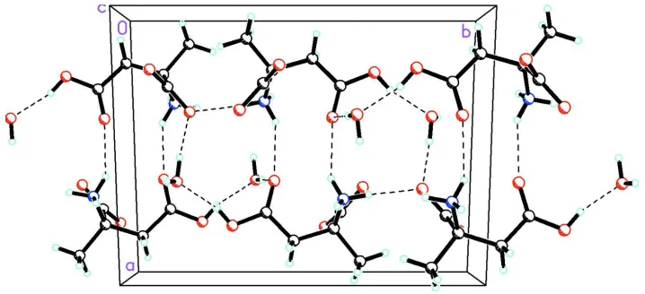
The title compound, C5H9NO4H2O, is an isomer of the -amino acid glutamic acid that crystallizes from water in its zwitterionic form as a monohydrate. It is not one of the 20 proteinogenic -amino acids that are used in living systems and differs from the natural amino acids in that it has an -methyl group rather than an-H atom. In the crystal, an O— H O hydrogen bond is present between the acid and water molecules while extensive N—H O and O—H O hydrogen bonds link the components into a three-dimensional array.
Related literature
For the eighty amino acids that have been detected in meteorites or comets, see: Pizzarello et al.(2006); Glavin & Dworkin, (2009); Burtonet al. (2012). For the role that crys-tallization plays in chiral separation, see: Blackmond & Klussmann (2007); Blackmondet al.(2008). For the role of the H atom on the-C atom in enhancing the rate of racemiza-tion, see: Yamada et al. (1983). For the mechanism of race-mization of amino acids lacking an-H atom, see: Pizzarello & Groy (2011). For the role that crystallization can play in the enrichment of l isovaline and its structure, see: Glavin & Dworkin (2009); Butcher et al. (2013). For normal bond lengths and angles, see: Orpen (1993). For the number of -methyl amino acids that have been observed with l -enanti-omeric excesses up to 20% that are not believed to be the result of contamination, see: Pizzarello & Cronin (2000); Glavin & Dworkin (2009); Glavinet al.(2011, 2012); Burtonet al.(2013).
Experimental
Crystal data
C5H9NO4H2O
Mr= 165.15
Monoclinic,P21=c
a= 9.9690 (6) A˚ b= 12.8677 (6) A˚ c= 5.8409 (3) A˚
= 106.491 (6)
V= 718.44 (7) A˚3
Z= 4
CuKradiation
= 1.20 mm1 T= 123 K
0.490.120.04 mm
Data collection
Agilent Xcalibur Ruby Gemini diffractometer
Absorption correction: multi-scan (CrysAlis PRO; Agilent, 2012) Tmin= 0.682,Tmax= 1.000
5544 measured reflections 1498 independent reflections 1436 reflections withI> 2(I) Rint= 0.038
Refinement
R[F2> 2(F2)] = 0.090 wR(F2) = 0.279
S= 1.20 1498 reflections 123 parameters
H atoms treated by a mixture of independent and constrained refinement
max= 0.50 e A˚
3
min=0.53 e A˚
[image:1.610.310.554.71.562.2]3
Table 1
Hydrogen-bond geometry (A˚ ,).
D—H A D—H H A D A D—H A
O4—H4O O1W 0.84 (8) 1.78 (8) 2.607 (4) 165 (7)
O1W—H1W1 O2i
0.89 (6) 1.83 (6) 2.705 (4) 168 (6) O1W—H1W2 O3ii 0.82 (6) 2.09 (6) 2.909 (4) 174 (5) N1—H1A O1iii
0.91 (6) 1.93 (6) 2.807 (5) 164 (5) N1—H1B O2iv
0.90 (7) 1.94 (7) 2.832 (4) 172 (5)
N1—H1C O3v 0.88 (6) 2.18 (6) 2.951 (4) 146 (5)
N1—H1C O3 0.88 (6) 2.50 (6) 3.033 (4) 119 (5)
Symmetry codes: (i)xþ1;yþ1 2;zþ
3
2; (ii) x;yþ 3 2;z
1
2; (iii) x;y;z1; (iv)
x;yþ1 2;z
1
2; (v)xþ1;yþ1;zþ1.
Data collection: CrysAlis PRO(Agilent, 2012); cell refinement:
CrysAlis PRO; data reduction: CrysAlis PRO; program(s) used to solve structure: SHELXS97(Sheldrick, 2008); program(s) used to refine structure:SHELXL97(Sheldrick, 2008); molecular graphics:
SHELXTL(Sheldrick, 2008); software used to prepare material for publication:SHELXTL.
RJB wishes to acknowledge the NSF–MRI program (grant CHE-0619278) for funds to purchase the diffractometer. GB wishes to acknowledge support of this work from NASA (NNX10AK71A).
Supplementary data and figures for this paper are available from the IUCr electronic archives (Reference: HG5362).
organic compounds
o1856
Breweret al. doi:10.1107/S1600536813032170 Acta Cryst.(2013). E69, o1856–o1857Acta Crystallographica Section E Structure Reports Online
References
Agilent (2012). CrysAlis PRO. Agilent Technologies UK Ltd, Yarnton, England.
Blackmond, D. G. & Klussmann, M. (2007).Chem. Commun.pp. 3990–3996. Blackmond, D., Viedma, C., Ortiz, J., Torres, T. & Izuma, T. (2008).J. Am.
Chem. Soc.130, 15274–15275.
Burton, A. S., Elsila, J. E., Hein, J. E., Glavin, D. P. & Dworkin, J. P. (2013). Meteorites Planet. Sci.48, 390–402.
Burton, A. S., Stern, J. C., Elsila, J. E., Dworkin, J. P. & Galvin, D. P. (2012). Chem. Soc. Rev.41, 5459–5472.
Butcher, R. J., Brewer, G., Burton, A. S. & Dworkin, J. P. (2013).Acta Cryst. E69, o1829–o1830.
Glavin, D. P., Callahan, M. P., Dworkin, J. P. & Elsila, J. E. (2011).Meteorites Planet. Sci.45, 1948–1972.
Glavin, D. P. & Dworkin, J. P. (2009).Proc. Natl Acad. Sci.106, 5487–5492. Glavin, D. P., Elsila, J. E., Burton, A. S., Callahan, M. P., Dworkin, J. P., Hilts,
R. W. & Herd, C. D. H. (2012).Meteorites Planet. Sci.47, 1347–1364. Orpen, G. A. (1993).Chem. Soc. Rev.22, 191–197.
Pizzarello, S., Cooper, G. W. & Flynn, G. J. (2006).The Nature and Distribution of the Organic Material in Carbonaceous Chondrites and Interplanetary Dust Particles in Meteorites and the Early Solar System II, edited by D. Lauretta, L. A. Leshin & H. Y. McSween Jr. University of Arizona Press, USA. Pizzarello, S. & Cronin, J. R. (2000).Geochim. Cosmochim. Acta,64, 329–338. Pizzarello, S. & Groy, T. L. (2011).Geochim. Cosmochim. Acta,75, 645–656. Sheldrick, G. M. (2008).Acta Cryst.A64, 112–122.
supporting information
sup-1
Acta Cryst. (2013). E69, o1856–o1857supporting information
Acta Cryst. (2013). E69, o1856–o1857 [doi:10.1107/S1600536813032170]
2-Methylaspartic acid monohydrate
Greg Brewer, Aaron S. Burton, Jason P. Dworkin and Ray J. Butcher
S1. Comment
The α-amino acids are essential for life as they are the building blocks of all proteins and enzymes. Nature uses almost
exclusively the L form of the nineteen common chiral amino acids. However, there are over eighty amino acids that have
been identified in meteorites (Pizzarello et al., 2006; Burton et al., 2012). One of these extraterrestrial non-proteinogenic
amino acids is 2-methylaspartic acid. The majority of meteoritic amino acids show little or no enrichment of one
enantiomer over the other. However, a number of alpha methyl amino acids have been observed with L-enantiomeric
excesses up to 20% that are not believed to be the result of contamination (Pizzarello & Cronin, 2000; Glavin &
Dworkin, 2009; Glavin et al., 2011; Glavin et al., 2012; Burton et al., 2013). An intriguing question is the process that
leads to the separation and enrichment of the L enantiomer over the D. There are several possible explanations for this
including the role that crystallization plays (Blackmond et al., 2007; Glavin et al., 2012). Only two of the twenty amino
acids used biologically crystallize in a chiral space group from a racemic solution, which allows for spontaneous
separation of enantiomers, at the level of the crystal (Blackmond et al., (2008).
Racemic 2-methylaspartic crystallizes from water in an achiral space group acid, forming a racemic compound, in
which there are equal numbers of D and L enantiomers in the unit cell. Thus, crystallization under these conditions would
not provide a mechanism for separation of enantiomers at the level of the crystal. Another important aspect in the
prebiotic chemistry of the amino acids is the role of racemization. All of the nineteen naturally occurring chiral amino
acids have a hydrogen atom on the alpha carbon atom, which enhances the rate of racemization (Yamada et al., 1983).
However, little is known about the mechanism of racemization of amino acids lacking an alpha hydrogen atom
(Pizzarello et al., 2011). We recently reported the structure of another non-proteinogenic amino acid, isovaline, which
crystallized as a racemic conglomerate from water in contrast to the present example which crystallizes in a
centro-symmetric space group and is thus a racemate as indicated above (Butcher et al., 2013). Resolved 2-methylaspartic acid
and the structure given here can be used as a starting point in mechanistic studies of racemization mechanisms of amino
acids lacking an alpha hydrogen atom.
In the structure of the title compound the amino acid is in the usual zwitterionic form involving the α carboxylate group
and all the the bond lengths and angles are in the normal range for such compounds (Orpen, 1993). There is extensive N
—H···O and O—H···O hydrogen bonding linking the zwitterions into a 3-D array.
S2. Experimental
2-Methylaspartic acid was purchased from Nagase and Co. Ltd. Crystals of the title compound were grown from slow
S3. Refinement
H atoms were placed in geometrically idealized positions and constrained to ride on their parent atoms with a C—H
distances of 0.98 and 0.99 Å. The protons on the N and O were refined isotropically with the O—H distances for the
[image:4.610.129.480.140.331.2]water H's constrained to be 0.82 Å and the H—O—H angle close to 104.5°.
Figure 1
Diagram of the title compound showing atom labeling. Atomic displacement parameters are at the 30% probability level.
Hydrogen bonds are shown as dashed lines.
Figure 2
Packing diagram of the title compound viewed along the c axis showing the extensive N—H···O and O—H···O hydrogen
bonds as dashed lines.
2-Methylaspartic acid monohydrate
Crystal data
C5H9NO4·H2O
Mr = 165.15
Monoclinic, P21/c
Hall symbol: -P 2ybc
a = 9.9690 (6) Å
b = 12.8677 (6) Å
c = 5.8409 (3) Å
β = 106.491 (6)°
V = 718.44 (7) Å3
Z = 4
F(000) = 352
[image:4.610.126.485.382.546.2]supporting information
sup-3
Acta Cryst. (2013). E69, o1856–o1857Cu Kα radiation, λ = 1.54178 Å Cell parameters from 3596 reflections
θ = 3.4–77.1°
µ = 1.20 mm−1
T = 123 K Plate, colourless 0.49 × 0.12 × 0.04 mm
Data collection
Agilent Xcalibur Ruby Gemini diffractometer
Radiation source: Enhance (Cu) X-ray Source Graphite monochromator
Detector resolution: 10.5081 pixels mm-1
ω scans
Absorption correction: multi-scan (CrysAlis PRO; Agilent, 2012)
Tmin = 0.682, Tmax = 1.000
5544 measured reflections 1498 independent reflections 1436 reflections with I > 2σ(I)
Rint = 0.038
θmax = 77.3°, θmin = 3.4°
h = −12→12
k = −15→16
l = −7→5
Refinement
Refinement on F2
Least-squares matrix: full
R[F2 > 2σ(F2)] = 0.090
wR(F2) = 0.279
S = 1.20 1498 reflections 123 parameters 0 restraints
Primary atom site location: structure-invariant direct methods
Secondary atom site location: difference Fourier map
Hydrogen site location: inferred from neighbouring sites
H atoms treated by a mixture of independent and constrained refinement
w = 1/[σ2(F
o2) + (0.171P)2 + 1.5626P]
where P = (Fo2 + 2Fc2)/3
(Δ/σ)max < 0.001
Δρmax = 0.50 e Å−3
Δρmin = −0.53 e Å−3
Special details
Geometry. All e.s.d.'s (except the e.s.d. in the dihedral angle between two l.s. planes) are estimated using the full
covariance matrix. The cell e.s.d.'s are taken into account individually in the estimation of e.s.d.'s in distances, angles and torsion angles; correlations between e.s.d.'s in cell parameters are only used when they are defined by crystal symmetry. An approximate (isotropic) treatment of cell e.s.d.'s is used for estimating e.s.d.'s involving l.s. planes.
Refinement. Refinement of F2 against ALL reflections. The weighted R-factor wR and goodness of fit S are based on F2,
conventional R-factors R are based on F, with F set to zero for negative F2. The threshold expression of F2 > σ(F2) is used
only for calculating R-factors(gt) etc. and is not relevant to the choice of reflections for refinement. R-factors based on F2
are statistically about twice as large as those based on F, and R- factors based on ALL data will be even larger.
Fractional atomic coordinates and isotropic or equivalent isotropic displacement parameters (Å2)
x y z Uiso*/Ueq
O1 0.1948 (3) 0.4358 (2) 0.7672 (5) 0.0230 (7)
O2 0.3545 (3) 0.3270 (2) 0.6994 (5) 0.0214 (6)
O3 0.3796 (3) 0.5820 (2) 0.5612 (5) 0.0212 (6)
O4 0.2105 (3) 0.6995 (2) 0.4131 (6) 0.0236 (7)
H4O 0.276 (7) 0.743 (6) 0.457 (13) 0.042 (17)*
O1W 0.3772 (3) 0.8584 (2) 0.5403 (6) 0.0246 (7)
H1W1 0.469 (7) 0.851 (5) 0.608 (12) 0.033 (15)*
H1W2 0.380 (6) 0.879 (4) 0.409 (11) 0.018 (12)*
N1 0.3151 (3) 0.3888 (3) 0.2516 (6) 0.0183 (7)
H1A 0.294 (6) 0.405 (5) 0.095 (11) 0.027*
H1C 0.393 (6) 0.422 (5) 0.325 (11) 0.027*
C1 0.2565 (4) 0.3913 (3) 0.6364 (7) 0.0182 (8)
C2 0.2010 (4) 0.4134 (3) 0.3646 (7) 0.0174 (8)
C3 0.0787 (4) 0.3399 (3) 0.2591 (7) 0.0198 (8)
H3A 0.0458 0.3495 0.0857 0.030*
H3B 0.0023 0.3552 0.3289 0.030*
H3C 0.1095 0.2679 0.2950 0.030*
C4 0.1559 (4) 0.5261 (3) 0.3097 (7) 0.0188 (8)
H4A 0.1373 0.5379 0.1360 0.023*
H4B 0.0671 0.5374 0.3502 0.023*
C5 0.2607 (4) 0.6045 (3) 0.4406 (7) 0.0183 (8)
Atomic displacement parameters (Å2)
U11 U22 U33 U12 U13 U23
O1 0.0262 (14) 0.0207 (14) 0.0234 (14) 0.0023 (11) 0.0091 (11) 0.0003 (11) O2 0.0223 (13) 0.0144 (13) 0.0254 (13) 0.0015 (10) 0.0033 (11) 0.0024 (10) O3 0.0178 (12) 0.0163 (12) 0.0275 (14) 0.0010 (10) 0.0032 (11) 0.0008 (11) O4 0.0228 (13) 0.0124 (12) 0.0333 (15) 0.0024 (11) 0.0041 (12) −0.0012 (11) O1W 0.0222 (14) 0.0216 (14) 0.0288 (16) −0.0019 (11) 0.0050 (12) 0.0042 (11) N1 0.0189 (16) 0.0142 (15) 0.0225 (16) −0.0011 (12) 0.0070 (12) −0.0011 (12) C1 0.0180 (17) 0.0103 (16) 0.0262 (19) −0.0053 (12) 0.0063 (15) −0.0004 (13) C2 0.0167 (17) 0.0118 (15) 0.0242 (18) 0.0009 (13) 0.0065 (14) −0.0002 (13) C3 0.0200 (17) 0.0149 (16) 0.0232 (18) −0.0032 (14) 0.0038 (14) −0.0029 (14) C4 0.0193 (17) 0.0141 (17) 0.0215 (16) 0.0034 (13) 0.0034 (14) 0.0005 (14) C5 0.0212 (17) 0.0106 (16) 0.0244 (18) 0.0014 (13) 0.0087 (14) −0.0005 (13)
Geometric parameters (Å, º)
O1—C1 1.247 (5) N1—H1C 0.88 (6)
O2—C1 1.253 (5) C1—C2 1.552 (5)
O3—C5 1.229 (5) C2—C4 1.525 (5)
O4—C5 1.313 (4) C2—C3 1.528 (5)
O4—H4O 0.84 (8) C3—H3A 0.9800
O1W—H1W1 0.89 (6) C3—H3B 0.9800
O1W—H1W2 0.82 (6) C3—H3C 0.9800
N1—C2 1.502 (5) C4—C5 1.497 (5)
N1—H1A 0.91 (6) C4—H4A 0.9900
N1—H1B 0.90 (7) C4—H4B 0.9900
C5—O4—H4O 110 (5) C3—C2—C1 108.1 (3)
H1W1—O1W—H1W2 99 (6) C2—C3—H3A 109.5
C2—N1—H1A 114 (4) C2—C3—H3B 109.5
C2—N1—H1B 112 (4) H3A—C3—H3B 109.5
H1A—N1—H1B 102 (5) C2—C3—H3C 109.5
C2—N1—H1C 111 (4) H3A—C3—H3C 109.5
H1A—N1—H1C 108 (5) H3B—C3—H3C 109.5
supporting information
sup-5
Acta Cryst. (2013). E69, o1856–o1857O1—C1—O2 127.0 (4) C5—C4—H4A 108.7
O1—C1—C2 116.6 (3) C2—C4—H4A 108.7
O2—C1—C2 116.3 (3) C5—C4—H4B 108.7
N1—C2—C4 108.8 (3) C2—C4—H4B 108.7
N1—C2—C3 108.0 (3) H4A—C4—H4B 107.6
C4—C2—C3 110.5 (3) O3—C5—O4 124.2 (3)
N1—C2—C1 108.5 (3) O3—C5—C4 123.6 (3)
C4—C2—C1 112.8 (3) O4—C5—C4 112.2 (3)
O1—C1—C2—N1 158.8 (3) N1—C2—C4—C5 −71.6 (4)
O2—C1—C2—N1 −24.4 (4) C3—C2—C4—C5 170.0 (3)
O1—C1—C2—C4 38.2 (4) C1—C2—C4—C5 48.8 (4)
O2—C1—C2—C4 −145.0 (3) C2—C4—C5—O3 8.0 (6)
O1—C1—C2—C3 −84.3 (4) C2—C4—C5—O4 −171.5 (3)
O2—C1—C2—C3 92.6 (4)
Hydrogen-bond geometry (Å, º)
D—H···A D—H H···A D···A D—H···A
O4—H4O···O1W 0.84 (8) 1.78 (8) 2.607 (4) 165 (7)
O1W—H1W1···O2i 0.89 (6) 1.83 (6) 2.705 (4) 168 (6)
O1W—H1W2···O3ii 0.82 (6) 2.09 (6) 2.909 (4) 174 (5)
N1—H1A···O1iii 0.91 (6) 1.93 (6) 2.807 (5) 164 (5)
N1—H1B···O2iv 0.90 (7) 1.94 (7) 2.832 (4) 172 (5)
N1—H1C···O3v 0.88 (6) 2.18 (6) 2.951 (4) 146 (5)
N1—H1C···O3 0.88 (6) 2.50 (6) 3.033 (4) 119 (5)