warwick.ac.uk/lib-publications
A Thesis Submitted for the Degree of PhD at the University of Warwick
Permanent WRAP URL:
http://wrap.warwick.ac.uk/102300
Copyright and reuse:
This thesis is made available online and is protected by original copyright.
Please scroll down to view the document itself.
Please refer to the repository record for this item for information to help you to cite it.
Our policy information is available from the repository home page.
THE INTERFERON SYSTEM IN THE DEVELOPING MOUSE
EMBRYO AND IN DIFFERENTIATING TERATOCARCINOMA
CELLS.
DENISE P. BARLOW
SUBMITTED FOR THE DEGREE OF DOCTOR OF PHILOSOPHY
UNIVERSITY OF WARWICK,
DEPARTMENT OF BIOLOGICAL SCIENCES.
CONTENTS
INTRODUCTION pp 1 - 73
What is interferon? p 4
Induction and production of interferon p 6
Assay of interferon yields p 11
Pleiotropic activities of interferon p 13
Alteration in cell surface composition p 16
and behaviour induced by interferon
Antiviral action of interferon p 18
Alteration of protein synthesis in p 24
interferon treated cells
Regulation of the immune response p 25
by interferon
Inhibition of cell replication p 29
by interferon
Possible role for interferon in the p 32
regulation of growth and differentiation
Mouse embryology: preface p 37
Cleavage stage embryo p 41
Blastocyst p 42
Development of the extra-embryonic p 43
membranes and placenta
rormation of the embryonic germ layers p 49
Ontogeny of the immune response p 50
Status of the foetal interferon system p 52
Teratocarcinoma and their use as in vitro p 54
analogues of development
Factors influenciro the formation of teratocarcinoma
p 56
P 57
D 59 Cellular origins of teratocarcinoma
iii
Comparison between embryonic and terato-
carcinoma stem cells P 62
Differentiation of teratocarcinoma cells P 63
MATERIALS AND METHODS PFi 74 - 90
Animals and cell lines P 74
Mycoplasma testing P 77
Cell media P 78
Passage of cell lines P 79
Differentiation of embryonal carcinoma cells P 80
INAS assay for interferon P 80
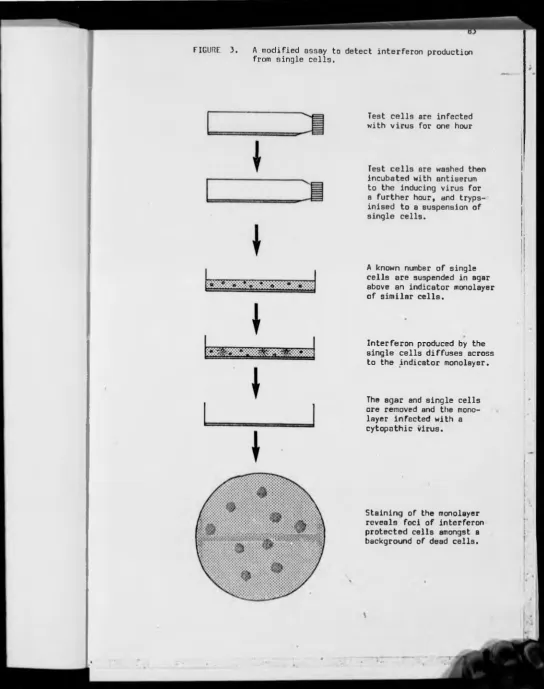
A modified single cell assay for interferon P 82
Growth of viruses P 84
Preparation of antiserum to NDV-F P 85
Induction of interferon synthesis
by virus infection P 86
Polyacrylamide gel electrophoresis
of cell lysates P 87
Extraction of cellular RNA and quantitation of interferon mRNA produced in virus infected cells
P 88
SECTION ONE — THE CHARACTERISATION OF THE
SINGLE CELL ASSAY PFi 91 - 149
The published single cell assays are inaccurate P 92
Can NDV-F cause the production of foci within the assay?
P 97
Does NDV-F replicate in or desorb from L929 cells?
P 99
Neutralisation of viral interference
produced by NDV-F infected cells P 103
Production of foci in the assay by virus infected cells which cannot produce interferon active upon the indicator monolayer
A modified single cell assay for interferon p 107
Optimisation of the modified single cell assay Q- •“H oi-H
Sensitivity of the assay to interferon p 124
Can the percentage of virus-induced interferon producers be increased?
p 129
Why cannot 100?o of culture cells be induced to produce interferon?
p 130
DISCUSSION p 137
SUMMARY p 148
SECTION TWO --- THE INTERFERON SYSTEM IN DIFFER ENTIATING TERATOCARCINOMA CELLS
pp 150 - 203
The interferon system in ' nullipotent1 ec and in differentiated teratocarcinoma cells
p 151
Why is the interferon system inactive in embryonal carcinoma cells
p 155
In vitro differentiation of 'nullipotent' teratocarcinoma cells
p 161
Effect of retinoic acid on the yield of interferon from differentiated cells
p 165
The kinetics of interferon production in 'nullipotent' ec cells treated with retinoic acid
p 168
The development of antiviral sensitivity to interferon in differentiating terato carcinoma cells
p 184
DISCUSSION p 187
SUMMARY p 202
SECTION THREE — THE INTERFERON SYSTEM IN THE DEVELOPING MOUSE EMBRYO
pp 204 - 231
Interferon inducibility in first and second trimester mouse embryo
p 205
DISCUSSION
\ p 219
SUMMARY p 229
FIGURES
1 — The early events in mouse embryogenesis
2 — Stages in mouse embryogenesis
3 — A modified assay to detect interferon production from individual cells
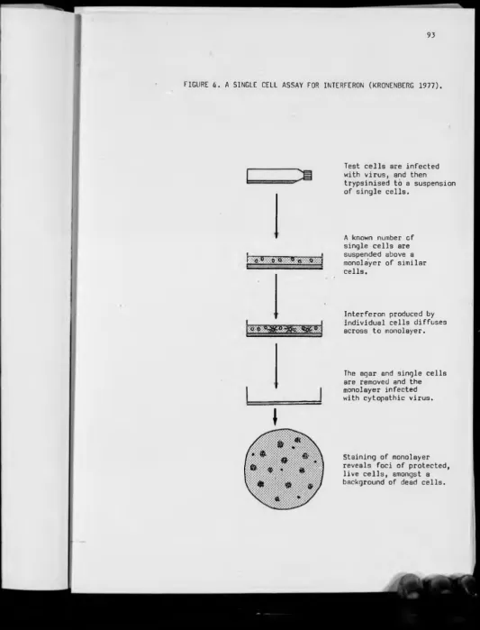
4 — A single cell assay for interferon (Kronenberg 1977)
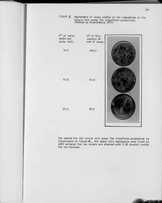
5 — Appearance of assay plates at the completion of the assay
6 — Single cell assay data (method as Kronenberg 1977).
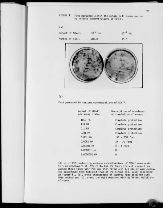
7 — Foci produced within the single cell assay by NDV-F
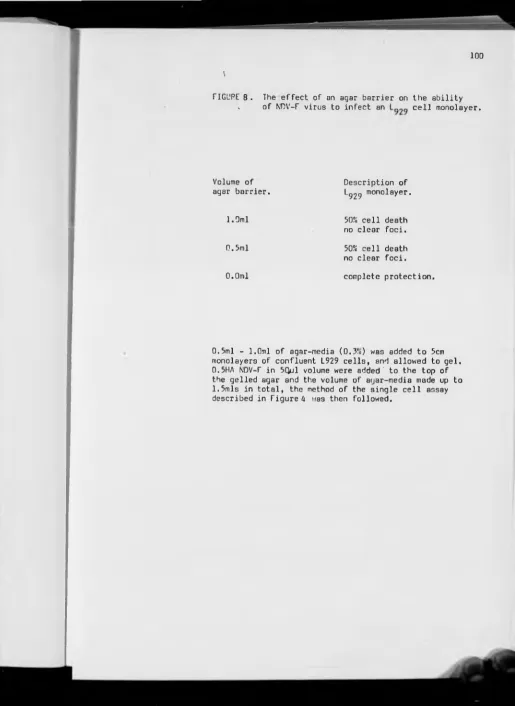
8 — The effect of an agar barrier on the movement of NDV-F
9 — The desorbtion of NDV-F from L929 cells
10— Neutralisation, by antiserum, of interference produced by NDV-F infected cells
11— Production of foci by virus infected cells which do not produce interferon active on the cells in the indicator monolayer
12— All foci produced in the modified assay are caused by interferon production from the single test cells
13— Variation in the percentage of interferon producers in L929 cells infected with NDV-F
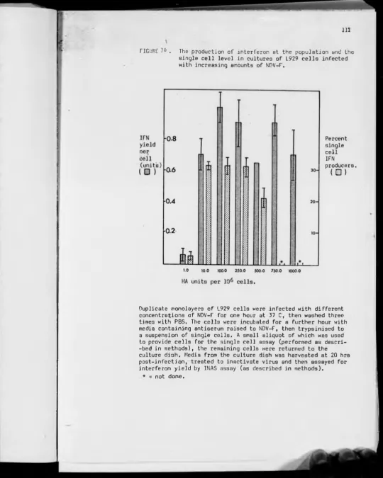
14— The production of interferon in L929 cells infected with increasing amounts of NDV-F
15— The viability of cells infected with NDV-F
16— The effect of cell density upon interferon yield
17— The yield of interferon in virus infected cells treated with antiserum to the infecting virus
18— Accuracy and repeatability of pipetting small cell numbers
P 39
pp 44 -
p 83
P 93
P 93
p 96
p 98
p 100
p 102
p 104
p 106
p 108
p 109
p 111
p 113
p 114
p 116
vii
19— Repeatability of results obtained from the single cell assay
p 119
20— The rate of interferon production in NDV-F infected L929 cells
p 120
21— Effect of cell density on the sensitivity of cells to the antiviral effect of IFN
p 122
22— Amount of IFN produced by cell populations infected with NDV-F
p 125
23— Sensitivity of the single cell assay p 126
24— Variation in foci size seen in the single cell assay
p 128
25— Effect of pretreatment of cells with small amounts of IFN, on subsequent induced yields of interferon
p 131
26— Effect of pretreatment of cells with butyrate, on subsequent induced yields of interferon
p 132
27— Interferon production in L929 cells synchronised with respect to cell culture
p 134
28— Percentage of single cell interferon producers in cloned L929 cell cultures
p 135
29— Status of the interferon system in undifferentiated and differentiated 'nullipotent' teratocarcinoma
p .152
30— Sensitivity of 'nullipotent' teratocarcinoma to the antiproliferative effects of interferon
p 154
31— The desorbtion of NDV-F from teratocarcinoma cells p 157
32— The kinetics of interferon production in virus infected teratocarcinoma cells
p 159
33— The production of IFN mRNA in differentiated teratocarcinoma cells
p 160
34a and 34b— Photographs of embryonal carcinoma cells treated with retinoic acid for various periods of time
p 163 - 4
35 and 36— Microdensitometer scans from autoradiographs of cell proteins from lysates of cells treated for various periods of time with retinoic acid
p 166 - 7
37— Production of interferon in differentiated teratocarcinoma cell lines
38 a,b and c— The kinetics of interferon production in differentiating 'nullipotent' teratocarcinoma cell lines
p 171 - 3
39— Ec cells induced to differentiate in retinoic acid show an increase in the yield of interferon per cell
p 175
AO— Interferon production in differentiating terato carcinoma cultures, trypsinised and reseeded prior to induction
p 176
41— Priming of differentiating teratocarcinoma cells
p 178
42— Relationship between interferon yield and length of exposure to retinoic acid
p 180
43— IFN production in stem teratocarcinoma cells treated with retinoic acid and DiButrylcAMP
p 182 - 3
44— Development of sensitivity to the antiviral effects of interferon in differentiating teratocarcinoma cells
p 185
45— The procedures involved in the assay for IFN production from embryo tissues
p 207
46— The control experiments run concurrently with the embryo 'single cell' assay
p 208
47 and 48 a & b — Interferon production in the early post-implantation mouse embryo
p 211 - 3
49 and 50— Interferon production in the mid-gestational mouse embryo
p 216 - 7
51— Depression of the antiviral action of interferon by the reproductive tissue of the female mouse
IX
ACKNOWLEDGEMENTS
I wish to thank my supervisors Professor D.C. Burke and
Dr. C.F. Graham for their consistent help and encouragement,
which has sustained me over the last three years, and also
for for their sound criticism of this manuscript.
I also thank Beverly Randle, for her help in undertaking
the work presented in section three; Dr.A. Colman and John
Shuttleworth for performing the oocyte injections, and Dr.J.
Morser for performing the rabbit reticulocyte lysate assays.
I am grateful to Dr. Alan Morris, Dr. Paul Bosely and
Dr. John Morser (Fellow members of the Interferon group)
for being available and willing to discuss problems and offer
advice. I am also indebted , in particular, to all members of
the Virus group, but also to the remaining members of the
Department of Biological Sciences at Warwick University, for
contributing to an interesting and challenging work environment.
Finally, I would also like to take this opportunity to
thank the Wellcome Trust for their financial support.
DECLARATION
The work presented in this thesis has not been accepted
in any previous form for a degree, and has been conducted by
myself except where specifically acknowledged. The work
presented in Section three was conducted jointly between
myself and Beverly Randle of the Department of Zoology, Oxford
University.
xi
SUMMARY
A modified assay to detect interferon production from individual cells has been designed which is more accurate than those already described. Use of this modified assay has demonstrated that the diff erence between cell lines that can be induced to produce high yields of interferon, and those which are only capable of producing low yields of interferon, resides in the number of individual cells able to prod uce interferon in that culture. Thus the apparent homogeneous response of a cell culture to an interferon inducing agent, masks the heterogene ous response of the individual cells which make up that culture. This modified assay is probably sensitive enough to detect all cells capable of producing interferon within a given cell population; and the data presented in section one suggests that this assay can be used with confidence to assay interferon production in cell systems which only produce small amounts of interferon.
Cloned 'nullipotent' embryonal carcinoma (ec) cells, like the pluri- potent ec cells described by Burke et al (1978), do not possess an active interferon system, and it is proposed that such cells lack the ability to produce interferon mRNA in response to an interferon-inducing agent. When these 'nullipotent' ec cells are treated with retinoic acid they show an activation of the interferon system which extends for approxima tely ten to fifteen days. The extent of activation seen varied between different embryonal carcinoma cell lines. In these differentiating cult ures there is a parallel increase, both in the percentage of individual cells able to produce interferon, and in the yield of interferon per producer cell. The percentage of single cells able to produce interferon always remained small compared to the non-producer cell3 in the culture. The pattern of development of interferon inducibility and sensitivity does not distinguish between the different types of endoderm-like cell generated by the various differentiating teratocarcinoma cell lines, nor can the amount of interferon produced by different cell lines be used to quantitate the extent of differentiation which has occurred. However, the activation of the interferon system; because it coincides with changes in morphology and in protein synthesis, can be used as an additional positive marker for the production of differentiated cells in this system.
The data presented in section three demonstrates that during the
first third of pregnancy the embryo is unable to produce interferon
in response to a virus infection, and furthermore suggests that the antiviral action of interferon may be non-specifically inhibited by the tissues of the reproductive system from the adult female mbuse. A functional interferon system develops during the seventh day of embry onic development , and the embryonic ectoderm and the visceral extra- embryonic endoderm are the last tissues to show a lack of interferon inducibility. Thus, the mouse embryo can be seen to become capable of mounting an interferon-based antiviral response during a period when it is unable to mount a humoral and cell mediated antiviral immune response. This factor may be of importance in the reduced susceptibility to the pathogenic effects of virus infections, which is a feature of the mid to late term mammalian embryo.
ABBREVIATIONS
AFP Alphafetoprotein
CPE Cytopathic effect
cAMP Cyclic adenosine 3'5' monophosphate
diBcAMP Dibutryl cyclic adenosine 3 ’5 ’ monophosphate.
DNA Deoxyribonucleic acid
dsRNA Double strand ribonucleic acid
ec Embryonal carcinoma cell
eIF2 Eukaryotic protein synthesis initiation factor 2
emb.ect. Embryonic ectoderm cells
emb.meso. Embryonic mesoderm
EMC Encephalomyocarditis virus
END Differentiated endoderm-like teratocarcinoma cell
epc Ectoplacental cone
ex.emb.ect. Extra-embryonic ectoderm cells
ex.emb.meso. Extra-embryonic mesoderm cells
HA (U) Haemagglutinin units
IFN Interferon
INAS Inhibition of viral Nucleic Acid Synthesis
mRNA Messenger ribonucleic acid
NDV-F Newcastle disease virus strain F
PAGE Polyacrylamide gel electrophoresis
PBS Phosphate buffered saline
PE Parietal endoderm cells
pfu Plaque-forming units
Poly rI:rC Poly ribo-ionsinic and poly ribo-cytidylic acid
xiii
RA Retinoic acid
RNA Ribonucleic acid
RRL Rabbit reticulocyte lysate protein synthesis assay
rRNA Ribosomal ribonucleic acid
SFV Semliki Forest virus
SV40 Simian virus type 40
TB Trophoblast cells
TE Trophectoderm cells
tris Tris(hydroxymethyl)aminomethane
tRNA Transfer ribonucleic acid
VE Visceral endoderm cells
v.emb.end Visceral embryonic endoderm cells
v.ex.emb.end. Visceral extra embryonic endoderm
VS V Vesicular Stomatitis virus
VYS Visceral yolk sac
2
\
INTRODUCTION
The role of interferon as an anti-viral agent has been known
and accepted for over two decades. However its pleiotropic effects
on cell function have only recently been described; and serve to
highlight how little is known of the extent and behaviour of the
interferon system in vivo. The molecular biology of this system
has yielded under a sustained assault over recent years, and it can
now be seen that interferon comprises multigene families whose
6
members are functionally heteroger^us and whose mode of action involves
both induction of specific enzymes and the alteration of cell surface
behaviour. Interferon is by definition an anti-viral agent, but
because it is an agent which has a wide range of non anti-viral effects
on cells it becomes necessary to have a more complete understanding
of the role and status of the interferon system in vivo.
Efforts in this direction have mainly centred on attempts to
gain an understanding of the role of interferon in in vivo pathogenesis,
with the aim of evaluating interferon as a possible therapeutic agent.
This aim, albeit worthy, has resulted in the neglect of the wider
potential of the interferon system.
It is generally thought that the interferon system is not
restricted to individual tissues or organs but can be expressed
throughout the whole of the animal. However very little is known
concerning its ontogeny and phylogeny even though such knowledge is
necessary if the understanding of the role of interferon is to extend
beyond its action 83 a component of the hosts' anti-viral response.
Interest in the developmental aspects of the interferon system
is inactive in undifferentiated teratocarcinoma cells but becomes
active when the teratocarcinoma cells are caused to differentiate.
The aim of this study is to examine the ontogeny of the interferon
system in the developing mouse embryo, and also to examine in more
detail the kinetics of the appearance of the interferon system in
teratocarcinoma cell cultures undergoing the transition from multi-
potent stem cell to a differentiated cell type. This study therefore
combines aspects of interest to two disparate fields and thus the aim
of this introduction is two-fold; to describe the potential role and
use as an experimental tool of the interferon system, and to describe
the concepts underlying both mouse embryogenesis and the use of
teratocarcinoma cells as in vitro analogues of development.
4
THE INTERFERON SYSTEM
What_is interferon ?
Interferon is defined a3 an anti-viral activity (see reviews in
Burke 1977, Baglioni 1979, Marx 1977a,b). It was first described
by Isaacs and Lindeman (1957) who demonstrated that cells exposed to
a virus released a substance which could be shown to protect other
cells from subsequent virus attack. The principle involved was named
interferon. In recent years other functions have been attributed to
the interferon molecule, e.g.; inhibition of cell growth (see review
in Taylor-Papidomitriou 1980) and regulation of the immune response
(see review in Eloom 1980). The discovery of these other functions
led to the description of interferon as a cellular mediator or
regulator (Clemens 1979, Cohen and Bigazzi 1980) or a pleiotropic
modifier of cellular function (Stewart 1979a)and even to the proposal
that interferon be classified as a hormone. This latter proposal is
based on three criteria; these are that interferon interacts with a
specific plasma membrane receptor as an initial step in affecting
cell function, that it can affect a range of cell functions in a
variety of cell types (Taylor-Papidomitriou 1980), and that one class
of human interferon strongly resembles the neuroendocrine hormones
and is thus capable of mediating some of the effects associated with
interferon through interaction with thi3 system (Blalock and Smith
1981). In addition to the proposed functions of interferon, the
molecular biology of interferon production and anti-viral sensitivity
provides a model system to study the induction and expression of a
eukaryotic gene (Havell 1977, Burke 198B).
considerable potential as an experimental tool and is thus of interest
to workers in several fields which includes virologists, cell and
cancer biologists and immunologists. However because interferon is
just one of a variety of biologically active molecules which can be
elicited under pathological conditions in vivo (Cohen and Bigazzi 1980),
it is necessary to define precisely what is meant by the term interferon.
The acceptance of a working definition will provide a coordinating
influence under which the numerous other proposed functions can be
examined. The Committee of Interferon Nomenclature (Stewart et al.
1980) have accepted the following definition for interferon; "to
qualify as an interferon, a factor must be a protein which exerts
virus non-specific anti-viral activity at least in homologous cells
through cellular metabolic processes involving synthesis of both
RNA and protein".
The term 'interferon system' used in this study is used to
denote both interferon production and sensitivity to its pleiotropic
effects. These are separate and distinct cellular phenomena and
occur by separate pathways. Most differentiated cell lines can produce,
and are sensitive to the effects of interferon. This is in contrast
to undifferentiated teratocarcinoma cells which cannot produce inter
feron and are insensitive to its anti-viral (Burke et^ cü. 1978) and
anti-proliferative effects (Wood and Hovanessian 1979). Some
differentiated cell lines, e.g.; African green monkey kidney (vero),
baby hamster kidney-21, and Chinese hamster ovary kl cannot produce
interferon but are sensitive to its effects (Desmyter et^ jjK 1968,
Taylor-Papidomitrious and Stoker 1971, Morgan 1976). Thus although
interferon production and sensitivity are properties which can both
reside in one cell they can be separated or temporally absent from
Induction and production of interferon
The presence of interferon cannot be detected in the normal
state in vivo and in vitro, but interferon production can be stimulated
by exposure of the cell to a specific inducer of interferon. The
inducer enters the cell and either directly or indirectly causes the
transcription of the interferon gene mRNA and the translation of it
into a secretory protein. Interferon production continues for a set
period of time which depends on the particular inducer/cell system,
and is then switched off (Burke 1980), and this shut-off of interferon
synthesis requires post-transcriptional modification of the mRNA
(Sehgal et al. 1977). Interferon production has been shown to be due
to cte novo transcription and translation and not merely activation of
pre-existing molecules by enucleation experiments which demonstrated
the necessity of the nucleus (Burke and Veomett 1977), and by the use
of inhibitors of transcription and translation, which were shown to
inhibit the production of interferon (see review in Burke 1980 and
Stewart 1979 p.90-109). There are numerous agents (called inducers)
which can stimulate the production of interferon. Most inducers are
viruses (Jameson 1977, Stewart 1979 p28-34) but non-viral substances
can also induce interferon. These include micro-organisms (Stewart
1979 p39-41) chemical polymers and low molecular weight compounds
(Stewart 1979 p47-48).
It is not yet known how the various inducers act within the cell
to activate the cellular gene for interferon, or whether thi3 happens
directly or indirectly. Because of the known ability of RNA viruses
to induce high yields of interferon (Meager and Burke 1971) and the
ability of natural and synthetic dsRNA to similarly induce high yields
1
of dsRNA can induce anti-viral resistance in a human fibroblast cell
in vitro) it has been proposed that viruses induce interferon via
synthesis of a dsRNA intermediate (Morser and Burke 1979).
Thus interferon is not seen in vivo and in vitro unless the
cells are first exposed to an inducer, and then its appearance is
transient. There are however pathological and other conditions under
which interferon is produced by cells which have had no apparent
contact with an inducer. This phenomenon may have one of two possible
explanations, either the interferon gene has altered and become
permanently expressed or an inducer is permanently present in that
cell. Since the true nature of the interferon inducer is not clear
these two possibilities cannot be distinguished, and the production
of interferon in cells not exposed to an inducer will be termed
spontaneous interferon production. This phenomenon will be described
later in this section.
Classes of interferon
Three main classes of interferon have been identified in all of
the animals so far studied. These classes are designated alpha, beta
and gamma, and interferons assigned to each on the basis of antigenic
similarity.
Class alpha and beta are produced transiently following the
exposure to non-mitogen inducers of most differentiated cells,
including cells of the immune system. The ratio of alpha to beta
interferons produced within a cell depends on the cell type and the
inducer, e.g.: human leukocytes produce predominantly alpha, and
8
immunocompetent lymphocytes following exposrue to a mitogenic
stimulus, e.g.: specific antigen or plant lectin, and it is thought
that T lymphocytes are the main source of gamma interferon (Epstein
1977). Since gamma interferon is produced from lymphocytes following
exposure to a mitogen it can also be termed a lymphokine (Cohen and
Bigazzi 1980).
Cloned human interferon genes
The concept of the interferon molecule has altered since the
availability of cloned interferon genes. So far only human alpha
(Nagata et^ £l^. 1980) and human beta (Derynck ejt ¿1. 1980) have been
cloned. The human alpha class has been shown to consist of a family
of different but homologous genes situated on the same chromosome,
which are capable of cross hybridisation at the nucleic acid level
and of cross neutralisation at the protein level. Up to ten alpha
genes, including one pseudogene, have so far been identified (Goeddel
£t a_l. 1981). These show little or no glycosylation (Allen and Fantes
1980), and do not contain introns either in the coding or non-coding
genomic sequences (Nagata et^ al_. 1980). The alpha multigene family
once cloned into E. coli can all be translated (except the pseudogene)
and therefore have the potential to be expressed within the cell, but
it is not yet clear how many members of this family are expressed in
cells treated with different inducers, or whether a particular
pattern of expression occurs within certain cells treated with certain
inducers. However it is thought that the alpha family are functionally
hetcrogcrjjjuo and this is supported by the observations of Streuli
et ni. (1981) on the different cross species reactivity amongst the
members of the alpha family. Cloned alpha 1 and 2, which differ in
only twenty nucleic acid positions, showed a large difference in
inducing anti-viral resistance in mouse cells. It has long been
known that some interferons can show reactivity on cell species
different to that which produced the interferon (Stewart 1979 pl36-42),
but interferons were thought in general to show broad species speci
ficity. The cloning studies have clarified this situation and it
appears the species restriction of sensitivity resides in specific
interferon molecules and is not a general property of interferons.
The human beta class is antigenically different to alpha inter
feron and also does not cross react at the nucleic acid level (Allen
and Fantes 1980). The total number and extent of the beta gene family
is not yet clear; Goeddel £t ajk (1981) found only one beta gene,
while Weissenbach et (1980) identified two beta interferon genes
in cell-free lysate translations of cloned DNA from poly rlrC induced
fibroblasts. These two interferons were identified serologically as
beta but did not cross-hybridise at the nucleic acid level, and
interestingly were not produced in intact cello; suggesting that a
sub-class of interferons are modified at a post-transcriptional level.
Sehgal et^ si. (1981) found a similar situation in fibroblast cells
induced in the presence of DRB (dichlororibofuranosylbenzimidazole),
and has suggested that a second, hetero-disperse, set of interferon
genes exist in both the alpha and beta classes. The second set are
proposed to contain introns which prevent nucleic acid cross
hybridisation.
Interferon was long thought to be a glycosylated protein, and
the varied functions and properties of interferon were thought to be
10
1977, 1979). The amount of glycosylation has still not been fully
resolved but Allen and Fantes (1980) and Derynck _et al_. (1980) have
both shown that alpha and beta interferons do not need to be glycosy
lated to be biologically active, although beta interferon is normally
synthesised as a glycosylated protein.
The biological role of the alpha multigene family has not been
clarified; the production of functionally heterogenous interferons may
explain the ability of interferon to protect cells from a wide
variety of viruses, or alternatively it may explain the large variety
of target cells which are susceptible to the .effects of interferon.
R. M. Friedman has suggested (personal communication) that the
different interferon species may be effective on different host tissues.
Mouse interferons
Full details of the structure of mouse interferons are not yet
available, since the genetic engineering program has for obvious reasons
concentrated on human interferon. However it is known that viral
induction of non-lymphoid mouse cells, e.g.: Erlich ascites cells and
C243, causes two species of interferono to be produced. The molecular
weights are approximately 22 and 35 kd. (De Maeyer-Guignard et al. 1978,
Kawakita £t al_. 1978, Iwakura j^t al_. 1978). The lower molecular weight
molecule shows some sequence homology with human alpha interferon.
The number of interferons produced in virus infected mouse cells doe3
vary; Knight (1975) found ten interferon polypeptides were produced
in MM virus-infected cells each with anti-viral activity. Heterogeneity
of glycosylation was also found by Knight (1975) and by De Maeyer-
resolved but Allen and ("antes (1980) and Derynck _et al^. (1980) have
both shown that alpha and beta interferons do not need to be glycosy
lated to be biologically active, although beta interferon is normally
synthesised as a glycosylated protein.
The biological role of the alpha multigene family has not been
clarified; the production of functionally heterogenous interferons may
explain the ability of interferon to protect cells from a wide
variety of viruses, or alternatively it may explain the large variety
of target cells which are susceptible to the .effects of interferon.
R. M. Friedman has suggested (personal communication) that the
different interferon species may be effective on different host tissues.
Mouse interferons
Full details of the structure of mouse interferons are not yet
available, since the genetic engineering program has for obvious reasons
concentrated on human interferon. However it is known that viral
induction of non-lymphoid mouse cells, e.g.: Erlich ascites cells and
C243, causes two species of interferons to be produced. The molecular
weights are approximately 22 and 35 kd. (De Maeyer-Guignard et^ al^. 1978,
Kawakita ct^al_.1978, Iwakura et al. 1978). The lower molecular weight
molecule shows some sequence homology with human alpha interferon.
The number of interferons produced in virus infected mouse cells doe3
vary; Knight (1975) found ten interferon polypeptides were produced
in MM virus-infected cells each with anti-viral activity. Heterogeneity
of glycosylation was also found by Knight (1975) and by De Maeyer-
Assay of interferon yields
A large area of this study is concerned with the production and
assay of interferon yield. The yield of interferon per cell is
subject to regulatory controls within the cell (on transcription rate,
half-life of mRNA and translation); but it is also regulated through
genetic controls not associated with the structural genes. De Maeyer
and co-workers (reviewed in De Maeyer and De Maeyer-Guignard 1979) have
obtained mice through breeding experiments which have the ability to
produce high or low levels of circulating interferon in response to
the same virus inducer. This genetic control of interferon yields is
only shown by macrophages _in vitro. A review of the intracellular
regulation of interferon can be found in Havell (1977) and Burke (1980),
Briefly it is proposed that expression of the interferon gene is
inhibited in normal conditions by the synthesis of a repressor
molecule and that interferon inducers act to disrupt this equilibrium
and allow expression of the interferon gene (Vilcek and Ng 1971).
As yet there is no direct confirmation of the existence of the
putative interferon repressor, or of any other eukaryotic gene
repressor.
The amount of interferon in a preparation is assayed indirectly
by measuring the effect that it can exert on cells. Since different
mouse cells vary in their sensitivity to interferon the definition of
a unit of interferon thus depends on its mode of assay. The effect
most commonly measured is that of antiviral resistance. This can be
assayed in a variety of ways including, virus RNA synthesis, yield
and plaque reduction and cytopathic effect; all of which vary in
\
This laboratory routinely employs the Inhibition of viral Nucleic Acid (INAS)
assay • (Atkins et^ al^. 1974). Units of interferon are reciprocals
of end point dilutions of an interferon preparation and the end point
of the INAS assay is the dilution at which virus nucleic acid synthesis
is reduced by fifty percent. These assays are all biological assays
and variations from many sources will occur; to eliminate these a
laboratory standard interferon preparation is included in all assays
and results corrected accordingly. To compare results between
different laboratories the laboratory standard interferon is calibrated
against an international research reference standard made available
from the National Institute for Biological Standards, London. Use of
a laboratory standard calibrated to the international research reference
standard enables interferon titrcs to be expressed in international
reference units.
The recent development of a monoclonal antibody to interferon
(Secher and Burke I960) has enabled non-biological assays to be
developed. Secher (1981) has devised an immunoradiometric assay which
although it has reduced sensitivity compared to the biological assays
is potentially useful since it can measure physical amounts of
interferon.
Single cell assay for interferon
The assays described above measure interferon yields from a
population of cells and thus represent the average response of many
cell3. Since the minimum number of cells required to produce on
interferon sample which can be used in these assays are approximately
13
small number of cells are available, e.g. from early embryo tissues.
» \
In addition it is possible that by measuring only the average response,
any heterogenous behaviour of either the cells or the inducer is
masked. Interferon production from either single cells or from small
groups of cells can be examined in a 'single cell assay'. Two types
of single cell assay have been developed both based on the biological
activity of interferon. Fleischman and Simon (1974) and Rodgers and
Merigan (1974) examined interferon production from single cells
isolated in microdrops, whilst Osbourn and Walker (1969) and Kronenberg
(1977) measured the percentage of cells able to produce interferon in
an assay analogous to the infectious centre assay for virus production
from single cells. This study employs a modified version of the
assay described by Kronenberg (1977).
PLEIOTROPIC ACTIVITIES OF INTERFERON \
Interferon is capable of exerting a vast and often bewildering
array of effects upon cellular structure and function (Stewart 1979
p224-231). These effects may be due to a single action of interferon
or more probably are effected through the concerted action of interferon
on many aspects of cell biology. The activities ascribed to interferon
mechanisms may be grouped into six major categories:
(a) Alteration in cell surface composition and behaviour.
(b) Antiviral actions against a wide range of viral pathogens.
(c) Alteration of induced and non-induced protein synthesis.
(e) Inhibition of cell replication in normal and tumour cells.
(f) Possible role in the normal control mechanisms of growth and
differentiation.
The order of the above groupings is arbitrary and serves to
focus attention on the main effects of interferon, whilst not ignoring
the fact that the causes of the observed phenomena may involve
several of the effects listed above. It is still not clear whether
or not all the activities listed above are secondary to the establish
ment of the anti-viral state within the cell.
The first non anti-viral effects of interferon were described by
Isaacs and Burke (1958) who demonstrated the cell multiplication
inhibitory effect. These early experiments were performed with impure
interferon preparations (probably less than l?o pure) and were open to
criticism that these non anti-viral effects were due to impurities in
the preparations. These criticisms have now been formally turned over
by the use of electrophoretically pure interferon, Gresser et al.
(1979) used mouse interferon of specific activity 10 units per mg.,
which was resolvable to two bands on polyacrylamide gel electrophoresis,
and clearly demonstrated that each band in this preparation could
produce all of the effects listed above.
Cell surface receptors?
The interferon molecule is not itself directly the cause of
the effects ascribed to interferon, for it is not active within the
cell in which it was synthesised but needs first to be externalised
(Vengris et^ jil_. 1975). Interferon does not need to enter the cell
15
the cell (Burke 1977). This suggests that sensitivity to the pleio-
tropic effects of interferon is mediated via the interaction between
the interferon molecule and the cell surface. Such a step could act
to amplify the action of interferon and might account for its high
biological activity.
It has been proposed (Stewart 1979 pl85, Cupples and Ian 1977,
Tan 1976, Revel et al. 1976) that the sensitivity of cells to the
effects of all the classes of interferon (alpha, beta and gamma) are
mediated initially by a common pathway regulated by a gene on
chromosome 21 in humans. The evidence for this is mainly based on
the increased sensitivity to human interferon shown by cells trisornic
for chromosome 21 and the complimentary decreased sensitivity shown
by cells monosomie for chromosome 21. Revel jet al_. (1976) proposed
that chromosome 21 codes for an interferon cell surface receptor, since
they were able to demonstrate that antisera raised to the cell
surface of human/mouse hybrids (which contained only chromosome 21
as its human contribution) blocked the action of interferon. Tan (1976)
proposed that chromosome 21 directs the production of an intermediate
substance (not a receptor) which mediates the action of interferon
within the cell. However contrary evidence has cast doubts on the
role of chromosome 21 at all. De Clercq et^ al. (1976) examined
interferon sensitivity in a wide range of cells both monosomie and
trisomie for chromosome 21 and found that the difference in interferon
sensitivities was not constant. Moreover they demonstrated that cells
monosomie, disomie and trisomie for chromosome 21 had equal ability
to remove interferon from culture fluids, suggesting that these cells
all bind equal amounts of interferon. However since the experiments
of De Clercq ct^ jü. (1976) were not performed on matched cells, the
general consensus (see review in Stewart 1979 pl85-190) still supports
a role for human chromosome 21 in mediating the effects of interferon.
The majority of work on interferons has involved the use of the
alpha and beta types because in contrast to gamma interferon they are
now available in relatively pure preparations; however it is considered
that all three classes of interferons possess a similar range of
pleiotropic effects.
Alteration in cell surface composition and behaviour
\
Interaction with or binding to the cell surface is a necessary
preliminary to the manifestations of the effects of interferon. This
reaction at the cell surface causes chemical, immunological and
morphological changes within the membrane before any of the other
effects of interferon can be demonstrated (Chang et^ al_. 1978,
Friedman 1979). These changes include inhibition of the binding of
specific low molecular weight molecules; the binding of cholera toxin
and thyroid stimulating hormone are inhibited by interferon whilst
that of human chorionic gonadotrophin, leutinising hormone and follicle
stimulating hormone are not. This interference with binding is not
thought to be due to direct competition for receptors but rather due
to conformational changes induced in the cell membrane by interferon.
This alteration also changes the expression of some cell surface
molecules; expression of cell surface gangliosides is decreased
(Friedman 1979) but expression of beta-microglobulin and the major
histocompatability antigens is increased (Lindahl et al^. 1972, 1973;
Heron et^ aJL 1979). Two general properties of the cell surface have
16
general consensus (see review in Stewart 1979 pl85-190) still supports
a role for human chromosome 21 in mediating the effects of interferon.
The majority of work on interferons has involved the use of the
alpha and beta types because in contrast to gamma interferon they are
now available in relatively pure preparations; however it is considered
that all three classes of interferons possess a similar range of
pleiotropic effects.
Alteration in cell surface_composition_and behaviour
Interaction with or binding to the cell surface is a necessary
preliminary to the manifestations of the effects of interferon. This
reaction at the cell surface causes chemical, immunological and
morphological changes within the membrane before any of the other
effects of interferon can be demonstrated (Chang at ¿1. 1978,
Friedman 1979). These changes include inhibition of the binding of
specific low molecular weight molecules; the binding of cholera toxin
and thyroid stimulating hormone are inhibited by interferon whilst §
that of human chorionic gonadotrophin, leutinising hormone and follicle
stimulating hormone are not. This interference with binding is not
thought to be due to direct competition for receptors but rather due
to conformational changes induced in the cell membrane by interferon.
This alteration also changes the expression of some cell surface
molecules; expression of cell surface gangliosides is decreased
(Friedman 1979) but expression of beta-microglobulin and the major
histocompatability antigens is increased (Lindahl et al. 1972, 1973;
Heron £t^ al_. 1979). Two general properties of the cell surface have
been found to alter: there is an increase in the number of
membranous granules of diameter 5-10um (Chang et al_. 1978), and an
increase in the overall net negative charge of the cell (Knight and
Korant 1977). This latter change may indicate that the membrane alters
in its permeability to certain molecules. The relevance of the
membrane changes to the effects of interferon is described later.
Friedman (1979) has suggested that changes in the cell surface may
be found to account for all of the non anti-viral effects of interferon.
The first detectable intracellular change after interferon
interacts with the cell surface is the elevation of the concentration
of the cyclic nucleotides adenine and guanosinc monophosphate. This
elevation has been reported only in those cells sensitive to the
effects of interferon (Friedman and Pastan 1969, Weber and Stewart
1975, Meldolesi et jal. 1977 and Tovey et al^. 1979). In non-interferon
treated cells, the levels of these cyclic nucleotides can be affected
by the rate of cell growth and cell density. Tovey et al.(1979)
developed a system in which L1210 cells cultured in a chemostat
produced constant levels of both cyclic nucleotides, and found that
after interferon treatment of these cells the levels of CGMP rose
within five minutes, but that in contrast, the levels of CAMP did
not rice until several hours later. They suggest that CGMP functions
as a membrane-nuclear signal. In addition Friedman and Pastan (1969)
and Meldolesi et ol. (1977) have provided further evidence that cyclic
nucleotides may be involved in the effects of interferon,by
demonstrating that the administration of CAMP added simultaneously
with interferon potientiated the degree of anti-viral resistance
achieved in the cells, but had no anti-viral action by itself.
18
ANTI-VIRAL ACTIONS AGAINST A WIDE RANGE OF VIRAL PATHOGENS
All six classes of animal viruses (i.e. enveloped and non-
enveloped, single strand and double strand, and DNA and RNA classes -
Matthews (1979) ) are sensitive to the inhibitory effects of interferon
to a lesser or greater extent (Stewart 1979 p202-206). The total
anti-viral response of an organism involves cells and other products
of the immune system, as well as the production of interferon within
infected cells (Roitt 1977 p201). Host humoral and cell-mediated
responses are both involved in the immune response to a virus infection;
B lymphocytes are involved in antibody production directed towards
virus antigens and T lymphocytes are directly involved in the recognition
and lysis of virus infected cells. It is of interest that T cell
recognition of virus-infected cells is directed through the major
histocompatability antigens and that interferon enhances the
expression of these antigens.
The production of interferon can be regarded as the first line
of defence against virus infection. Gresser et al. (1976 a and b) have
demonstrated the role of interferon in in vivo virus infections. They
treated mice infected with a variety of viruses with antiserum to
interferon, and found that in most cases, the disease showed a more
rapid onset and greatly increased mortality. An interesting exception
was that of Influenza type A in which the course of the disease was
not affected.
Treatment of cells with interferon will induce nnti-viral
resistance within the cell, but this resistance is not apparent until
the cell is exposed to virus or to dsRNA (Burke 1977, Marx 1979, Revel
1979). This covert antiviral state may be a mechanism to protect host
cell synthesis. The development of this interferon-induced covert
anti-viral state requires the presence of the cell nucleus (Radke
et al. 1974) and requires de novo RNA and protein synthesis of molecules
previously termed 'anti-viral' proteins (Burke 1977). Although
interferon preparations can show broad species specificity the anti
viral proteins can be transferred between, and are active in heterolo
gous cells which are not responsive to the interferon molecule itself
(Blalock and Baron 1977).
Interferon is able to inhibit the growth of a wide variety of
viruses to varying degrees (Stewart 1979 p202-206), and this may
reflect the ability of the interferon-induced anti-viral state to
raise many and diverse barriers to virus replication. Interferon has
been shown to inhibit uncoating, transcription and translation, and
assembly and release of the virion (Revel 1979). The primary effect
of the anti-viral state upon virus replication will depend on the
particular virus/cell system under study, however effects upon
translation are most commonly observed. Revel (1979) has shown that
SV40 replication is inhibited at the early and late stages in inter
feron treated cells affecting uncoating and DNA replication and later
assembly and release of the virions. Strachen et^ a L (1977)
demonstrated that interferon can prevent expression of mouse mammary
tumour virus in steroid induced cells probably acting at both trans
cription and translation. Joklik and Merigan (1966) have shown that
translation is inhibited in interferon treated vaccinia virus infected
cells because of the disruption of the association between viral mRNA
and host cell ribosomes.
A further example of the multiphasic antiviral state induced by
interferon is seen in Murine Leukaemia virus infected cells. The
20
production of virus RNA and protein is little affected but the yield
of infective virus is considerably reduced due to a defect in the
budding and release of virions on the host membrane (reviewed in
Friedman 1979). Maheshwari and Friedman (1979) and Maheshwari et al.
(1980) have demonstrated the same phenomena in interferon treated
vesicular stomatitis virus infected cells which are released from the
cell by a mechanism similar to that used by murine leukaemia virus.
Specific inhibition of virus synthesis
It has been suggested that virus protein synthesis is preferentially
inhibited in interferon treated cells, and whilst this is not certain
there may be physical reasons why this is so. Garry and Waite (1979)
have shown that lytic viruses induce alterations in the Na+ and K+
concentrations within the cell, and that under these new conditions
host cell synthesis is inhibited but virus synthesis is not affected
and neither is the synthesis of interferon itself. In addition, Garry
and Waite have demonstrated that experimental alteraton of the intra
cellular concentration of these ions prolongs the synthesis of
interferon, probably by inhibiting the synthesis of the host protein
responsible for shut-off. In such cells virus synthesis will constitute
the bulk of the cells synthetic effort and any inhibitors of protein
synthesis will have a proportionally greater effect on virus synthesis.
In addition Baglioni (1979) suggests that since the antiviral state
in an interferon treated cell is not activated until the cell is
exposed to virus the inhibitory effect may be localised to the
immediate area of the virus. An alternative to these views is the
suggestion that exposure of an interferon treated cell to a virus may
cause the destruction of that cell and thus destruction of the virus
(Burke 1777).
Mechanism of antiviral action
\
As mentioned above, interferon induces de novo synthesis of
anti-viral proteins. The nature of these proteins was elucidated by
examining the antiviral action of lysates from interferon-treated and
control ce 11s .Kerr et si (1977),Kerr and Brown (1978) and Farrell et al.
(1978) demonstrated that lysates from interferon treated cells showed
enhanced sensitivity to the protein synthesis inhibitory effects of
dsRNA. This was shown to be due to two dsPNA dependent enzymes;
elevated levels of inactive forms of these enzymes formed in interferon-
treated cells being converted to an active form by exposure of the
cell to dsRNA (or to virus). These enzymes were a cAMP dependent
protein kinase and an oligonucleotide synthetase, termed 2,5 oligo A
synthetase by Kerr et ail (1977).
(a) cAMP dependant protein kinase
In view of the reported elevation of cAMF by interferon (Tovey et al.
1979) it is of interest that this enzyme is cAMP dependent. The
protein kinase phosphorylâtes two cell proteins of molecular weights
approximately 35Kd and 67Kd (Farrellet^ al. 1978). The smaller
molecular weight protein is the alpha sub unit of cIF2 (eukaryotic
initiation factor-2) (Hovanessian ¿1_. 1980). The nature of the
larger protein is not clear;0htsuki et al. (1980) consider that it is
a ribosomal protein that may act as an intermediate between the
protein kinase and phosphorylation of eIF2. The phosphorylation of
eIF2 prevents the initiation of protein synthesis (Lenz and Baglioni
22
\
(b) 2,5 Oligo A synthetase
This catalyses the formation of a low molecular weight oligonucleotide
of the general formula pppA(2'p5'A)n where 'n' could be from 1-10 but
most frequently the trinucleotide was observed (Kerr and Brown 1978).
The trinucleotide consists of three adenosine residues linked by
two phosphodiester bonds at the V 5' positions and linked to three
phosphate groups; i.e.
A A A
\
D[\ [
P P P v ^ ^ ^ r
\l \ f
OH (2')
OH
(51)
The trinucleotide activates an endonuclease which can degrade viral
and cellular mRNA, and thus acts to inhibit virus replication at the
level of transcription. Further actions of V 5' oligo A synthetase
and the trinucleotide have been proposed; Thang et al. (1981)
demonstrated that 2'5' oligo A synthetase catalyses the aderylation
of tRNA and NAD+ . The function of this is not clear but this process
does limit the size of oligo A nucleotide formed, in the absence of
tRNA and NAD+ long chain oligo A polymers are synthesised. Wallach
and Revel (1980) have suggested that the trinucleotide as well as
activating the endonuclease, may have a more direct anti-viral role,
since they detected the 2'5' A trinucleotide bound to the ribonuclear-
protein core of VSV and MLV virions released from interferon-treated
cells.
The 2'5' A trinucleotide is itself degraded by an interferon
enhanced phosphodiesterase and Revel (1979) proposes that this PDE acts
83 additional pathway to inhibit virus replication by inhibiting directly the amino acylation of tRNA and thu3 inhibiting peptide chain
The'role of 2'5' oligo A synthetase and the cAMP dependant
protein kinase in mediating the antiviral action of interferon
has been confirmed by many experiments which have correlated
levels of these enzymes with both the degree of antiviral resis-
tamce and the level of interferon. Williams and Reed (1981),
Krishnan and Baglioni (1980) and Hovanessian et_£l_ (1981) have
demonstrated this correlation in in vivo pathogenesis, and Thang
elt al^ (1981) have found this to be true in interferon treated
cells infected with MLV in vitro. In addition Hovanessian et al
(1981) have detected elevated levels of 2'5' oligo A synthetase
in the sera of human volunteers treated with poly rA:rU, and
also in the mononuclear cells of humans suffering from virus
infection.
Role_of_'antiviral' enzymes inmediating the non-antiviral
effects_of interferon
The interaction between the interferon molecule and the
establishment of the covert antiviral state within the cell
reflects a profound effect on the cells' biology, and because
of this it may be considered that the other non-antiviral effects
attributed bo interferon are secondary events and do not represent
a series of distinct non-antiviral functions of interferon. Des
pite intensive searches a further series of interferon-induced
enzymes specific to other effects have not been found, and it is
therefore possible that the induction of antiviral enzymes and
changes at the cell surface mediate the other effects of inter feron.
If this is so, it should be possible to demonstrate that the
interferon-induced antiviral enzymes can produce the non-antiviral
24
that 2'5' oligo A trinucleotide can confer anti-vira.1 resistance
against a wide variety of viruses, has shown that this trinucleotide
can inhibit DMA synthesis in a variety of human and mouse cells and
enhance the activity of macrophages and natural killer cells; and
thus can have anti-proliferative and immunoregulatory effects. The
anti-proliferative effects of 2'5' oligo A trinucleotide have also
been demonstrated by Baglioni (1979) and Kimchi et al. (1981)
who used this trinucleotide to inhibit the mitogen-induced prolifer
ation of both serum starved 3T3 mouse fibroblasts and mouse
lymphocytes.
An interesting adjunct to the role of 2'5' oligo A has come from
studies on cells insensitive to the action of interferon. Wood and
Hovanessian (1980) have shown that in stem cell teratocarcinoma cells
which are insensitive to the actions of interferon, that levels of
2'5' oligo A synthetase arc elevated by exposure to interferon but
those of the cAIIP dependent protein kinase are not. Epstein et al.
(1981) have shown that in a mouse fibroblast line insensitive to
interferon the levels of both the 2'5' oligo A synthetase and the
protein kinase are enhanced by interferon hut no endonuclease
activity can be detected. These experiments suggest that other
factors are necessary for the functioning of 2*5' oligo A and also
that it may be too simplistic on interpretation to propose this
molecule os the common pathway through which interferon exerts its effects
Alteration of induced and non-inducedgrotein synthesis
Interferon is known to influence the synthesis of specific
induced and non-induced proteins as well as changing the overall bio
* \
differential stability of cell mRNAs to the intracellular changes
induced by interferon, or even the alteration in uptake of component
molecules caused by changes in the cell membrane.
Treatment of cells with high or low concentrations of interferon
can respectively depress or enhance the production of interferon
itself, phenomena celled blocking and priming (Stewart 1979 p233-238).
Interferon i3 also known to enhance the synthesis of factors
associated with the immune response, e.g. IgE-induced histamine
release is enhanced (Bloom I960) and so is the synthesis of prosta
glandin E (PG-E) (Yarron et^^l. 1977), PG-E is known to be involved
in the inflammatory process associated with infection. The release
of molecules such as histamine and PG-E may contribute to the febrile
illness often associated with interferon therapy (Gresser 1961).
Levels of DNA, RNA and protein synthesis are generally depressed
irt interferon-treated cells but in some cell systems the synthesis
of specific proteins is depressed more than this background level,
e.g. globin synthesis in interferon treated differentiating Friend
leukaemia cells (Possi £t £l_. 1977a,b), and the enzyme ornithine
decarboxylase in mitogen treated cells (Taylor-Papidomitriou 1980).
Regulation of_humoral and_cellular mediated immune responses
Interferon has effects on all aspects of the immune response
including cell mediated and humoral reactions and the production of
soluble factors, e.g. complement. Many of the recent papers (see
reviews in Bloom 1980, Ortaldo e^t al_. 1981 and Oe Maeyer 1981)
and by this is meant that an opposite effect can be seen if the dosage
or timing of administration is altered. It is not clear why these
effects occur but they may represent a combination of direct effects
upon the cell and the induced release of other mediators of the immune
response. The influence of interferon upon the immune response will
first be described end then its mechanism of action considered.
Cell mediated immunity
These responses are carried out via T lymphocyts, and they include the
lysis of virus infected cells, delayed hypersensitivity (OHS) and
allograft rejection. Interferon has been shown to inhibit mitogen-
stimulated T cell proliferation (Matheson £t al_. 1981), and to depress
DHS as measured by its in vitro counterpart, leukocyte migration
inhibition (Sziegeti et ¿1. 1980). However it has also been shown
that if the timing of interferon treatment is altered to after the
administration of the antigen stimulus, then the DHS response can be
enhanced in vivo (reviewed in Gresser 1981). A similar dual response
is seen in allograft rejection, low concentrations of interferon
accelerate allograft rejection (Lindahl 1973) and high concentrations
prolong survival.
Humoral immunity
a
Production of antibody from antigen stimul^ed B lymphocytes is
altered by treatment with interferon but the direction depends on the
timing of treatment; cells treated at the same time an the antigen
stimulus show decreased production (reviewed in Stewart 1979 p254-256).
It has been thought that interferon depressed mitogen-induced pro
liferation in all lymphocytes but recently llatheson £t^ a l . (1981)
of mitogen-stimulated B cells. Whilst this is contrary to the known
cell multiplication inhibition of interferon, it may represent
elicitation of other factors which cause mitogenesis; Blomgren and
Einhorn (1981) have shown that interferon enhances the release of
lymphokines which contain mitogen factors.
Antibody-dependent cell-mediated cytotoxicity is enhanced by
interferon. This response does not primarily involve B cells, but
requires circulating antibody, complement and macrophages (Heberman
et al. 1979).
Macrophage and monocyte response
Interferon can enhance or restore phagocytic ability to both monocytes
and macrophages, and this effect can be seen in both in vivo and
in vitro (Gresser 1981). However Degree et _al. (1981) report that
this enhancement is only seen if low levels of interferon are used; at
high levels the response is depressed. Hovi et_ al. (1981) have also
shown that interferon at low to moderate concentrations can inhibit
the differentiation of monocytes to macrophages, whilst at the same
time increasing the phagocytic ability of the monocytes.
Natural killer cells (NK cells)
These are non-T and non B-lymphocytes, which although cytotoxic for
all cells show enhanced cytotoxicity for neoplastically transformed
cells (Bloom 1980). There is a positive correlation between resistance
to transplanted tumours and the level of NK activity (Gidlund et al.
1978). Many workers have shown that interferon increases the level
of activity of NK cells in vivo and in vitro (Wigzell 1981, Gidlund
et a K 1978, Zarling ejt al. 1979, Heberman et al_. 1979 and Huddlestone
28
al_. 1979). The interferon-enhanced activity of NK cells ha3
two causes; there is an increase in the number of functional NK cells
by promotion of differentiation of precursor N'K cells (Bloom 1980,
Ortaldo et^ al^ 1981), and an increase in the lytic activity of pre
existing NK cells (Ortaldo 1981).
From the involvement of interferon in the immune responses
described above, it is evident that interferon can influence the
overall immune response in a variety of ways. Friedman et_ aJL (1981)
has suggested that since the majority of the immunoregulatory effects
of interferon only require brief treatment, and do not require DNA
synthesis or differentiation (NK cells are an exception), then such
effects could be mediated via changes in cell surface antigens. It
is known that interferon enhances expression of the major histo
compatibility antigens on all cells including lymphocytes (Gresser
1981). Fridman et al. (1981) have demonstrated that expression of
Fc-gamma receptors, which mediate many immune functions, are enhanced
by interferon.
It should be appreciated that although application of exogenous
interferon can have profound effects upon the immune system, the
production of interferon itself by mitogen or antigen stimulated
lymphocytes is part of the immune response itself (Roitt 1977, Cohen
and Bigazzi 1980). This close involvement of interferon in the
immune response and the fact that not only can interferon promote
differentiation of NK cells but may be essential to their normal
differentiation in vivo (Heberman et al. 1979) does strongly suggest
that interferon has a natural role in the regulatory machinery of
the immune response of the host to virus infection and to tumouri-
genesis.