addenda and errata
Acta Cryst.(2006). E62, e5 doi:10.1107/S1600536806000754 #2006 International Union of Crystallography
e5
Acta Crystallographica Section E
Structure Reports Online
ISSN 1600-5368
(
RS
)-2,3-Dibromosuccinic acid. Erratum
Margareta Eriksson,aAndreas Fischer,a* Johan Lindband A˚ sa Zazzia
aInorganic Chemistry, School of Chemical
Science and Engineering, Royal Institute of Technology, 100 44 Stockholm, Sweden, and bNuclear Chemistry, School of Chemical
Science and Engineering, Royal Institute of Technology, 100 44 Stockholm, Sweden
Correspondence e-mail: [email protected]
#2006 International Union of Crystallography Printed in Great Britain – all rights reserved
In the paper by Eriksson, Fischer, Lind & Zazzi [Acta Cryst. (2006), E62, o200–o201], the correct name of the title compound is ‘(2R,3S)-2,3-Dibromosuccinic acid’.
organic papers
o200
Erikssonet al. C4H4Br2O4 doi:10.1107/S1600536805040493 Acta Cryst.(2006). E62, o200–o201
Acta Crystallographica Section E Structure Reports
Online
ISSN 1600-5368
(2
R
,3
S
)-2,3-Dibromosuccinic acid
Margareta Eriksson,aAndreas Fischer,a* Johan Lindband A˚ sa Zazzia
aInorganic Chemistry, School of Chemical
Science and Engineering, Royal Institute of Technology, 100 44 Stockholm, Sweden, and
bNuclear Chemistry, School of Chemical
Science and Engineering, Royal Institute of Technology, 100 44 Stockholm, Sweden
Correspondence e-mail: [email protected]
Key indicators
Single-crystal X-ray study T= 298 K
Mean(C–C) = 0.013 A˚ Rfactor = 0.045 wRfactor = 0.102
Data-to-parameter ratio = 18.1
For details of how these key indicators were automatically derived from the article, see http://journals.iucr.org/e.
#2006 International Union of Crystallography Printed in Great Britain – all rights reserved
Crystals of the title compound, C4H4Br2O4, were grown from an aqueous solution. The structure features centrosymmetric molecules, each of which forms hydrogen bonds with two adjacent acid molecules, yielding long chains.
Comment
Some time ago, the structure of racemic 2,3-dibromosuccinic acid, which had been obtained by an electrophilic reaction between maleic acid and bromine, was determined (Bolte & Degen, 2000). The structure features a complex pattern of hydrogen bonds between carboxy groups of adjacent acid molecules. Inspired by the fact that the melting points of the racemic and the meso compounds are extremely different (racemate: 444 K; meso compound: 528 K), we expected very different hydrogen-bonding patterns in the two phases and decided therefore to determine the structure of the meso compound. From a reaction between bromine and fumaric acid, we obtained single crystals of the meso compound, (I), whose structure is described here. The molecule lies about an inversion centre located at the mid-point of the C2—C2ibond [symmetry code: (i)12x,
1
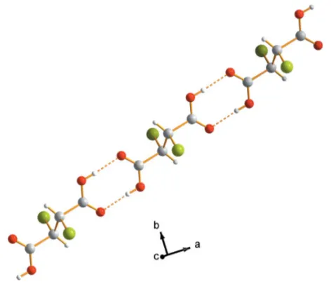
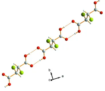
2y, 1z)]. The geometry of the molecule is essentially the same as in the structure of pyri-done–(RS)-2,3-dibromosuccinic acid (1:1) (Aakero¨y et al., 2000). In the crystal structure, the carboxy groups link pairs of molecules, forming an inversion-related closed hydrogen-bonding loop and infinite chains along theaaxis (Fig. 2).
Experimental
An aqueous solution (2.5 ml) containing 0.69 mol l1of fumaric acid, 2.1 mol l1of KBr and 1.9 mol l1of Br
2was placed in a boiling water bath. To avoid precipitation of KBr, the volume of the solution was kept constant by addition of deionized water. After 10 min, crystals of (I) were vacuum-filtered and placed in a heated cabinet at 373 K for 1 h.
Crystal data
C4H4Br2O4
Mr= 275.89
Monoclinic,C2=c a= 14.244 (1) A˚ b= 5.1664 (6) A˚ c= 11.3736 (8) A˚ = 117.684 (9)
V= 741.17 (13) A˚3 Z= 4
Dx= 2.477 Mg m 3
MoKradiation Cell parameters from 37
reflections = 4.3–21.0
= 10.91 mm1 T= 298 K Block, colourless 0.330.300.27 mm
Data collection
Bruker–Nonius KappaCCD diffractometer
’scans
Absorption correction: numerical HABITUS(Herrendorf & Ba¨rnighausen, 1997) Tmin= 0.091,Tmax= 0.137
5265 measured reflections
849 independent reflections 679 reflections withI> 2(I) Rint= 0.039
max= 27.5
h=15!18 k=6!6 l=14!14
Refinement
Refinement onF2
R[F2> 2(F2)] = 0.045
wR(F2) = 0.102
S= 1.08 849 reflections 47 parameters
H-atom parameters constrained
w= 1/[2(F
o2) + (0.0299P)2
+ 8.0961P]
whereP= (Fo2+ 2Fc2)/3
(/)max< 0.001
max= 1.14 e A˚
3
min=0.73 e A˚
3
Table 1
Selected bond lengths (A˚ ).
Br1—C2 1.969 (8)
O1—C1 1.229 (10)
O2—C1 1.244 (9)
C1—C2 1.605 (11)
C2—C2i
1.403 (16)
Symmetry code: (i)xþ1 2;yþ
1 2;zþ1.
Table 2
Hydrogen-bond geometry (A˚ ,).
D—H A D—H H A D A D—H A
O1—H1 O2ii
0.82 1.83 2.650 (7) 175
Symmetry code: (ii)x;y;zþ1.
The H atoms were located in a difference Fourier map and were refined using a riding model, with C—H = 0.96 A˚ and Uiso(H) =
1.2Ueq(C), and with O—H = 0.82 A˚ andUiso(H) = 1.5Ueq(O). The highest peak is located 0.97 A˚ from atom C2.
Data collection: COLLECT (Nonius, 1999); cell refinement:
DIRAX/LSQ(Duisenberget al., 2003); data reduction:EVALCCD
(Duisenberg, 1992); program(s) used to solve structure:SHELXS97
(Sheldrick, 1997); program(s) used to refine structure:SHELXL97
(Sheldrick, 1997); molecular graphics: DIAMOND (Brandenburg, 2005); software used to prepare material for publication:MAXUS
(Mackayet al., 1999).
The Swedish Research Council (VR) is acknowledged for providing funding for the single-crystal diffractometer.
References
Aakero¨y, C. B., Beatty, A. M., Nieuwenhuyzen, M. & Zou, M. (2000). Tetrahedron,56, 6693–6699.
Bolte, M. & Degen, A. (2000).Acta Cryst.C56, e410.
Brandenburg, K. (2005).DIAMOND.Release 3.1. Crystal Impact GbR, Bonn, Germany.
Duisenberg, A. J. M. (1992).J. Appl. Cryst.25, 92–96.
Duisenberg, A. J. M., Kroon-Batenburg, L. M. J. & Schreurs, A. M. M. (2003). J. Appl. Cryst.36, 220–229.
Herrendorf, W. & Ba¨rnighausen, H. (1997). HABITUS. Universities of Giessen and Karlsruhe, Germany.
Mackay, S., Gilmore, C. J., Edwards, C., Stewart, N. & Shankland, K. (1999). MAXUS. Bruker–Nonius, The Netherlands, MacScience, Japan, and The University of Glasgow, Scotland.
Nonius (1999).COLLECT. Nonius BV, Delft, The Netherlands.
Sheldrick, G. M. (1997). SHELXS97 and SHELXL97. University of Go¨ttingen, Germany.
organic papers
Acta Cryst.(2006). E62, o200–o201 Erikssonet al. C
[image:3.610.319.555.69.280.2]4H4Br2O4
o201
Figure 2
[image:3.610.105.229.75.181.2]The chains formed by hydrogen bonds in (I). Hydrogen bonds are indicated by dashed lines.
Figure 1
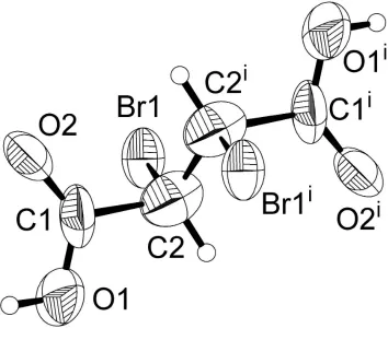
The (R,S)-2,3-dibromosuccinic acid molecule. Displacement ellipsoids are drawn at the 50% probability level [symmetry code: (i)1
2x, 1
supporting information
sup-1
Acta Cryst. (2006). E62, o200–o201
supporting information
Acta Cryst. (2006). E62, o200–o201 [doi:10.1107/S1600536805040493]
(
RS
)-2,3-Dibromosuccinic acid
Margareta Eriksson, Andreas Fischer, Johan Lind and
Å
sa Zazzi
S1. Comment
Some time ago, the structure of racemic 2,3-dibromosuccinic acid, which had been obtained by an electrophilic reaction
between maleic acid and bromine, was determined (Bolte & Degen, 2000). The structure features a complex pattern of
hydrogen bonds between carboxy groups of adjacent acid molecules. Inspired by the fact that the melting points of the
racemic and the meso compounds are extremely different (racemate: 444 K; meso compound: 528 K), we expected very
different hydrogen-bonding patterns in the two phases and decided therefore to determine the structure of the meso
compound. From a reaction between bromine and fumaric acid, we obtained single crystals of the meso compound, (I),
whose structure is described here. The molecule lies about an inversion centre located at the mid-point of the C2—C2i
bond [symmetry code: (i) 1/2 − x, 1/2 − y, 1 − z). The geometry of the molecule is essentially the same as in the structure
of pyridone.(R,S)-2,3-dibromosuccinic acid (1:1) (Aakeröy et al., 2000). In the crystal structure, the carboxy groups link
pairs of molecules, forming an inversion-related closed hydrogen-bonding loop and infinite chains along the a axis (Fig
2).
S2. Experimental
An aqueous solution (2.5 ml) with the respective concentrations 0.69 mol l−1 of fumaric acid, 2.1 mol l−1 of KBr and 1.9
mol l−1 of Br
2 was placed in a boiling water bath. To avoid precipitation of KBr, the volume of the solution was kept
constant by addition of deionized water. After 10 min, crystals of (I) were vacuum-filtered and placed in a heated cabinet
at 373 K for one hour.
S3. Refinement
The H atoms were located in a difference Fourier map and were refined using a riding model, with C—H = 0.96 Å and
supporting information
sup-2
[image:5.610.129.483.71.382.2]Acta Cryst. (2006). E62, o200–o201 Figure 1
The (R,S)-2,3-dibromosuccinic acid molecule. Displacement ellipsoids are drawn at the 50% probability level [symmetry
supporting information
sup-3
[image:6.610.122.489.73.393.2]Acta Cryst. (2006). E62, o200–o201 Figure 2
The chains formed by hydrogen bonds in (I). Hydrogen bonds are indicated by dashed lines.
(I)
Crystal data
C4H4Br2O4
Mr = 275.89
Monoclinic, C2/c
Hall symbol: -C 2yc
a = 14.244 (1) Å
b = 5.1664 (6) Å
c = 11.3736 (8) Å
β = 117.684 (9)°
V = 741.17 (13) Å3
Z = 4
Dx = 2.477 Mg m−3
Mo Kα radiation, λ = 0.71073 Å Cell parameters from 37 reflections
θ = 4.3–21.0°
µ = 10.91 mm−1
T = 298 K Cube, colourless 0.33 × 0.30 × 0.27 mm
Data collection
Bruker–Nonius KappaCCD diffractometer
Radiation source: fine-focus sealed tube
φ scans
Absorption correction: numerical
HABITUS (Herrendorf & Bärnighausen, 1997)
Tmin = 0.091, Tmax = 0.137
5265 measured reflections
849 independent reflections 679 reflections with I > 2σ(I)
Rint = 0.039
θmax = 27.5°, θmin = 4.5°
h = −15→18
k = −6→6
supporting information
sup-4
Acta Cryst. (2006). E62, o200–o201
Refinement
Refinement on F2
Least-squares matrix: full
R[F2 > 2σ(F2)] = 0.045
wR(F2) = 0.102
S = 1.08 849 reflections 47 parameters 0 restraints
Primary atom site location: structure-invariant direct methods
Secondary atom site location: difference Fourier map
Hydrogen site location: inferred from neighbouring sites
H-atom parameters constrained
w = 1/[σ2(F
o2) + (0.0299P)2 + 8.0961P]
where P = (Fo2 + 2Fc2)/3
(Δ/σ)max < 0.001
Δρmax = 1.14 e Å−3
Δρmin = −0.73 e Å−3
Special details
Geometry. All e.s.d.'s (except the e.s.d. in the dihedral angle between two l.s. planes) are estimated using the full covariance matrix. The cell e.s.d.'s are taken into account individually in the estimation of e.s.d.'s in distances, angles and torsion angles; correlations between e.s.d.'s in cell parameters are only used when they are defined by crystal symmetry. An approximate (isotropic) treatment of cell e.s.d.'s is used for estimating e.s.d.'s involving l.s. planes.
Refinement. Refinement of F2 against ALL reflections. The weighted R-factor wR and goodness of fit S are based on F2,
conventional R-factors R are based on F, with F set to zero for negative F2. The threshold expression of F2 > σ(F2) is used
only for calculating R-factors(gt) etc. and is not relevant to the choice of reflections for refinement. R-factors based on F2
are statistically about twice as large as those based on F, and R- factors based on ALL data will be even larger.
Fractional atomic coordinates and isotropic or equivalent isotropic displacement parameters (Å2)
x y z Uiso*/Ueq
Br1 0.15304 (5) 0.36080 (15) 0.28052 (6) 0.0568 (3) O1 0.0918 (5) −0.1270 (12) 0.4481 (7) 0.0808 (17)
H1 0.0395 −0.1590 0.4580 0.121*
O2 0.0784 (4) 0.2541 (10) 0.5270 (5) 0.0636 (13) C1 0.1201 (5) 0.0978 (18) 0.4820 (8) 0.065 (2) C2 0.2153 (7) 0.1680 (17) 0.4492 (8) 0.080 (2)
H2 0.2491 0.0147 0.4392 0.096*
Atomic displacement parameters (Å2)
U11 U22 U33 U12 U13 U23
Br1 0.0415 (3) 0.0827 (5) 0.0441 (4) 0.0038 (3) 0.0182 (3) 0.0114 (3) O1 0.077 (4) 0.073 (4) 0.130 (5) 0.002 (3) 0.079 (4) 0.007 (3) O2 0.052 (3) 0.057 (3) 0.074 (3) −0.021 (2) 0.023 (2) −0.004 (3) C1 0.035 (3) 0.083 (6) 0.084 (5) 0.003 (4) 0.033 (3) 0.025 (4) C2 0.089 (6) 0.074 (6) 0.069 (5) 0.016 (4) 0.030 (4) −0.003 (4)
Geometric parameters (Å, º)
Br1—C2 1.969 (8) C1—C2 1.605 (11)
O1—C1 1.229 (10) C2—C2i 1.403 (16)
O1—H1 0.8200 C2—H2 0.9600
O2—C1 1.244 (9)
supporting information
sup-5
Acta Cryst. (2006). E62, o200–o201
O1—C1—O2 126.1 (6) C1—C2—Br1 107.1 (5)
O1—C1—C2 109.2 (7) C2i—C2—H2 113.1
O2—C1—C2 124.5 (7) C1—C2—H2 111.5
C2i—C2—C1 107.1 (9) Br1—C2—H2 108.9
Symmetry code: (i) −x+1/2, −y+1/2, −z+1.
Hydrogen-bond geometry (Å, º)
D—H···A D—H H···A D···A D—H···A
O1—H1···O2ii 0.82 1.83 2.650 (7) 175