CENTROSOME, CELL GEOMETRY AND TISSUE TOPOLOGY.
Audio Logic: Department of Bioengineering and Mathematical Modeling, Via Francesco Ferrucci 6, 20145 Milano (Italy)
ARTICLE INFO ABSTRACT
Despite decades of studies
model explaining anisotropy andbilateral symmetryof Metazoa development: the main problem consists in the transition from a linear (1D) genetic code to spatial (3D)cells and tissueso
Here,a simple metazoan developing systemis attentively reviewed, sea urchin pluteus skeleton formation.
propertiesfor programming and controlling anisotropy, bilat
necessary to solve the topological problems of developing tissues in Metazoa: it is possible to conclude that, by a topological point of view, without an intrinsic cellular spherical (3D) reference system (the centrosome
orientation and positioning of receptor developmental program
Copyright ©2014 Marco Regolini. This is an open access article distributed under the Creative Commons Attribution License, which permits unrestricted use, distribution, and reproduction in any medium, provided the original work is properly cited.
INTRODUCTION
"Once upon a time ... a zygote began to divide": this is not a fairy tale but the story of each organism; the end, again and again, is always the same: depending on the species a new organism is born with the morphological characteristics of its species.From simple structures, composed of a few cells and then more understandable, we have a lot of things to learn: a little number of dermal cells stimulates few epiderm
realize a hair follicle, whose orientation with the sagittal plane and the plane of the epidermis shows predetermined values; in human eyebrows the orientation of hairs is accurately patterned so that different portions of the eyebrow show diff (stereotypical) angles in respect to the anterior
cranio-caudal) axis. Similarly sea urchin larva (pluteus) possesses a characteristic skeleton whose bilaterally symmetric shape (angles between spicules) does not change
experimentally perturbed.
Sea urchin skeleton
After fertilizationthe first and second cleavage of the sea urchin zygote(Hardin, 1996)occurmeridionally (division planes are parallel to the animal-vegetal axis). The next cleavage is equatorial (division plane perpendicular to the animal
axis: animal is “North” or anterior pole, vegetal
*Corresponding author: Marco Regolini
AudioLogic, Department of Bioengineering and Mathematical Modeling, Via Francesco Ferrucci 6, 20145 Milano (Italy).
ISSN: 0975-833X
Article History:
Received 22nd August, 2014
Received in revised form 29th September, 2014
Accepted 24th October, 2014
Published online 18th November,2014
Key words:
Centrosome, Organogenesis, Morphogenesis.
RESEARCH ARTICLE
LL GEOMETRY AND TISSUE TOPOLOGY. THE BIOLOGICAL 3D REFERENCE
SYSTEM OF METAZOA
*Marco Regolini
Department of Bioengineering and Mathematical Modeling, Via Francesco Ferrucci 6, 20145 Milano (Italy)
ABSTRACT
Despite decades of studies and researches, morphologists have no good ideas to propose a theoretical model explaining anisotropy andbilateral symmetryof Metazoa development: the main problem consists in the transition from a linear (1D) genetic code to spatial (3D)cells and tissueso
Here,a simple metazoan developing systemis attentively reviewed, sea urchin pluteus skeleton formation. During this process chemical gradients (morphogens) do not show the geometrical propertiesfor programming and controlling anisotropy, bilateral symmetry and 3D orientation necessary to solve the topological problems of developing tissues in Metazoa: it is possible to conclude that, by a topological point of view, without an intrinsic cellular spherical (3D) reference system (the centrosome, with its orthogonal enantiomorphouscentrioles) capable of driving the orientation and positioning of receptors, junctions, cell division planes
developmental programs cannot be performed.
is an open access article distributed under the Creative Commons Attribution License, which permits unrestricted use, distribution, and reproduction in any medium, provided the original work is properly cited.
"Once upon a time ... a zygote began to divide": this is not a fairy tale but the story of each organism; the end, again and the species a new organism is born with the morphological characteristics of its species.From simple structures, composed of a few cells and then more understandable, we have a lot of things to learn: a little number of dermal cells stimulates few epidermal cells to realize a hair follicle, whose orientation with the sagittal plane and the plane of the epidermis shows predetermined values; in human eyebrows the orientation of hairs is accurately patterned so that different portions of the eyebrow show different (stereotypical) angles in respect to the anterior-posterior (or caudal) axis. Similarly sea urchin larva (pluteus) possesses a characteristic skeleton whose bilaterally symmetric does not change if size is
After fertilizationthe first and second cleavage of the sea urchin zygote(Hardin, 1996)occurmeridionally (division planes are vegetal axis). The next cleavage is perpendicular to the animal-vegetal axis: animal is “North” or anterior pole, vegetal
Department of Bioengineering and Mathematical Modeling, Via
is “South” or posterior): so four cells (the animal blastomeres) stand above fourso called “vegetal blastomeres”. Later theanimal blastomeres dividemeridionally, forming a tier of 8 cells (mesomeres), whereas the vegetal blastomeres divide equatorially and asymmetrically: four large macromeres are under the ring of mesomeres, four small micromeres lie under the macromeres. Also these micromeres divide asymmetrically arising four smallmicromeres at the vegetal pole and four large micromeres (skeletogenic cells) above
blastomere divides, the embryo reaches a stage of a “ball” (blastula: a cavity, the coelom, inside one layer of superficial cells) composed of 60 cells arranged in rings of 16 or 8 or 4 cells whose fates are fixed.We are interested
large micromeres (McClayet al
mesenchyme cells(PMCs)which enter inward (Wessel 2004) for building the skeleton, migrate to form two ventrolateral clusters (Armstrong
ring and finally fuse to form a syncytium (Boehm
Animal blatsomeres, derived from the mesomeres, form the ectoderm. Sea urchin larval skeleton, composed of calcite (CaCO3), shows a shape similar to a chair skeleton with a back
and four legs: depending on species, there are differences in the number of legs (4, 6 or even 8) (Rahman
shape of connectors between the legs and in the inclination of the spicules forming the back. Many proteins (about 50, obviously DNA coded) called sp
associated with spicules and involved in biomineralization and formation of spicules. Skeleton formation (Okazaki, 1960) starts in the two bilaterally symmetric ventrolateral clusters of
International Journal of Current Research
Vol. 6, Issue, 11, pp.9685-9689, November,2014
THE BIOLOGICAL 3D REFERENCE
Department of Bioengineering and Mathematical Modeling, Via Francesco Ferrucci 6, 20145 Milano (Italy)
and researches, morphologists have no good ideas to propose a theoretical model explaining anisotropy andbilateral symmetryof Metazoa development: the main problem consists in the transition from a linear (1D) genetic code to spatial (3D)cells and tissuesorganization. Here,a simple metazoan developing systemis attentively reviewed, sea urchin pluteus skeleton During this process chemical gradients (morphogens) do not show the geometrical eral symmetry and 3D orientation necessary to solve the topological problems of developing tissues in Metazoa: it is possible to conclude that, by a topological point of view, without an intrinsic cellular spherical (3D) reference ith its orthogonal enantiomorphouscentrioles) capable of driving the s, cell division planes and extracellular matrix fibers,
is an open access article distributed under the Creative Commons Attribution License, which permits unrestricted use,
or posterior): so four cells (the animal blastomeres) stand above fourso called “vegetal blastomeres”. Later theanimal blastomeres dividemeridionally, forming a tier of 8 cells (mesomeres), whereas the vegetal blastomeres divide cally: four large macromeres are under the ring of mesomeres, four small micromeres lie under the macromeres. Also these micromeres divide asymmetrically arising four smallmicromeres at the vegetal pole and four large micromeres (skeletogenic cells) above them. Because not every blastomere divides, the embryo reaches a stage of a “ball” (blastula: a cavity, the coelom, inside one layer of superficial cells) composed of 60 cells arranged in rings of 16 or 8 or 4 cells whose fates are fixed.We are interested in considering the
et al., 1992), called primary
mesenchyme cells(PMCs)which enter inward (Wessel et al. 2004) for building the skeleton, migrate to form two ventrolateral clusters (Armstrong et al.,1993) and an equatorial finally fuse to form a syncytium (Boehm et al., 2010). Animal blatsomeres, derived from the mesomeres, form the ectoderm. Sea urchin larval skeleton, composed of calcite ), shows a shape similar to a chair skeleton with a back ing on species, there are differences in the number of legs (4, 6 or even 8) (Rahman et al., 2012), in the shape of connectors between the legs and in the inclination of the spicules forming the back. Many proteins (about 50, obviously DNA coded) called spicule matrix proteins are associated with spicules and involved in biomineralization and formation of spicules. Skeleton formation (Okazaki, 1960) starts in the two bilaterally symmetric ventrolateral clusters of
PMCs where a “triradiate” structure appears (so two bilaterally symmetric triradiate-spicules are the primordia of the skeleton). One of these three arms (directed backwards or dorsally) elongates 10 µm, divides and bends at right angles originating two arms with the same 3D orientation but opposite direction: one, named body rod,elongates posterior-ward, the second, termed postoral rod, elongates (on the same line)but anterior-ward. The second arm of the triradiate primordium extends ventrally and is called the transverse rod: both the left and right transverse rods converge and, depending on the species, can join together. The last arm, the dorsoventral connecting rod, grows ventrally directed and then curves and elongates anterorlaterally (anterolateral rod).
1) When PMCs are cultured in vitro, in the absence of ectodermal cells (Kitajimaand Urakami, 2000), skeleton formation begins with the appearance of some triradiate spicules, that develop into a correctly shaped,although incomplete, 3D skeleton like in living plutei, only lacking dorsoventral and anterorlateral rods. PMCs are able to assemble spicule in the absence of ectodermal cues.
2) The PMCs during migration and skeleton formation extend filopodia in every direction (Guss and Ettelsohn, 1997), that are believed to provide an input of information to the PMCs (Kaneko et al., 2005): after receiving patterning cues from the ectoderm, PMCs build the skeleton by themselves, independently from external signals, using their own genotypic information. In fact, after transplantation of single PMCs, added into different positions in the PMC ring, any PMC is capable of producing any part of the skeleton, whose size and shape remains unaltered. Furthermore (Lyons et al., 2012) cross transplants between close species
(Tripneustesesculentus and Lytechinusvariegatus)
demonstrate the genotypic nature of patterning; the two species produce differently shaped skeletons and when the entire set of PMCs from one species(donor) is transferred to replace all the PMCs of the other embryo (host), PMCs form a skeleton corresponding to the genotype of PMCs: the host embryo does not have its typical skeletonbut the skeleton typical of the donor. This experiment indicates that PMCs are programmed (DNA code) to build a particular type of skeleton with its proper angle between the arms: the ectoderm supplies only spatial information, signals telling to the PMCs where they are: as a response, PMCs reorient themselves and correctly orient the arms of the skeleton in respect to the gastrula polarity.
3) If more supernumerary PMCs are experimentally added(50-100, i.e. a very large number because during gastrulation, in L. variegatusonly 32 cells ingress and
divide to produce 64, whereas in
Strongylocentrotuspurpuratus16 cells ingress and divide
to produce 32 PMCs), the size of the skeleton does not change and, above all,spiculetilting and orientation remain unaltered.
4) In half and quarter-sized embryos (obtained by separating the first 4 blastomeres) there are, depending on the species, 32 or 16 PMCs as usual, that produce
half or quarter-sized skeletons: spicule-rod size changes, angles between spicules does not (homothety).
Now it is clear that PMCs are not patterned or driven by ectodermal cells: from ectoderm they obtain only position information useful for positioning correctly themselves in order to build the skeleton, precisely orienting it in respect to the whole organism (anus, mouth, archenteron). This implies that PMCs are finely polarized, much more than a simple up-down front-rear left-right polarity, but something like an icosahedron with many different and identifiable (by different receptors) faces (compartments). ECM fibers, produced in the Golgi system and carried in large vesicles to the cell membrane can be oriented along the line that joins two identifiable different receptors located in two different close compartments: thus exocytic vesicles can be aligned along many different directions (through such fine-tuned polarity) and drive the correct orientation of skeletal fibers (Hodoret al., 2000); the intrinsic program (stored in DNA) imposes from time to time the proper orientation of skeletal fibers to obtain 4,6,8 armed plutei.
From 1D linear DNA molecule to 3D (bilateral symmetric) spatial organization of metazoan tissues and organs
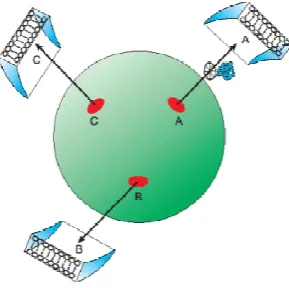
[image:2.595.359.504.591.735.2]Centrosomes are absent in Plants that are only ableto realize simple laminar tissues, i.e. 2D-structures,or, at the most, cylindrical structures (apparently 3D, but originated, by rolling and wrapping,from 2D-laminar sheets); similarly,Planaria, Platelmyntes lacking centrosomes, are (as their name “Platelmyntes” suggests) flat like leaves and cannot even perform gastrulation. On the contrary all the other Metazoa possess centrosomes (one per cell, save multiciliated cells), gastrulate and build very complex organ (the heart, the eye, the middle and inner ear for instance). Centrosomes are made up of two orthogonal (during S, G2 and M phases of the cell cycle) centrioles, disposed like the capital letter “L”: an axial ”Mother” Centriole(MC) and an eccentric “Daughter” Centriole (DC) embedded in an (apparently) amorphous protein matrix named PeriCentriolar Material, responsible for anchoring and nucleation of microtubules (MTs): an “aster” of non-intersecting robust MTs irradiates radially from the centrosomal γ-TuRCs (the structures that assemble each microtubule) toward the cell cortex, like, from the central square of a city, many streets irradiate toward (and connect with) the periphery (Fig. 1). Each centriole is composed of 9 orderly spaced-out MT parallel blades named “triplets”.
Small ellipses represent γ-TuRCs on the centrosome (large sphere): each one is identified by its own private receptor specific for its longitude and latitude(capital letters: A, B, C) which recognizes only the corresponding targeting sequence. Each γ-TuRC has the orientation of the plane which, in that point, is tangent to the centrosome “spherical” surface. MTs (arrows) are nucleated with directions imposed by the orientation of the corresponding γ-TuRC: like orientation, like direction (one “discrete” orientation, one “discrete” direction). So, a molecular complex (twisted tube) through its “geometric” targeting sequence, recognizes and links exclusively the receptor (A or B or C) which marks theγ-TuRCthat has the correct orientation to nucleate a microtubule directed to the desired (corresponding: A or B, or C) destination, reached through a kinesin carrier (wheel): one targeting sequence, one γ-TuRC receptor, one cortical compartment: one-to-one univocal correspondence. (From: M. Regolini Centrosome: a geometrical model Lambert Academic Publishing Germany 2014)
The two orthogonal centrioles possess 9-fold symmetry and circumferential polarity (non-equivalenceof triplets, different from each other: Beisson and Jerka-Dziadosz, 1999): the centrosome, by its aster of MTs, is a geometric organelle with the structure of a tool capable of realizing a high-resolutionmap of position within the cell (Regolini, 2013).Centrioles are the
only cell organelles which show a real
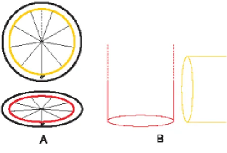
[image:3.595.45.275.469.624.2]geometricalorganization, an intriguing 9-fold symmetric structure very resembling to a protractor: effectively, a pair of 9-fold symmetric cylinders, oriented at right angles to each other and capable of irradiating an aster of radially directed MTs, combined to the molecular non-equivalence of triplets (molecular “labels” or “address receptors”) constitutes aspherical reference system organizer, based on two biological 9-marks protractors (Fig. 2).
Figure 2. Centrosome theoretical geometrical model: a spherical reference system composed of two orthogonal
protractors/goniometers
A: frontal view of two orthogonal protractors/goniometers, subdivided into nine sectors, which schematizes the two orthogonal centrioles: the first (horizontal) represents the base of the MC, arranged on the equatorial “x y“ plane; its “0° ” mark is used to orient the protractor/centriole; the second, the DC (vertical, orthogonal to the first), is closer to the reader: both “0°“ marks coincide; it is convenient to consider the
second protractor divided, by its “vertical” diameter crossing the “0°” mark, into two halves (two opposite symmetrical hemiprotractors). B: schematic lateral view of the proximal end of both centrioles (during S, G2) to show the respective position of the above two sections. (From: M. Regolini Centrosome: a geometrical model Lambert Academic Publishing Germany 2014)
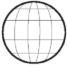
[image:3.595.321.547.565.672.2]One 360°-protractor (a complete ring of 9 different marks) is responsible for longitude (9 meridians) (Fig. 3), the other, orthogonal to the first, is responsible for latitude (4 parallels)and is composed of two symmetric 180° half-protractors facing each other to compose a complete ring(a globe is normally equipped with only one 180° vertical half-protractor): together, they divide the space into nine meridian wedges and five parallel sectors (two polar caps and three parallel disks). In the centrosome the MC is responsible for longitude (9 meridian wedges), the DC for latitude (5 parallel sectors). These nine wedges and fivesectors subdivide the centrosome surface into 45 small areas (scaffolds for γ-TuRCs) each oriented in correspondence to its position (Fig. 4): their inclination is the result of the addition of two inclinations, one imposed by theMC(longitude) and the other by the DC(latitude). As on a globe, longitude covers the entire circumference (2π; 9 different meridians or 9 different 40° wedges) while latitude covers (symmetrically) only half circumference (π: from North to South pole; 2 caps and 3 parallel discs). This “two-protractors instrument” is sufficient to subdivide the space into 45 pyramidal frustums with the little base at the center, each one identifiable by its own longitude and latitude: the large base (subtending a vertex solid angle of 4π /45 steradians) has an extension of 4π r2/45, thus, in a cell with a diameter of 10 μm (radius: 5 μm; surface: 4π r2 approximately 314 μm2) it corresponds to a cell cortex extension of about 7 μm2 (a circle with a diameter of 3 μm, or a square with a side of 2.6 μm). These dimensions together with the physical properties of the MTs (bending-resistance and rigidity) give an idea about the interesting order of magnitude of the noise-resistance of this system and of its fine-tuned precision,much better than that of chemical gradients: 45 cell cortexcompartments (or rather poles) are much more than six poles(anterior, posterior, dorsal, ventral, left and right)(Regolini, 2013).
Figure 3. Centrosome theoretical geometrical model: from 2D rotational polarity to 3D spherical polarity
annulus, weakly coloured), whose γ-TuRCs (small bars) acquire an inclination parallel to the corresponding centriolar blade; each MC blade faces one meridian wedge. In each wedge, all the γ-TuRCs have the same longitudinal inclination. B: after the intervention of the DC, that imposes a rotational inclination corresponding to that of its blades, each γ-TuRC acquires also the latitude inclination which is added to that of longitude. There is a double inclination: firstly each γ-TuRC is parallel to the corresponding blade of the MC, then it acquires the inclination parallel to the corresponding DC blade; the eccentric positioned DC is responsible for “latitude” (two opposed spherical caps and three parallel spherical disks): this second centriole/protractor is composed of two symmetric hemi-protractors/goniometers. C: all the γ-TuRCs contained in the same cap or disc (coloured circles) whatever their longitudinal orientation, are rotated to acquire the same latitudinal orientation, identical in the same cap or disk. So, two 2D circumferential-rotational polarities are merged to realize a 3D spherical polarity. (From: M. Regolini Centrosome: a geometrical model Lambert Academic Publishing Germany 2014)
Figure 4. Centrosome theoretical geometrical model: discrete subdivision of the centrosome surface
Nine meridians and four parallels subdivide the centrosome surface into 45 small areas (scaffolds for oriented γ-TuRC, which include SAS-4/CPAP, CNN, Asl and Pericentrin), each oriented in correspondence to its position: their inclination is the result of the addition of two inclinations, one imposed by the MC (longitude) and the other by the DC (latitude). As on a globe, longitude covers the entire circumference (2π; 9 different meridians or 9 different meridian 40° wedges) while latitude covers (symmetrically) only half circumference (π; 2 caps and 3 parallel discs). (From: M. Regolini Centrosome: a geometrical model Lambert Academic Publishing Germany 2014)
Control of spatial disposition of junctions, receptors, extracellular fibers and spindle poles
The centrosome, as the main microtubule organizing center and because of the 9-fold symmetry of its centrioles, their (transient) orthogonal arrangement and, above all, their circumferential polarity (non-equivalence of triplets), may play the role of a biological discrete and noise resistant interface, built on two orthogonal protractors, shaped like a polyhedron of 45 faces (each equipped with its own longitude-latitude recepteors), that recognizes (receptor-ligand interaction) and decodes morphogenetic instructions, or, more generally, topogenic molecular targeting signals (frequently present at the
N-terminus of newly synthesized proteins or in the 3’UTR of mRNAs) and translates them by delivering each targeted molecular complex (polarity and adhesion factors, transmembrane receptors for extracellular matrix fibers) into its expected 3D real location in the cell: like an interface or a wiring device, the centrosome connects each targeting sequence with the corresponding correctly-oriented microtubule: in this way morphogenetic geometric (DNA linearly 1D coded) instructions are translated by the centrosome into actual 3D locations in cells to build 3D tissues and organs. Centrosome molecular geometry and architecture imply its function: through the centrosome and its aster of robust MTs, DNA can draw, build and “label” the intrinsic 3D grid line of the cell. Centrosome, aster and primary cilium (its basal body is a centriole of the centrosome) constitute the “hardware” of an interactive cross-talking system that manages geometrical communication inside the cell and between cells and establishes in tissues coordinated and shared cell polarity. Targeting sequences and related receptors constitute the “software”.Centriole and centrosome duplication follow a unique and characteristic process: it is part of the mechanism by which the cytoskeleton of the daughter cell (centrosome and aster) is patterned in respect to that of the mother to maintain a coordinated and shared polarity.
The MC, before disengagement, transmits to its old DC the information of orientation, and physically orients it in respect to the cytoskeleton; effectively also in Ciliates the process of centriole duplication occurs at right angle and utilizes a pre-existing centriole as a platform to orientate the arising centriole polarity in order to insert it correctly in the complex cytoskeleton, something like a new trolley-bus (whose two sprung trolley poles must be correctly connected to the two polarized electric wires) is orientated and correctly (front-rear) positioned in respect to the “electric city-skeleton” made of aerial-suspended wires; so the cells in a tissue become co-ordinately polarized by co-co-ordinately oriented centrosomes: in Metazoa the centrosome is the “intrinsic” (no external cues) reference system; plants, fixed in the ground, use an “extrinsic” reference system (light and gravity), just as a compass uses (extrinsic) Earth magnetism and a GPS utilizes (extrinsic) satellites: as an “extrinsic” reference system is common to each receiver, similarly an “intrinsic” reference system must be the same (identically oriented) in each cell; multicellular organisms must possess a mechanism to transmit, share and co-ordinate their inside points of reference (as we have seen, centrosomes increase cell polarity up to 45 poles) and this function is performed through the orientation imprinted by the MC to its “old” DC before disengagement, so that two co-oriented MCs build two co-co-oriented centrosomes before cell division: so, all the cells of a tissue have the same points of reference to correctly build complex 3D organs. This unique behaviour of centrosomes supports the idea that their main role is the translation of the virtual (DNA coded) cell geometry into an actual real cell wiring system. When a new procentriole arises (orthogonally and near the MC) the cartwheel is formed: 9-fold symmetry and chiral non-equivalence of triplets (fundamental, in the geometrical model of centrosome functioning, for left-right patterning) are established co-ordinately and corresponding with those of the MC.
Bilateral symmetry
The centrosome (the most or, rather, the only chiral, enantiomorphous cell structure) can play a geometric key role in left-right patterning: two globes are bilaterally symmetric if their longitude shows reversed orientation, i.e. in one globe the values from 0° to 360° grow following a clockwise direction, whereas in the mirror symmetric globe longitude values grow in the opposite, reversed (counter-clockwise) direction. Thus MC circumferential polarity, if reversely oriented, constitutes a likely easy base for building bilaterally symmetric organisms: genetic programs must not be changed because their coded geometrical instructions can be carried out by bilateral symmetric centrosomes and so translated in a bilaterally symmetric way: one program -> two chiral executive tools (centrosomes) -> two bilaterally symmetric structures (Regolini, 2013).
REFERENCES
Armstrong R., Hardin J.,McClay D.R. 1993. Cell-cell interactions regulate skeleton formation in the sea urchin embryo.Development, 119, 833-840
Beisson J., Jerka-Dziadosz M. 1999. Polarities of the centriolar structure: morphogenetic consequences. Biol. Cell., 91: 367-378.
Boehm B., Westerberg H., Lesnicar-Pucko G., Raja S., Rautschka M., Cotterell J., Swoger J., Sharpe J. 2010. The Role of Spatially Controlled Cell Proliferation in Limb Bud Morphogenesis.PLoS Biology 8 : 7 e1000420. Guss K.A., Ettensohn C.A. 1997. Skeletal morphogenesis in
the sea urchin embryo: regulation of primary mesenchyme gene expression and skeletal rod growth by ectoderm-derived cues. Development,124, 1899-1908.
Hardin J. 1996. The Cellular Basis of Sea Urchin Gastrulation.Current Topics in Dev. Biol., Vol. 33. Hodor P.G., Illies M.R., Broadley S., Ettensohn C. A. 2000.
Cell-Substrate Gastrulation: Interact with That Contain and Multiple Interactions during Sea Urchin Migrating Primary Mesenchyme Cells and Align Extracellular Matrix Fibers ECM3, a Molecule with NG2-1ike Calcium-Binding Domains. Dev. Biol., 222, 181.-194.
Kaneko H., Okai M., Murabe N., Shimizu T., Ikegami S.,Dan-Sohkawa M. 2005. Fibrous Component of the Blastocoelic Extracellular Matrix Shapes Epithelia in Concert with Mesenchyme Cells in Starfish Embryos Dev. Dynam. 232:915–927.
KitajimaT., Urakami H. 2000. Differential distribution of spicule matrix proteins in the sea urchin embryo skeleton.
Develop. Growth Differ., 42, 295–306.
Lyons D.,Kaltenbach S., McClay D.R. 2012. Morphogenesis in sea urchin embryos: linking cellular events to gene regulatory network states. Dev Biol., 1(2): 231–252. McClay D.R., Armstrong A., Hardin J. 1992 Pattern formation
during gastrulation in the sea urchin embryo. Development
Supplement, 33-41
Okazaki H. 1960. Skeleton formation in sea urchin larvae. II Organic Matrix of the spicules. Embryologia., 5 (3) 283-320.
Okazaki H., Inouè S. 1976.Crystalpropertyof the larval sea urchin spicules.Develop., Growth and Differ., 1 (18) No.4, RahmanM.A.,Yusoff F.,Arshad A., Shamsudin M., Amin
S.M.N. 2012. Embryonic, Larval, and Early Juvenile
Development of the Tropical Sea Urchin,
Salmacissphaeroides(Echinodermata: Echinoidea). The
ScientificWorld Journal Article, ID 938482
Regolini M. 2013. Centrosome: is it a geometric, noise resistant, 3D interface that translates morphogenetic signals into precise locations in the cell? Italian Journal of
Anatomy and Embryology, 118 (1), 19-66
Regolini M. 2014. Centrosome: The Cell Spherical Reference System Organizer. Indian Journal of Research, 3 (6), 84-87
Regolini M. 2014.Centrosome:a geometrical model. Lambert Academic Publishing
Regolini M. 2014. The Spherical Reference System of Metazoan Cells. J.Phys.Chem.Biophys., 4 (151), 2161-0398.1000151
Wessel G.M., Katow H. Regulation of epithelial-to-mesenchymal transition in sea urchin embryos. 2004 in: Rise and fall of epithelial phenotype. Edited by Pierre Savagner Eureka.com and Kluwer Academics/Plenum Publisher
Yajima M., Kiyomoto M. 2006. Study of Larval and Adult Skeletogenic Cells in Developing Sea Urchin Larvae. Biol.
Bull., 211: 183–192.