Case Report
Unusual anatomy of a maxillary second molar: a second
mesiobuccal canal or a second palatal canal?
Meng Ding1, Du Chen1, Weidong Yang2
1Nanjing Stomatological Hospital, Medical School of Nanjing University, 30# Zhongyang Road, Xuanwu District,
Nanjing, China; 2Department of Caries and Endodontics, Nanjing Stomatological Hospital, Medical School of
Nan-jing University, 30# Zhongyang Road, Xuanwu District, NanNan-jing, China
Received August 27, 2017; Accepted July 2, 2018; Epub October 15, 2018; Published October 30, 2018
Abstract: This study presents a maxillary second molar (MSM) with an unusual root canal morphology confirmed by cone-beam computed tomography (CBCT). The tooth had two fused mesiobuccal (MB) roots with 2 separate canals, 1 normal distobuccal (DB) root with 1 canal, and 1 bulky palatal root with 1 canal. The second mesiobuccal (MB2) canal orifice, located in the mesiopalatal (MP) corner, was considered a second palatal (P2) canal when the cham-ber floor was examined, but it was verified as an MB2 based on CBCT. The described case reveals the complexity of MSM variations and emphasizes the importance of using advanced imaging modalities such as CBCT to confirm the 3-dimensional (3D) anatomy of teeth during endodontic treatments. Additionally, an in-depth evaluation of this case based on CBCT could provide new insights into the definition of MB2.
Keywords: Canal morphology, cone-beam computed tomography, second mesiobuccal canal
Introduction
Thorough knowledge of tooth anatomy and root canal morphology is essential for successful root canal treatment. If any portion of the root canal is missed, it cannot be properly cleaned, shaped and sealed, inducing endodontic fail-ure. Therefore, the clinician must be aware of typical anatomical morphologies and certain unique variations during diagnosis and treat-ment [1].
Previous studies have indicated that the maxil-lary molars have the most complicated root canal configurations, especially the second maxillary molars, given their diverse root num-bers and shapes [2, 3]. Several studies, includ-ing in vivo and in vitro studies, have investigat-ed the configuration of the root canal systems of these teeth [4-6], which typically have the following three roots: (1) mesiobuccal (MB), (2) distobuccal (DB) and (3) palatal (P). However, the MB root often has many variations. Kulild and Peters stated that the presence of an MB2 canal in the maxillary first and second molars is fairly common [7]. Some case reports have
described other maxillary second molar (MSM) variations, such as an MSM with additional pal-atal canals and/or roots; however, most of these reports were based on radiographs taken both pre- and postoperatively [8-12].
This paper reports a case of an MSM with four root canals and reviews the literature on MB2 and P2 root canals in MSMs. The vagueness of the morphology of the extra root canal in this case contributes to the difficulties associated with its definition. Clinicians should consider CBCT imaging when such anatomical differenc-es are encountered during treatment.
Case report
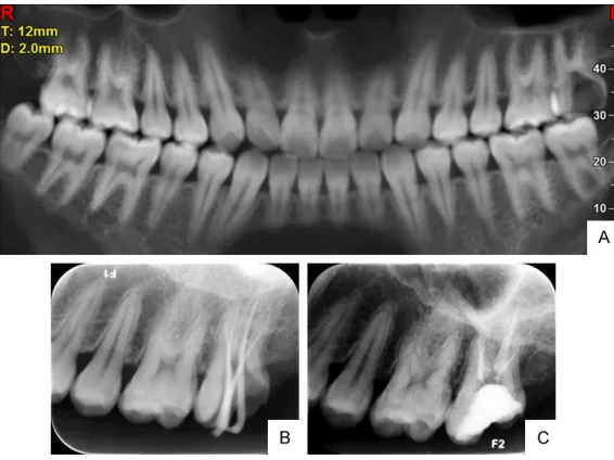
A 22-year-old male patient was directed to the Department of Caries and Endodontics at Nanjing Stomatological Hospital and presented with a history of spontaneous pain in the maxil-lary left second molar. The spontaneous pain had steadily increased in intensity from moder-ate to acute over the preceding few days. The patient’s medical history was noncontributory. Clinical inspection revealed deep occlusal car-ies with severe pain upon probing with an explorer. These findings and sensitivity tests led to a diagnosis of acute irreversible pulpitis requiring root canal therapy. A preoperative panoramic radiograph was taken, and the over-lapping images of the roots alerted us to the existence of a morphological variation (Figure 1A).
To completely understand this complex canal system, a preoperative CBCT image was obtained and analyzed. The CBCT images were obtained using a NewTom VG scanner (QR SRL,
Vertucci) (Figure 2). The geometric location of the MB2 canal was found in relation to the MB1 and P canals. The central points of each canal were located (PMB1, PMB2 and PP), and straight lines were projected between them (PMB1-PP and PMB1-PMB2). A third line was drawn (PMB2-PT) perpendicular to the PMB1-PP line (PT point) according to the protocol described by Betancourt et al. [4]. The distanc-es between the points were measured in milli-meters (Figure 3B).
The root shape characteristics of the involved tooth were represented in detail using a 3D reconstructed image (Figure 4). The image revealed the presence of two mesiobuccal (MB) roots fused from the coronal segment to the apical segment; however, the canals were sep-arate (type I Vertucci). Consequently, two differ-ent canal orifices for the MB roots (one located mesiobuccally and the other located adjacent to the P orifice), two separate foramens in the apical part, and a small apical furcation in the fused root were present.
The patient received 2% lidocaine with 1:100,000 epinephrine for local anesthesia, and the maxillary left second molar was isolat-ed under a rubber dam (Hygenic Coltene, Ohio, USA). After carefully analyzing the CBCT imag-es, a modified trapezoidal endodontic access opening was created using a low-speed dia-mond round bur with the assistance of an
oper-Figure 1. A. Preoperative panoramic radiograph of tooth No. 27. B. Working length determination. C. After root canal obturation.
[image:2.612.90.373.70.282.2]ating microscope (OPMI Pico, Zeiss, Ober- kochen, Germany) to expose all the canals. Clinical examination of the chamber floor with an endodontic explorer (DG-16, Ash Instr- uments, Dentsply, Gloucester, UK) revealed four root canal orifices, including two on the buccal side and two on the palatal side (Figure 3A).
The working lengths of each canal were esti-mated with an electronic apex locator (Root ZX, Morita, Tokyo, Japan) and confirmed with a peri-apical radiograph. Coronal flares were per-formed using Gates-Glidden burs No. 2 and 3 (Dentsply Maillefer, Ballaigues, Switzerland). The root canals were cleaned and shaped using NiTi rotary protapers (Dentsply Maillefer, Ballaigues, Switzerland) with the crown-down technique and were enlarged to the size of a No. 25 file.
The canals were irrigated with 5% and 2.5% sodium hypochlorite between each file during instrumentation. Calcium hydroxide-based in- tracanal dressing was applied to all the canals,
After completing the root canal treatment, the tooth was restored with a posterior composite filling (Z350; 3M Dental Products, St. Paul, MN).
Discussion
Maxillary molars, particularly second molars, have the most complicated root and canal mor-phologies [5]. Zhang et al. reported that among MSMs, 10% had one root, 8% had two roots, and 81% had three roots. Of the MB roots, 22% had two canals and the others had one [14]. This illustrates that the most common MSM morphology is three roots (MB, DB, and P), with one canal in each root. However, many studies have reported abnormal maxillary molars with additional root canals, and the most common anatomic variation is related to the root canal configuration of the MB root. Pablo Betancourt used CBCT to study 225 permanent MSMs and reported that the MB root MB2 canal was pres-ent in 48% of the cases [4]. However, various studies have reported large differences in the detection of the MB2 canal in the MSM
[image:3.612.91.522.71.204.2]accord-Figure 2. A-C: The different CBCT scanning levels of tooth No. 27 before root canal treatment.
Figure 3. A. The chamber floor revealed 4 root canal orifices: MB1, MB2, DB and P. B. Axial view of tooth No. 27. PMB1 (center of the mesiobuccal canal), PMB2 (center of the MB2 canal), and PP (center of the palatal canal).
and a sterile cotton pellet was placed in the pulp cham-ber. The tooth was then fitt- ed with a temporary Cavit filling (ESPE, Seefeld, Ger- many).
[image:3.612.90.376.245.368.2]ing to the technique used; the in vivo preva-lence may range from 19.7% to 73% [15, 16]. The substantial discrepancies in the results among these studies are mostly due to the interpretation of morphological variations using different methods.
Methods used to examine root and canal mor-phologies include canal staining and tooth clearing, serial sectioning, conventional and digital radiography, microscopic evaluation, CT, spiral CT, and CBCT [17-21]. Neelakantan et al. compared five methods (the modified canal staining and clearing technique, CBCT, periph-eral quantitative CT, spiral CT, and plain and contrast medium-enhanced digital radiogra-phy) to investigate their accuracy in studying root canal morphology and found that CBCT imaging was a useful diagnostic tool for this purpose [21]. CBCT uses a cone-shaped beam and multiple exposures around an object to reveal its internal structure. This method has many advantages, including low radiation expo-sure, faster image acquisition without requiring chemicals, and high-resolution images in mul-tiple planes of space while eliminating superim-position of surrounding structures. CBCT has
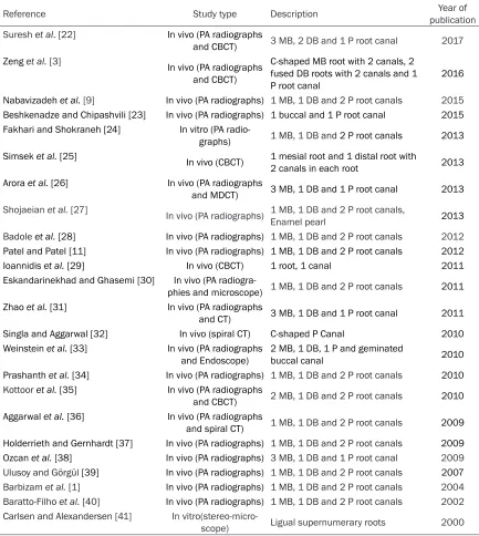
(MP) corner was considered a P2 canal from the coronal view under direct vision; however, CBCT scanning revealed that it was an MB2 canal. The tooth had four roots and four canals, including two fused MB roots with one canal in each, a DB root with one canal, and a bulky palatal root with one canal (Figures 2 and 4). For a better understanding, 24 cases of MSMs with unusual root canal anatomy reported in the literature were compared, as shown in Table 1. Most previous studies used radiogra-phy, whereas more recent studies tended to use CT or CBCT to improve the accuracy of investigation. Among these studies, 14 papers were related to the palatal root, where the pres-ence of two canals has most often been report-ed. Nevertheless, the majority P2 cases were inspected by 2-dimensional radiographs, which cannot provide exact information about the extra root canal. Whether the “claimed P2 root canals” of the reported cases were truly P2 root canals or the same variation as in our case is uncertain.
[image:4.612.88.376.73.362.2]The geometric location of the MB2 canal in the present case is shown in Figure 3B. The length of the PMB1-PP line was 6.76 mm. The length
Figure 4. A-D: 3D reconstructed image of tooth No. 27. This tooth had 4 roots: 2 fused MB roots, 1 DB root, and 1 palatal root.
improved clinical success with respect to diagnosing, locat-ing, and treating complex root canals. However, in daily clini-cal practice, the application of CBCT is limited due to its high cost and relatively complex operation. Conventional radio-graphs, from periapical and panoramic radiography, re- main the best method for helping dentists understand the main problems associated with teeth, periodontal tissue, and the jaw. CBCT can be rec-ommended only when these 2D images are insufficient to evaluate unusual anatomic variations.
of PMB1-PMB2 was 4.29 mm, and the length of PMB2-PT was 1.58 mm. This result is quite different from those of previous studies. In an in vitro study, Görduysus et al. observed that the MB2 canal was located mesially to the MB1 canal and 1.81 mm from the MSM using an operating microscope, and Degerness & Bowles located this canal 1.78 ± 0.6 mm from the MB1 canal using a stereomicroscope [42, 43]. In an in vivo study, Pablo Betancourt et al.
[image:5.612.90.523.85.569.2]reported that the MSM MB2 canal was located 2.41 ± 0.64 mm palatally and 0.98 ± 0.33 mm mesially to the MB1 canal using CBCT [44]; using the same technique, Betancourt et al. reported that the canal was 2.2 ± 0.54 mm palatally and 0.98 ± 0.32 mesially to the MB1 canal [4]. This discrepancy between in vivoand in vitrostudies may be explained by the loss of the anatomical relations in the in vitro studies or the use of various magnification tools with Table 1. Review of cases reports on maxillary second molars with unusual canal morphology
Reference Study type Description publicationYear of
Sureshet al.[22] In vivo (PA radiographs
and CBCT) 3 MB, 2 DB and 1 P root canal 2017
Zenget al.[3] In vivo (PA radiographs
and CBCT)
C-shaped MB root with 2 canals, 2 fused DB roots with 2 canals and 1
P root canal 2016
Nabavizadeh et al.[9] In vivo (PA radiographs) 1 MB, 1 DB and 2 P root canals 2015
Beshkenadze and Chipashvili[23] In vivo (PA radiographs) 1 buccal and 1 P root canal 2015 Fakhari and Shokraneh [24] In vitro (PA
radio-graphs) 1 MB, 1 DB and 2 P root canals 2013
Simsek et al.[25] In vivo (CBCT) 1 mesial root and 1 distal root with
2 canals in each root 2013
Arora et al. [26] In vivo (PA radiographs
and MDCT) 3 MB, 1 DB and 1 P root canal 2013
Shojaeian et al. [27] In vivo (PA radiographs) 1 MB, 1 DB and 2 P root canals,
Enamel pearl 2013
Badole et al.[28] In vivo (PA radiographs) 1 MB, 1 DB and 2 P root canals 2012
Patel and Patel[11] In vivo (PA radiographs) 1 MB, 1 DB and 2 P root canals 2012
Ioannidis et al. [29] In vivo (CBCT) 1 root, 1 canal 2011
Eskandarinekhad and Ghasemi [30] In vivo (PA
radiogra-phies and microscope) 1 MB, 1 DB and 2 P root canals 2011
Zhao et al. [31] In vivo (PA radiographs
and CT) 3 MB, 1 DB and 1 P root canal 2011
Singla and Aggarwal [32] In vivo (spiral CT) C-shaped P Canal 2010
Weinstein et al.[33] In vivo (PA radiographs
and Endoscope) 2 MB, 1 DB, 1 P and geminated buccal canal 2010 Prashanth et al.[34] In vivo (PA radiographs) 1 MB, 1 DB and 2 P root canals 2010
Kottoor et al.[35] In vivo (PA radiographs
and CBCT) 2 MB, 1 DB and 2 P root canals 2010
Aggarwal et al.[36] In vivo (PA radiographs
and spiral CT) 1 MB, 1 DB and 2 P root canals 2009 Holderrieth and Gernhardt [37] In vivo (PA radiographs) 1 MB, 1 DB and 2 P root canals 2009
Ozcan et al. [38] In vivo (PA radiographs) 3 MB, 1 DB and 1 P root canal 2009
Ulusoy and Görgül [39] In vivo (PA radiographs) 1 MB, 1 DB and 2 P root canals 2007
Barbizam et al.[1] In vivo (PA radiographs) 1 MB, 1 DB and 2 P root canals 2004
Baratto-Filho et al.[40] In vivo (PA radiographs) 1 MB, 1 DB and 2 P root canals 2002
Carlsen and Alexandersen [41] In
microscopy that distort the images. With CBCT, the resolution of the resulting image is isotropic on every spatial axis. Consequently, we select-ed the more efficient and accurate method, CBCT, to identify the location of the MB2 canal. However, a discrepancy between our result and those of other CBCT studies remains, possibly due to sample limitations in previous reports. Assisted by CBCT, our results expand upon pre-vious knowledge of the MB2 location and sug-gest that the MB2 canal may be located more palatally to the MB1 canal.
A recent study on the anatomy of four-rooted MSMs classified 25 molars into three types (I-III) according to their root divergence consid-ering that fusion may occur at the different lev-els of all roots [45]. In type I molars, the palatal roots were widely divergent and were often lon-ger and more tortuous than the buccal roots, which were less divergent and often “cow-horn” shaped. In type II molars, the roots had blunt apices, ran almost parallel to one another and were often shorter than those in type I teeth. In type III molars, the palatal roots were less divergent and were often shorter than the buc-cal roots, which were widely divergent. In the present study, CBCT scanning and 3D recon-struction indicated that the two MB roots and the DB root were shorter, ran in parallel, and exhibited blunt root apices (Figure 4). The two MB roots were fused together and leaned towards the mesiobuccal area. The P root revealed a pronounced elongated shape that stood alone and diverged from the other roots. This type of root morphology did not belong to any of the three previously described classifications.
In addition to the variation in the number or morphology of the root/canal, fusion is a com-mon deviation observed in maxillary molars. However, no uniform definition of fusion is cur-rently available, and researchers have widely divergent viewpoints on fusion. Some consider fusion to be full roots fusing together, whereas others define fusion as one-third or less of the roots fusing together. Some researchers do not even provide a definition for fusion [46]. Fusion occurs more frequently in MSMs than in MFMs, which may explain the increased variation in MSMs. According to a previous study, 42.25% of MSMs had fused roots. Of the 42.25% of MSMs with fused roots, 27.85% and 7.59%
exhibited partial and complete canal merging, respectively [47]. Marco Aurelio Versiani et al. examined the anatomy of four-rooted MSMs using CBCT and reported that root fusion was evident in 44% of the samples [45]. Sabala et al. studied 500 dental records and found that root fusion occurred more often in MB roots, with a 0.4% prevalence and 100% bilateral presence [48]. These studies demonstrated that the four-rooted MSM with two fused MB2 roots identified in the present case is not a rare phenomenon. The main reason for the limited reports on this type of morphological variation may be the variability in the definition of MB2/ P2 or limitations due to the examining me- thods.
Conclusion
The current study challenges previous studies claiming the existence of two P roots or canals without any CBCT analysis and provides new data regarding the location of MB2. Our study emphasizes that an accurate radiographic technique and proper interpretation are essen-tial for sound diagnosis and treatment.
Acknowledgements
This work was funded by the Jiangsu Provincial key research and development plan BE- 2016623.
Disclosure of conflict of interest
None.
Address correspondence to: Weidong Yang, De- partment of Caries and Endodontics, Nanjing Stomatological Hospital, Medical School of Nanjing University, 30# Zhongyang Road, Xuanwu District, Nanjing, China. Tel: 86-025-83620213; Fax: 86- 025-83620100; E-mail: [email protected]
References
[1] Barbizam JV, Ribeiro RG and Tanomaru Filho M. Unusual anatomy of permanent maxillary molars. J Endod 2004; 30: 668-671.
[3] Zeng C, Shen Y, Guan X, Wang X, Fan M and Li Y. Rare root canal configuration of bilateral maxillary second molar using cone-beam com-puted tomographic scanning. J Endod 2016; 42: 673-677.
[4] Betancourt P, Navarro P, Cantin M and Fuentes R. Cone-beam computed tomography study of prevalence and location of MB2 canal in the mesiobuccal root of the maxillary second mo-lar. Int J Clin Exp Med 2015; 8: 9128-9134. [5] Li L, Zhan FL and Jin YW. Preliminary study on
root canal morphology of maxillary second mo-lars. Shanghai Kou Qiang Yi Xue 2014; 23: 179-183.
[6] Domark JD, Hatton JF, Benison RP and Hilde-bolt CF. An ex vivo comparison of digital radiog-raphy and cone-beam and micro computed to-mography in the detection of the number of canals in the mesiobuccal roots of maxillary molars. J Endod 2013; 39: 901-905.
[7] Kulild JC, Peters DD. Incidence and configura-tion of canal systems in the mesiobuccal root of maxillary first and second molars. J Endod 1990; 16: 311-317
[8] Christie WH, Peikoff MD and Fogel HM. Maxil-lary molars with two palatal roots: a retrospec-tive clinical study. J Endod 1991; 17: 80-84. [9] Nabavizadeh M, Abbaszadegan A, Mirhadi H
and Ghahramani Y. Root canal treatment of a maxillary second molar with two palatal ca-nals: a case Report. J Dent (Shiraz) 2015; 16: 371-373.
[10] Paul B and Dube K. Endodontic treatment of a maxillary second molar with two separate pala-tal roots: a case report. J Clin Diagn Res 2013; 7: 3127.
[11] Patel S and Patel P. Endodontic management of maxillary second molar with two palatal roots: a report of two cases. Case Rep Dent 2012; 2012: 590406.
[12] Scarparo RK, Pereira L, Moro D, Grundling G, Gomes M and Grecca FS. Morphologic varia-tions of maxillary molars palatal root and the importance of its knowledge for endodontic practice: a case series. J Contemp Dent Pract 2011; 12: 138-142.
[13] Blattner TC, George N, Lee CC, Kumar V and Yelton CD. Efficacy of cone-beam computed to-mography as a modality to accurately identify the presence of second mesiobuccal canals in maxillary first and second molars: a pilot study. J Endod 2010; 36: 867-870.
[14] Zhang R, Yang H, Yu X, Wang H, Hu T and Dum-mer PM. Use of CBCT to identify the morphol-ogy of maxillary permanent molar teeth in a Chinese subpopulation. Int Endod J 2011; 44: 162-169.
[15] Al-Fouzan KS, Ounis HF, Merdad K and Al-He-zaimi K. Incidence of canal systems in the
mesio-buccal roots of maxillary first and sec-ond molars in Saudi Arabian population. Aust Endod J 2013; 39: 98-101.
[16] Parker J, Mol A, Rivera EM and Tawil P. CBCT uses in clinical endodontics: the effect of CBCT on the ability to locate MB2 canals in maxillary molars. Int Endod J 2017; 50: 1109-1115. [17] Weine FS, Healey HJ, Gerstein H and Evanson
L. Canal configuration in the mesiobuccal root of the maxillary first molar and its endodontic significance. Oral Surg Oral Med Oral Pathol 1969; 28: 419-425.
[18] Silva EJ, Nejaim Y, Silva AI, Haiter-Neto F, Zaia AA and Cohenca N. Evaluation of root canal configuration of maxillary molars in a Brazilian population using cone-beam computed tomo-graphic imaging: an in vivo study. J Endod 2014; 40: 173-176.
[19] Zheng QH, Wang Y, Zhou XD, Wang Q, Zheng GN and Huang DM. A cone-beam computed tomography study of maxillary first permanent molar root and canal morphology in a Chinese population. J Endod 2010; 36: 1480-1484. [20] Pattanshetti N, Gaidhane M and Al Kandari
AM. Root and canal morphology of the mesio-buccal and distal roots of permanent first mo-lars in a Kuwait population-a clinical study. Int Endod J 2008; 41: 755-762.
[21] Neelakantan P, Subbarao C and Subbarao CV. Comparative evaluation of modified canal staining and clearing technique, cone-beam computed tomography, peripheral quantitative computed tomography, spiral computed to-mography, and plain and contrast medium-en-hanced digital radiography in studying root ca-nal morphology. J Endod 2010; 36: 1547-1551. [22] Suresh M, Karthikeyan K and Mahalaxmi S.
Maxillary second molar with fused root and six canals- a case report. J Clin Diagn Res 2017; 11: ZD35-ZD37.
[23] Beshkenadze E and Chipashvili N. The maxil-lary second molar - anatomical variations (case report). Georgian Med News 2015; 35-38.
[24] Fakhari E and Shokraneh A. A maxillary second molar with two separate palatal roots: a case report. J Dent (Shiraz) 2013; 14: 87-89. [25] Simsek N, Keles A and Bulut ET. Unusual root
canal morphology of the maxillary second mo-lar: a case report. Case Rep Dent 2013; 2013: 138239.
[26] Arora A, Acharya SR, Saraswathi MV, Sharma P and Ather A. Dilemmas pertaining to three ca-nals in the mesiobuccal root of a maxillary sec-ond molar: a case report. Restor Dent Endod 2013; 38: 172-177.
pala-tal root canals and a furcal enamel pearl. Iran Endod J 2013; 8: 37-39.
[28] Badole GP, Bahadure RN, Warhadpande MM and Kubde R. A rare root canal configuration of maxillary second molar: a case report. Case Rep Dent 2012; 2012: 767582.
[29] Ioannidis K, Lambrianidis T, Beltes P, Besi E and Malliari M. Endodontic management and cone-beam computed tomography evaluation of seven maxillary and mandibular molars with single roots and single canals in a patient. J Endod 2011; 37: 103-109.
[30] Eskandarinezhad M and Ghasemi N. Nonsurgi-cal endodontic retreatment of maxillary sec-ond molar with two palatal root canals: a case report. J Dent Res Dent Clin Dent Prospects 2012; 6: 75-78.
[31] Zhao J, Li Y, Yang ZW, Wang W and Meng Y. Three-dimensional computed topography anal-ysis of a patient with an unusual anatomy of the maxillary second and third molars. Int J Oral Sci 2011; 3: 225-228.
[32] Singla M and Aggarwal V. C-Shaped palatal ca-nal in maxillary second molar mimicking two palatal canals diagnosed with the aid of spiral computerized tomography. Oral Surg Oral Med Oral Pathol Oral Radiol Endod 2010; 109: e92-95.
[33] Weinstein T, Rosano G, Del Fabbro M and Tas-chieri S. Endodontic treatment of a geminated maxillary second molar using an endoscope as magnification device. Int Endod J 2010; 43: 443-450.
[34] Prashanth MB, Jain P and Patni P. Maxillary right second molar with two palatal root ca-nals. J Conserv Dent 2010; 13: 94-96. [35] Kottoor J, Hemamalathi S, Sudha R and
Velm-urugan N. Maxillary second molar with 5 roots and 5 canals evaluated using cone beam com-puterized tomography: a case report. Oral Surg Oral Med Oral Pathol Oral Radiol Endod 2010; 109: e162-165.
[36] Aggarwal V, Singla M, Logani A and Shah N. Endodontic management of a maxillary first molar with two palatal canals with the aid of spiral computed tomography: a case report. J Endod 2009; 35: 137-139.
[37] Holderrieth S and Gernhardt CR. Maxillary mo-lars with morphologic variations of the palatal root canals: a report of four cases. J Endod 2009; 35: 1060-1065.
[38] Ozcan E, Aktan AM and Ari H. A case report: Unusual anatomy of maxillary second molar with 3 mesiobuccal canals. Oral Surg Oral Med Oral Pathol Oral Radiol Endod 2009; 107: e43-46.
[39] Ulusoy OI and Gorgul G. Endodontic treatment of a maxillary second molar with 2 palatal roots: a case report. Oral Surg Oral Med Oral Pathol Oral Radiol Endod 2007; 104: e95-97. [40] Baratto-Filho F, Fariniuk LF, Ferreira EL, Pecora
JD, Cruz-Filho AM and Sousa-Neto MD. Clinical and macroscopic study of maxillary molars with two palatal roots. Int Endod J 2002; 35: 796-801.
[41] Carlsen O and Alexandersen V. Radix mesiolin-gualis and radix distolinmesiolin-gualis in a collection of permanent maxillary molars. Acta Odontol Scand 2000; 58: 229-236.
[42] Gorduysus MO, Gorduysus M and Friedman S. Operating microscope improves negotiation of second mesiobuccal canals in maxillary mo-lars. J Endod 2001; 27: 683-686.
[43] Degerness R and Bowles W. Anatomic determi-nation of the mesiobuccal root resection level in maxillary molars. J Endod 2008; 34: 1182-1186.
[44] Betancourt P, Navarro P, Munoz G and Fuentes R. Prevalence and location of the secondary mesiobuccal canal in 1,100 maxillary molars using cone beam computed tomography. BMC Med Imaging 2016; 16: 66.
[45] Versiani MA, Pecora JD and de Sousa-Neto MD. Root and root canal morphology of four-rooted maxillary second molars: a micro-com-puted tomography study. J Endod 2012; 38: 977-982.
[46] Kim Y, Lee SJ and Woo J. Morphology of maxil-lary first and second molars analyzed by cone- beam computed tomography in a korean popu-lation: variations in the number of roots and canals and the incidence of fusion. J Endod 2012; 38: 1063-1068.
[47] Zhang Q, Chen H, Fan B, Fan W and Gutmann JL. Root and root canal morphology in maxillary second molar with fused root from a native Chinese population. J Endod 2014; 40: 871-875.