University of Warwick institutional repository: http://go.warwick.ac.uk/wrap
A Thesis Submitted for the Degree of PhD at the University of Warwick
http://go.warwick.ac.uk/wrap/59800
This thesis is made available online and is protected by original copyright. Please scroll down to view the document itself.
Anticancer Complexes
A Thesis Submitted for the Degree of
Doctor of Philosophy
By
Evyenia Shaili,
M. Sc.
University of Warwick
Molecular Organisation and Assembly in Cells
Doctoral Training Centre
To my parents, Evangelos and Lenia, for their unconditional
Acknowledgments ... i
Declaration ... iii
Conferences Attended ... iv
Courses Attended ... v
Abstract ... vi
Abbreviations ... viii
Chapter 1 Introduction ... 1
1.1 Cancer ... 2
1.2 Cisplatin ... 2
1.3 Cisplatin drawbacks and development of second and third generation Pt(II) agents... 5
1.4 Non-conventional Pt(II) agents ... 7
1.5 Pt(IV)-prodrugs ... 10
1.6 Photodynamic therapy (PDT) ... 11
1.7 Light Delivery ... 14
1.8 Photophysical and photochemical properties of metal complexes ... 15
1.9 Non-platinum metal-based photoactivated drugs ... 19
1.10 Photoactivity of Pt(II) complexes ... 21
1.11 Photoactivity of Pt(IV) complexes ... 24
1.11.1 Pt(IV)-diiodo and Pt(IV)-diazido complexes ... 25
1.12 Aims ... 31
2.1 Nuclear Magnetic Resonance spectroscopy (NMR) ... 41
2.2 Mass Spectrometry ... 42
2.2.1 Electrospray mass spectrometry (ESI-MS) ... 42
2.2.2 LC-MS (Liquid Chromatography-Mass Spectrometry) ... 43
2.3 HPLC (High Performance Liquid Chromatography) ... 44
2.4 X-Ray crystallography ... 44
2.5 UV-VIS spectroscopy ... 45
2.6 pH measurements ... 46
2.7 Irradiation methods and devices... 46
2.8 Light measurements ... 47
2.10 DFT and TDDFT calculations ... 48
2.11 Cell culture ... 48
2.12 Photo-dark toxicity testing ... 49
2.12.1 Neutral Red assay ... 50
2.12.2 MTT assay ... 50
References ... 52
Chapter 3 Axial ligand derivatisations of Pt(IV)-diazido compounds.. 53
3.1 Introduction ... 54
3.2 Experimental ... 59
3.2.1 Materials ... 60
3.2.2 Methods and Instrumentation ... 60
3.2.2.4 Electron Paramagnetic Resonance ... 62
3.2.2.5 Computational details ... 62
3.2.2.6 Cell uptake studies ... 62
3.2.2.7 Fluorescence measurements ... 63
3.2.2.8 Synthesis and characterisation ... 64
3.3 Results ... 70
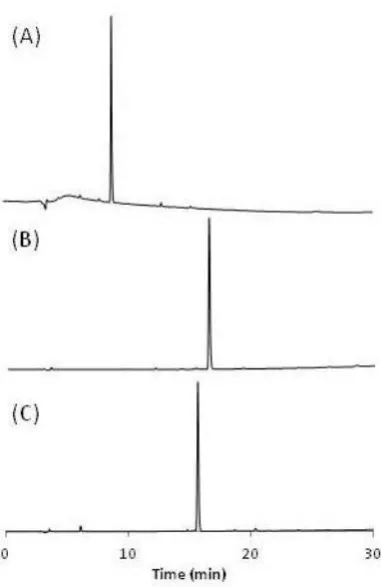
3.3.1 HPLC purity test ... 71
3.3.2 X-Ray diffraction ... 72
3.3.3 Aqueous solubility and stability in the dark ... 83
3.3.4 pKa* for complex 4 ... 87
3.3.5 Extinction coefficient studies ... 88
3.3.6 Photoirradiation studies followed by UV-Vis ... 90
3.3.7 Photoirradiation studies followed by EPR ... 94
3.3.8 NMR irradiation studies ... 97
3.2.8.1 Photoirradiation studies of complex 4 trans, trans, trans-[Pt(N3)2(OH)(Succ)(pyr)2] ... 98
3.2.8.2 Photoirradiation studies of complex 6 trans, trans, trans-[Pt(N3)2(OH)(N-MI)(pyr)2] ... 103
3.3.9 High resolution LC-MS studies ... 106
3.2.9.1 LC-MS studies of complexes [Pt(N3)2(OH)(Succ)(pyr)2] 4 and [Pt(N3)2(OH)(Succ-(RGD)f)(pyr)2] 7 ... 107
3.2.9.2 LC-MS study of [Pt(N3)2(OH)(N-MI)(pyr)2] 6 ... 113
3.3.10 DFT and TDDFT calculations ... 117
3.4.2 Aqueous solubility and stability in the dark ... 132
3.4.3 Photoirradiations ... 133
3.4.3.1 UV-Vis studies ... 133
3.4.3.2 EPR studies ... 135
3.4.4 NMR and LC-MS studies ... 137
3.4.4.1 NMR and LC-MS studies of complexes [Pt(N3)2(OH)(Succ)(pyr)2] 4 and [Pt(N3)2(OH)(Succ-(RGD)f)(pyr)2] 7 ... 137
3.4.4.2 NMR and LC-MS studies of complex [Pt(N3)2(OH)(N-MI)(pyr)2] 6 ... 141
3.4.5 Fluorescence studies ... 143
3.4.6 Cell studies ... 144
3.5 Conclusions ... 146
References ... 148
Chapter 4 Photoactivatable Pt(IV)-diazido complexes bearing aromatic N-heterocyclic ligands ... 153
4.1 Introduction ... 154
4.2 Experimental ... 156
4.2.1 Materials ... 157
4.2.2 Methods ... 157
4.2.2.1 HPLC ... 157
4.2.2.2 Extinction coefficient determination ... 158
4.2.2.3 X-Ray Diffraction ... 158
4.3.1 Characterisation and purity test ... 172
4.3.2 X-Ray diffraction ... 172
4.3.3 Solubility and stability ... 177
4.3.4 Extinction coefficient determinations ... 177
4.3.5 Photoirradiation of complexes followed by UV-Vis ... 178
4.3.6 1H-NMR of complexes cis-[Pt(I)2(2-pic)2] 10 and cis-[Pt(Cl)2(2-pic)2] 11 ... 182
4.3.7 DFT-TDDFT for complex 18 ... 183
4.4 Discussion ... 186
4.4.1 Synthesis, characterization and solubility ... 187
4.4.2 X-Ray crystallography ... 193
4.4.3 Photoirradiations monitored by UV-Vis ... 196
4.5 Conclusions ... 198
References ... 199
Chapter 5 Nucleobase binding and cellular behaviour of Pt(IV)- diazido complexes ... 202
5.1 Introduction ... 203
5.2 Materials ... 205
5.3 Methods ... 206
5.3.1 5’-GMP binding studies ... 206
5.3.2 Phototoxicity testing ... 206
5.3.3 Light dose dependence on cell survival (Action spectra) ... 207
5.4.1 5’-GMP binding studies ... 211
5.4.2 Phototoxicity testing ... 216
5.4.3 Cell survival in Schizosaccharomyces pombe in response to Pt(IV)-diazido drug ... 218
5.4.4 Live-cell confocal microscopy ... 220
5.5 Discussion ... 223
5.5.1 5’-GMP binding studies ... 223
5.5.2 Phototoxicity and structure-activity relationships ... 224
5.5.3 Cell survival assessed in S.pombe ... 226
5.5.4 Live-cell confocal microscopy ... 230
5.6 Conclusions ... 232
References ... 234
Chapter 6 Conclusions and future outlook ... 237
6.1 Conclusions ... 238
6.2 Future Work ... 243
6.2.1 Axial ligand modifications ... 243
6.2.2 Further work on ligand modifications ... 246
References ... 247
i
Firstly, I would like to thank Professor Peter Sadler for his guidance and
supervision during my PhD. I greatly appreciate the opportunity I was given to be a
member of his large research group and his exciting research.
EPSRC and MOAC are acknowledged for the funding. It has been a great
experience to be a part of the MOAC DTC science community. A special
acknowledgement is given to Prof Alison Rodger for her advice and
encouragement throughout my PhD.
I would like to thank from the bottom of my heart all the PJS members (past and
present). Special thanks to Dr Nicky Farrer for all her guidance especially at the
beginning of my PhD, Miss Jennifer S. Butler for the EPR experiments, Dr Isolda
Romero-Canelón for the help with the (crazy-hour!) biological work, Dr Yao Zhao
for all the useful discussions, Dr Carlos Sánchez-Cano for the thesis proof-reading,
the advice (and the office fun!), Dr Nicolas Barry for the general support, Miss
Ruth McQuitty and Dr Ana Pizarro for the help with the HPLC. Also I would like
to acknowledge our collaborators (Dr Vicente Marchan’s group, University of
Barcelona) for the peptide work.
Special thanks to Dr Julie Woods for the phototoxicity studies and also I am
particularly grateful for allowing me to visit and work in her research facility in
Ninewells Hospital (Dundee, UK). Many thanks to Dr Jacob Dalgaard and his
group for the help with the yeast project and Dr Keith Leppard for the assistance
with the confocal microscopy work.
I would also like to thank especially Dr Ivan Prokes for his help with the NMR,
Dr Lijiang Song and Mr Phil Aston for their assistance with the mass spectrometry
experiments and Dr Guy Clarkson for the X-ray crystallography.
To mi amiga, Dr María José Romero Castro, I don’t think I have enough space
here to express my gratitude. Thanks for listening, teaching, guiding and
supporting especially towards the end where I needed you the most. You have been
an impeccable friend to me and I will never forget you.
To Dr Luca Salassa (aka, the coach) thank you for the help with the DFT
ii
To Dr Abraha Habtemariam, I would like to say massive thanks for always
advising on chemistry and non-chemistry related matters, the interesting
discussions and for always being supportive. Thank you for having faith in me and
for always seeing the best in people.
Furthermore, I would like to thank Dr Julie Ann Lough for the help with the
thesis, for the encouragement and for the hugs (virtual and actual).
To Khatija Bhayat, honey bee: thank you for being a shoulder to cry on, for the
words of encouragement and for all those moments and memories that I will
cherish.
To Joan Josep Soldevila Barreda thank you for believing in me, for the endless
nights we spent in the chemistry department, your help throughout my PhD and
your friendship.
To Adam Millett (aka homie) thank you for listening to my constant moaning
but also the countless funny office moments!
To Nichola Smith, thank you for being a photoactivatable buddy and for
allowing me to keep the radio on in our “dark side” of the lab and also joining in
for magnificent musical duets!
To Luke Taylor and Vicky Marlow: thank you for being true friends from the
MSc year. You have left me with wonderful memories.
To Andrew-what can I say! Thank you for your patience, your kindness, your
support, your loyalty, your love, for listening to my uninteresting science moans
and for never doubting me. Thank you for being here in every step of the way.
Last but not least I would like to thank my parents to whom this thesis is
dedicated to. Without a doubt, your love has been a constant fuel through the
difficult times that I have encountered. To my sister Ioanna and my friends Polina,
Stephanie, Margarita and Natalie: you are undoubtedly the best friends a girl could
ask for. Our long-lasting friendship is something that I can certainly rely on
iii
I hereby declare that except where specific reference is made to other sources, the
work contained in this Thesis is the original work of the author. It has been
composed by myself and has not been submitted, in whole or in part, for any other
degree, diploma, or other qualification.
Evyenia Shaili
iv
1) Warwick Chemistry postgraduate symposia (University of Warwick,
Coventry, UK):
June, 2012 (Oral presentation)
May, 2011 (Poster presentation)
May 2010 (Attendance)
2) MOAC Annual Conferences:
July, 2012 (Poster Presentation, Ravenstor, Peak District, England),
July, 2011 (Poster Presentation, Arnside, Lake District, England),
May, 2010 (Poster Presentation, Arnside, Lake District, England)
3) Inter-DTC symposia between Institute of Chemical Biology DTC
(Imperial College London), White Rose DTC (University of Sheffield) and
MOAC (University of Warwick):
June, 2009 (Imperial College London, Attendance)
April, 2010 (Warwick University, Poster Presentation)
4) Photoactivatable metals: from theory to therapy (June 2012, London,
Attendance)
5) Photoactivatable metal complexes: exciting potential in biotechnology and
medicine? (June 2012, Chicheley, Poster and Oral Presentation)
6) Inorganic Chemistry 2012-A joint meeting of Dalton Division Interest
Groups (April 2012, University of Warwick, Poster Presentation)
7) 5th EuCheMS Conference on Nitrogen Ligands (September 2011, Granada,
v
1) Transferable skills courses in MOAC
2) Weekly MOAC seminars
3) Weekly Chemical Biology cluster talks
Research Stays
August 2011: One month stay at the Photobiology Unit of Ninewells Hospital
vi
[Pt(N3)2(OH)(OCOR)(pyr)2] (where OCOR is a carboxylate axial ligand) and
[Pt(N3)2(OH)2(L1)(L2)] (where L1 and L2 are aromatic N-heterocyclic ligands)
have been synthesised and characterised. The chemical and photochemical
properties of these complexes, as well as their photobiological behaviour, have
been studied in order to check their potential as photoactivatable anticancer drugs.
Four trans-diazido Pt(IV) complexes with general formula trans, trans, trans
-[Pt(N3)2(OH)(OCOR)(pyr)2] (OCOR= succinate, 4-oxo-4-propoxybutanoate,
N-methylisatoate and succinate-(RGD)f peptide ligands) have been obtained by axial
derivatisation of one hydroxido ligand from trans, trans, trans
-[Pt(N3)2(OH)2(pyr)2]. The crystal structures of three axially-derivatised complexes
have been determined by X-ray diffraction. Photoirradiation studies have shown
an improved photoactivity of the carboxylate versus the dihydroxido complexes at
the longer wavelengths. Release of the axial ligands was observed in the studied
complexes. This fact is especially relevant in the case of the Pt(IV)-(cRGD)f
complex, where the RGD was incorporated as a tumour cell targeting moiety.
DFT-TDDFT calculations performed on the complex trans, trans,
trans-[Pt(N3)2(OH)(Succ)(pyr)2] showed dissociative transitions at longer wavelength,
which could explain the photolability observed in these carboxylate derivatives.
Studies of photoactivation of the diazido Pt(IV) complexes in the presence of
5’-GMP indicate the formation of a mono-5’-GMP Pt(II) adduct as main photoproduct,
therefore DNA could be considered a potential target site for these anticancer
compounds. Additionally, EPR studies showed that azidyl radical release was
observed when complexes bearing the succinate and 4-oxo-4-propoxybutanoate
ligands were irradiated with green light. No such result was obtained for the
dihydroxo precursor showing that these complexes could be phototoxic with
longer wavelength light activation.
Seven trans-diazido Pt(IV) complexes, trans, trans, trans
-[Pt(N3)2(OH)2(L1)(L2)] (where L1 and L2 are pyridine, 2-picoline, 3-picoline,
4-picoline, thiazole or 1-methylimidazole ligands), have been obtained by oxidation
vii
sterically demanding ligand, e.g. trans, trans, trans-[Pt(N3)2(OH)2(2-pic)(pyr)],
greatly enhances the photoactivity in these complexes. DFT-TDDFT calculations
are in agreement with these results, since higher intensity transitions were
observed for such complex at longer wavelength.
Phototoxicity studies carried out on A2780, A2780cis and OE19 cell lines
with the trans, trans, trans-[Pt(N3)2(OH)2(pyridine)(n-picoline)] family concluded
that steric hindrance close to the platinum centre does not favour phototoxicity.
Most of the complexes were equally potent in cisplatin resistance against the
ovarian cancer cell line (A2780cis), except [Pt(N3)2(OH)2(3-pic)2] and
[Pt(N3)2(OH)2(4-pic)2] which exhibited some cross resistance. All of the
complexes tested in both OE19 and A2780 cell lines have shown less sensitivity
to OE19 than to A2780. Studies in S. pombe yeast strains (WT and ΔRad3) with
trans, trans, trans-[Pt(N3)2(OH)2(pyr)2] suggest that DNA is potentially an
important target for this type of compounds, although other targets are not
excluded. Furthermore, live-cell confocal microscopy was performed on A2780
cells treated with the complex trans, trans, trans-[Pt(N3)2(OH)2(pyr)2] and
irradiated with a low dose of blue light. The cell death, monitored by propidium
viii
Å Angstrom
ACN Acetonitrile
ca. circa
CDDP Cisplatin
CDI 1,1'-carbonyldiimidazole
COSY Correlation Spectroscopy
δ chemical shift
d doublet
Da Dalton
dd doublet of doublets
DFT Density Functional Theory
DMF N, N´-dimethylformamide
DMPO 5, 5-dimethyl-pyrroline N-oxide
DMSO Dimethylsulfoxide
DNA Deoxyribonucleic acid
D2O Deuterated water
EDTA Ethylenediaminetetraacetic acid
EPR Electron Paramagnetic Resonance
eq equivalent
ESI-MS Electrospray Ionization Mass Spectrometry
FDA U.S. Food and Drug Administration
ix
HOMO Highest Occupied Molecular Orbital
HPLC High Performance Liquid Chromatography
HSQC Heteronuclear Single-Quantum Correlation
IC50 50% growth inhibition concentration
ICP Inductively Coupled Plasma
ICP-MS Inductively Coupled Plasma Mass Spectrometry
i.e. id est
IL Inter-ligand transitions
J Coupling constant
Wavelength
LC-MS Liquid Chromatography-Mass Spectrometry
LED Light-Emitting Diode
LUMO Lowest Unoccupied Molecular Orbital
m multiplet
MeOD Methanol-d4
MLCT/LMCT Metal-to-ligand/Ligand-to-metal Charge Transfer
mol eq molar equivalents
m/z mass/charge
nm nanometers
N-MIA N-methylisatoic acid
NMR Nuclear Magnetic Resonance
x
PI Phototoxic Index
ppb parts per billion
ppm parts per million
ROS Reactive Oxygen Species
rpm revolutions per minute
RPMI Roswell Park Memorial Institute medium
s singlet
t triplet
TDDFT Time-dependent Density Functional Theory
Tempol 4-hydroxy-2, 2, 6, 6-tetramethyl-piperidine-1-oxyl
TFA Trifluoroacetic acid
UV Ultraviolet
UVA Ultraviolet A
Chapter 1
2 1.1 Cancer
There are more than 200 types of cancer which have as a common characteristic
the uncontrolled cell growth. These are generated from a series of errors in vital
signalling pathways which eventually generate a survival advantage over
neighbouring cells. Subsequent cell division leads to a modified group of cells
(cancer) which divide autonomously since they do not obey to any anti-growth
signals. This uncontrolled cell growth allows the formation of tumours, which
together with the capacity to spread across different tissues and organs, can be
fatal.1 Statistics show that more than 1 in 3 people will develop some form of
cancer in their lifetime, indicating an enormous prevalence.2 It is the leading cause
of death in developed countries and the second cause of death in developing
countries.3
At the moment there are three main streams for the treatment of cancer: surgery
(removal of solid tumour when localized in a specific tissue), radiotherapy
(radiation with X-ray beam) and chemotherapy (use of antiproliferative drugs).
Amongst the drugs used in the last category, platinum drugs are used to treat over
40% of all cancer patients.4,5 For this reason, new and more advanced treatments
involving platinum and other metallodrugs are likely to be greatly beneficial in
enhancing the survival chances of cancer patients.
1.2 Cisplatin
Cisplatin (cis-[PtCl2(NH3)2], CDDP) was the first example of a platinum
anticancer drug. Its antiproliferative effects were first reported by Rosenberg et al.
in 1968.6,7 The drug was approved for clinical use by the FDA (Food and Drug
3 testicular cancer from 10% to greater than 90%. Currently, it is routinely used for
the treatment of bladder, advanced cervical cancer, non-metastatic non-small cell
lung, ovarian carcinoma, malignant mesothelioma, head and neck squamous cell
carcinoma and testicular cancer.8
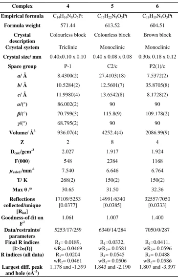
After cisplatin is administered (in a saline solution), via blood perfusion, it
remains in the neutral form due to high Cl- concentration in the blood stream
(>100 mM) which suppresses hydrolysis of the chlorides. Cellular uptake occurs
through passive and active transport (partly via copper transporters).9 Once inside
a cell, where the chloride concentration drops significantly (ca. 20 mM), aquation
occurs, leading to DNA binding (Figure 1.1).
It is widely accepted that the main target of cisplatin is cellular DNA to which it
can bind to two proximal nucleobases forming intra and interstrand crosslinks.
Most crosslinks are 1,2-GpG intrastrand (60-65%), whereas 1,2-ApG intrastrand
account for another 20-25%. Less frequent are the 1,3-GpXpG intrastrand
crosslinks (where there is another base between the two platinated guanines), the
G-G interstrand crosslinks as well as monofunctional adducts.10,11 Formation of
the 1,2-intrastrand crosslinks can kink the DNA structure, inhibiting DNA
replication and transcription. These platinum-DNA lesions can be recognized by a
number of nuclear proteins such as High Mobility Group-domain (HMG) proteins
that protect them from nucleotide excision repair upon binding, finally leading to
cell death.9 Recently, Lippard et al. illustrated that interstrand crosslinks, although
low in abundance, have profound effects since they can inhibit transcription as
efficiently as 1,2-intrastrand cross links and also inhibit DNA-histone sliding thus
4 greater role in the antiproliferative capacities of cisplatin than previously
suspected.
Other pathways might also be responsible for the cytotoxicity of the drug. CDDP
causes an increase in the levels of ROS (Reactive Oxygen Species). This effect is
caused by the reaction of cisplatin with thiols (–SH containing molecules), which
maintain the redox homeostasis within the cell.14 Furthermore, CDDP is able to
block the enzyme Thioredoxin Reductase (TrxR), responsible for the reduction of
disulfide bonds, and can alter the mitochondrial functioning, causing NADPH
depletion, thus leading to an increase in OH· and O2·- species.15
[image:23.595.131.526.333.684.2]5 1.3 Cisplatin drawbacks and development of second and third generation
Pt(II) agents
Despite the clinical success of the drug, there are many disadvantages associated
with cisplatin. One of the main drawbacks of CDDP, and other Pt(II) drugs, is the
development of resistance, which can be inherent or acquired. The former is a
result of spontaneous mutations occurring during cell division as part of intrinsic
genetic instability and the latter is a result of initial exposure to the
chemotherapeutic agent.15 The three main causes of acquired resistance are as
follows:
(a) Decreased drug uptake and/or increased efflux. It is generally accepted that
active transport (e.g. via copper transporter) plays a major role in cisplatin uptake
(Figure 1.1). Downregulation of these proteins or upregulation of the efflux
proteins (e.g. ATP7A/B) leads to decreased sensitivity to the drug.16,17
(b) Increased DNA repair (especially nucleotide excision repair which is the main
repair mechanism for 1,2-GG intrastrand crosslinks) or bypassing DNA-Pt
adducts during replication leads to a decrease in apoptotic response.10
(c) Increased drug deactivation through elevated cellular amounts of glutathione
and/or metallothioneins. The high affinity of platinum for sulphur makes the drug
reactive towards the sulphur-containing molecules in the cell (e.g. GSH).
Formation of the inactive adducts prohibits the drug from reaction with the DNA.
Furthermore cisplatin produces serious side effects, including nephrotoxicity,
nausea, renal toxicity, vomiting, hair loss and asthenia.18 Ultimately the
therapeutic window is narrowed, thus making necessary the development of
alternative treatments. Lastly cisplatin is not orally bioavailable and it needs to be
6 Other strategies for the development of new Pt drugs have also been explored
leading to the discovery of the second (changing the leaving group) and third
generation (changing ammines) anticancer drugs. From all the newly-tested
complexes, carboplatin, oxaliplatin, nedaplatin, heptaplatin and lobaplatin (Figure
1.2) have found clinical approval, from which the former two are globally
employed whereas the latter three are mainly used in Asia.19 Carboplatin is used
for the same spectrum of cancers as cisplatin, but with the advantage of fewer side
effects, whereas oxaliplatin responds to a different spectrum of cancers and has
found a main applicability in colorectal cancer.10 Nevertheless, these platinum
drugs still suffer from disadvantages, mainly the development of resistance and
severe dose limiting side-effects.5
Figure 1.2: Pt(II)-anticancer drugs approved for clinical use.
Other strategies to potentiate platinum drugs are: increase delivery of the drugs to
cancer cells via targeting, combination therapy, use of platinum resistance
7 1.4 Non-conventional Pt(II) complexes
The aforementioned Pt(II) complexes exert their mode of action by the formation
of coordination bonds with the DNA, which are mainly 1,2-intrastrand
crosslinks.18 In this section the mode of action of non-conventional Pt(II) drugs
will be described, with a main focus on the monofunctional and trans-Pt(II)
complexes.
The term monofunctional implies that the complex can bind to DNA through only
one coordination site (Figure 1.3 A). Although some monofunctional complexes,
such as [Pt(NH3)3Cl]+ and [Pt(dien)Cl]+ are inactive, it is believed this inactivity is
because they fail to alter DNA synthesis via inhibition of DNA polymerase,20
Hollis and co-workers reported that replacement of one chloride with a
N-heterocyclic ligand (pyridine, purine, pyrimidine, aniline) could afford
compounds showing significant cytotoxicity.21 Further studies by Lippard et al.
illustrated that the complex cis-[Pt(NH3)2(pyr)Cl]+, pyriplatin, is able to stall RNA
polymerase II and has a different spectrum of activity than cisplatin.22 This
molecule is uptaken by cells via Organic Cation Transporters (OCTs 1 and 2) and
forms monofunctional adducts which are able to stall RNA polII.9, 23 These
adducts can be repaired via NER mechanism (similarly to cisplatin) but less
efficiently.24 The interesting properties of pyriplatin spurred on the synthesis of
other monofunctional complexes, of which phenanthriplatin (Figure 1.3 B) was
found to be the most promising, with a 4-40 fold lower IC50 compared with
8 Figure 1.3: (A) Scheme illustrating the monofunctional DNA adduct formed by pyriplatin, with green and cyan representing the nondamaged and damaged strands, respectively. Taken from reference 26. (B) Structure of phenanthriplatin, developed by Lippard et al.
Similarly to the monofunctional Pt(II) complexes, trans-Pt(II) complexes were
also thought to be inactive according to the initial structure-activity relationships
due to inability to form 1,2-intrastrand crosslinks.
The mechanism of action for trans complexes follows the same principle as for
the cis compounds, in which aquation of the chlorides needs to occur. Hydrolysis
of the first chloride is fast in comparison to the second, as it is opposite to H2O,
which has a weak trans effect.
Replacement of one or both ammines with iminoethers, aliphatic amines or
heterocyclic aliphatic amines (Figure 1.4) generates a wide variety of trans (A)
[image:27.595.131.454.85.393.2]9 antitumor complexes which display cytotoxicity in cisplatin-resistant cell
lines.27,28
Figure 1.4: Examples of trans-[PtCl2(L)(L)’] containing iminoether, aliphatic heterocyclic amines and aromatic amines which show in vivo efficacy.29,30,31
These complexes display a different mode of DNA binding than transplatin, but
also differences amongst themselves, depending on the type and the steric bulk of
the ammine. For example, trans-[PtCl2(NH3)(thiazole)] generates an almost equal
number of monofunctional, interstrand and intrastrand crosslinks, but the
piperidine and piperazine complexes produce more intrastrand crosslinks. The use
of two iminoether ligands, on the other hand leads to mainly monofunctional
adducts.28 In the case of trans-[Pt(pyridine)2Cl2], which can be considered as a
prototype of trans complexes with planar aromatic ligands, it was shown that it is
very efficient in forming interstrand crosslinks, despite the steric bulk created by
the two pyridine ligands.32
The interstrand crosslinks formed by these kinds of complexes are usually
between two adjacent GC base pairs, resembling those formed by cisplatin,
whereas transplatin’s interstrand adducts are formed between G-C adjacent pairs
10 Depending on the adduct type, protein recognition (e.g. by the HMG-group
family) can be different, which ultimately affects downstream effects (e.g. repair).
Figure 1.5: Types of interstrand DNA cross links formed by transplatin (left), cisplatin (middle) and trans complexes with a planar aromatic amine, using trans -[PtCl2(NH3)(isoquinoline)] as an example. Taken from reference 28.
1.5 Pt(IV)-prodrugs
A different strategy used for the production of drugs which could potentially
decrease the side effects shown by the conventional Pt(II) drugs is the
development of Pt(IV) prodrugs. A prodrug is a “derivative of a drug that is
metabolized or activated in the body to release or generate the active drug”.33 The
status of Pt(IV) prodrugs can be justified by the fact that these complexes enter
the cells as Pt(IV) and reduction to Pt(II) is essential before any substitution
reaction takes place.34,35
The kinetic inertness exhibited by Pt(IV) complexes renders them less prone to
undergo reactions on route to the tumour, enabling a higher therapeutic index.35
The presence of two additional ligands compared to Pt(II) also allows
modification of important chemical properties of the drug (e.g. lipophilicity,
11 targeting features such as nanoparticles and peptides. Furthemore, there is also the
capability of a dual mode of action by the incorporation of biologically active
axial ligands which can be released in the cell upon reduction.9
Four Pt(IV) drugs have so far entered clinical trials (Figure 1.6): iproplatin,
tetraplatin, satraplatin and LA-12. The first two have been abandoned. Iproplatin
showed less cytotoxicity than cisplatin and tetraplatin was too toxic36, whereas
satraplatin is now being trialled for combination therapy.37 LA-12 is currently
under phase I clinical trials.38
Figure 1.6: Pt(IV) complexes that have entered clinical trials.
Although the examples discussed above entail chemical activation, of special
interest is the use of light to photoactivate a prodrug, which is the main concept
behind photodynamic therapy.
1.6 Photodynamic therapy (PDT)
PDT is a clinical treatment for cancer based on the administration of a
photosensitizer and subsequent activation using light at a wavelength where the
12 state, causing Type I and Type II reactions. The former entails a redox reaction
with subsequent formation of radicals and the second involves energy transfer to
ground state molecular oxygen (3O2) to produce the highly reactive 1O2 (singlet
oxygen).39 This species is toxic to cells and causes death via oxidative stress
mechanisms, DNA damage, protein destruction and cell lysis.40,41 The short life
time of singlet oxygen in water (3 μs) and the maximum diffusion distance of up
to 100 nm within a cell, gives rise to localized cellular damage.42 Further to this
mechanism of action, PDT can damage the vasculature associated with the tumour
and also trigger an immune response.40
The photosensitizer ideally should be activated by longer wavelength light for
deeper penetration into the tissue, have good aqueous solubility, no dark toxicity,
preferential accumulation in the cancer tissue, and finally a long enough half-life
in the blood to reach the tumour cells in adequate concentrations.43,44 Most of the
photosensitizers are highly conjugated macrocycles (e.g. porphyrins, chlorins,
bacteriochlorins, phthalocyanines, naphtlaocyanines), although research on
metal-based photosensitizers has shown that they are already promising candidates for
PDT.45 Notable examples (Figure 1.7) include aluminium sulfonated
phthalocyanine (Photosense), which is approved for clinical use in Russia,46 the
Motexafin Lutetium (Lu-trin or Lu-Tex) and the palladium bacteriopheophorbide
(Tookad soluble), both of which are evaluated as a treatment for prostate
13 Figure 1.7: PDT agents containing metals which are either used clinically (A, Motexafin Lutetium) or under clinical evaluation (B, aluminium sulfonated phthalocyanine and C,palladium bacteriopheophorbide).
The medical benefits of PDT for the treatment of cancer and pre-cancerous
conditions are the follows: (a) There is little effect on the non-living connective
tissue, allowing healing without a risk of the mechanical integrity of hollow
organs.48 (b) There are no total-dose limitations, as in the case of radiotherapy.43
(c) It allows excellent spatial control therefore avoiding damage to healthy cells.
PDT’s main drawbacks lie in the fact that it is oxygen-dependent and oxygen is
scarce in hypoxic tumour environments.49 Furthermore, porphyrin aggregation,50
poor-solubility51 and photosensitivity after treatment52 are some other
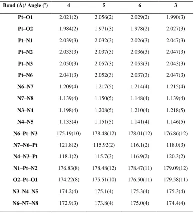
[image:32.595.119.507.93.374.2]14 1.7 Light Delivery
Although light penetration into a specific tissue is dependent upon the competitive
absorption of endogenous chromophores (e.g. melanin and haemoglobin), the
optimum range is 620 nm – 850 nm. Light in the range of 600-700 nm penetrates
50-200 % deeper than in the 400-500 nm, therefore is preferentially used in the
clinic (Figure 1.8).45 However, shorter wavelength of light can be used if the
target lesion is thin and the tissue surface is easily accessible.43
Figure 1.8: Light penetration through the tissue. Adapted from reference 53.
LED lights are used for dermatological purposes and recent advances in laser
technology have enabled physicians to perform PDT in internal organs (e.g. lungs,
brain, bladder, cervix and colon) via the use of optical fibres to deliver laser
light.43,54 The use of advanced high power and tightly focused femtosecond laser
systems may also enable PDT via two photon excitation in the future. This
[image:33.595.106.509.281.541.2]15 600 nm, to achieve excitation at shorter wavelength (approximately half).55 As a
result, metal complexes with the ability to absorb two photons (usually large with
highly conjugated donor and acceptor ligands) can be activated with longer and
clinically relevant wavelengths. In a recent report, Zhao et al. presented the first
example of a Pt(II) complex containing the π-conjugated ligand
4-[2-(4-methoxyphenyl)ethynyl]pyridine for which activation with laser pulses between
600 – 740 nm yielded the same photoproducts as UV activation.56 Furthermore,
research has been carried out on the development of upconversion nanoparticles in
PDT. A photosensiziter which is activated with UV or short visible wavelength is
loaded onto the nanoparticle which is composed of material capable of photon
upconversion (usually lanthanides, such as NaYF4: Yb, Er 57) and upon irradiation
with near-infrared light, shorter wavelength light is produced which is able to
activate the photosensitizer coated on the outer shell of the NP.58
1.8 Photophysical and photochemical properties of metal complexes59,60, 61,62 Photochemistry is concerned with the effects of light in chemical systems and will
be discussed here with respect to metal coordination compounds. Absorption of a
photon by a molecule can cause excitation if the energy gap between the excited
and the ground state matches the energy of the photon. The possibility of an
electronic transition occurring depends on the transition dipole which in turn is
dependent on the two following selection rules.
(a) Spin selection rule: transitions with a change of multiplicity are not
allowed (ΔS = 0). This selection rule can be relaxed by the coupling of the
spin and orbital angular momenta. Heavier atoms have a greater spin-orbit
16 (b) Laporte selection rule: in a centrosymmetric molecule the only allowed
transitions are those accompanied by a change in parity (e.g. g ↔ u). This
rule can be relaxed when the complex’s symmetry departs from the perfect
centrosymmetry, through natural asymmetry or through distortions
occurring during vibrations.
The fact that these transitions are forbidden suggest that experimentally they will
occur with low probability, hence the extinction coefficients are small.
Absorption of radiation by a molecule can lead to the population of singlet excited
states, which then follow various deactivation pathways, as illustrated in the
Jablonski diagram (Figure 1.9). When the population of higher electronic states
(e.g. S1, S2) occurs, then return to a lower singlet level can occur without radiation
via internal conversion, vibrational relaxation, quenching or via a radiative
process, fluorescence. Otherwise, intersystem crossing can lead to population of a
triplet state which can give rise to phosphorescence (radiative process) upon
return to the ground state (S0). Phosphorescence has a longer life time than
fluorescence (10-3–1 s vs 10-9–10-6 s),41 as the former is a spin forbidden process.
A decrease in the intensity of fluorescence is commonly observed when transition
metals are in close proximity to the fluorophore because they increase the rate of
Intersystem-Crossing to the triplet state due to an increase in the spin-orbit
coupling. This radiationless process competes with the S1→S0 transition,
responsible for fluorescence.63
The deactivation of an excited state often does not go through a physical process
17 dissociation, redox, substitution). Nevertheless, the population of the triplet state,
occurring through intersystem crossing from the singlet state, is an important step
that dominates a lot of the routes of electronic excitation.
Figure 1.9: A Jablonski diagram to summarise all potential deactivation pathways of complex in an excited state. Reproduced and modified from reference 64.
Prior to discussing the various types of the excited states in a d6 metal complex, it
is important to describe the typical molecular orbital diagram for this class of
compounds. In an octahedral d6 metal complex (e.g. PtIV), the splitting of the
orbitals is such that the dxy, dyz, dxz are lower in energy than the dz2 and d
x2-y2
orbitals, as described by crystal field theory. The energy of the ligand orbitals is
lower than the metal orbitals and therefore the lower energy bonding orbitals are
ligand centred, whereas the lowest energy antibonding orbitals are predominantly
metal-based.
The excited state reactivity of a metal complex, as induced by light, is
summarized by the following (Figure 1.10).
1) Metal-centred transitions (d-d transitions). These are Laporte forbidden
1-18 1000 M-1cm-1. Such transitions typically lead to the occupation of
antibonding orbitals, often leading to bond elongation and ligand
substitution.
2) Charge transfer transitions (ligand-to-metal, metal-to-ligand or to-solvent)
are fully allowed, giving rise to intense absorption bands. A MLCT may
result if the metal is electron rich and the ligand has low lying empty
orbitals. A LMCT occurs when the ligand is easy to oxidize and the metal
easily reduced. A LMCT leading to a chemical change can cause change in
the oxidation state of the metal, as well as ligand release by a homolytic
bond cleavage to liberate radicals.
3) Ligand-centred transitions mainly occur in aromatic ligands with extended
π and π*
orbitals and they are allowed by both spin and Laporte selection
19 Figure 1.10: A simplified MO diagram of an octahedral d6 complex, assuming strong crystal field splitting. The bonding orbitals π1 and π2 are predominantly ligand in character, whereas the t2 are metal in character. The blue arrows represent electrons with the associated spin in the ground state, whereas the coloured arrows represent electrons involved in a transition. In the singlet and triplet states the spins are up (↑↑↑↑) or down (↓↓↓↓), respectively. Reproduced and modified from Farrer et al.44
1.9 Non-platinum metal-based photoactivated drugs
Many transition metals (e.g. Ti, Cr, Mn, Cu, Co, Pd, Ir, Fe, V) show medically
interesting photochemical properties which have been studied. A few notable
examples are the oxovanadium(IV) curcumin complex [VO(cur)(dppz)Cl] and the
Fe(L)(cat)NO3, where L= 9-[(2,2’-dipicolylamino)methyl]anthracene and cat=
catecholate. The former was shown to have a lower IC50 than the FDA approved
PDT agent photofrin when irradiated with visible light (400-700 nm) and its
efficacy is thought to be due to its DNA photocleavage ability.65 The latter
20 ligand is responsible for the fluorescent and intercalative abilities of the complex,
whereas the catecholate ligand provides IR absorption.66
Ru(II) and Rh(III) complexes have been studied the most over the years. A very
prominent family is that compromising Ru-polypyridyl complexes which not only
show intercalative properties via the ligand but also because the photoexcitated
state is a strong oxidant, capable of oxidizing DNA nucleobases.67 Especially
when tridentate ligands (e.g. 2-pyridyldipyrido-[3,2-a;2’,3’-c]phenazine) were
employed, quantum yields of singlet oxygen production were exceptionally
high.68 Ru-polypiridyl complexes which contain monodentate ligands can also
exhibit ligand release of one or two ligands which is exploited for the uncaging of
bioactive molecules (e.g. amino acids, nucleotides, neurotransmitters and genetic
inducers). The photosubstitution in these complexes is enabled by the population
of a d-d state from the 3MLCT, a transition which is temperature dependent.69
Light-induced ligand substitution and subsequent nucleobase binding was also
shown by Sadler and coworkers in the case of Ru(II)-arene complexes containing
a pyridine monodentate ligand.70 Furthermore, the use of polyazaaromatic ligands
(e.g. 1,4,5,8,9,12-hexaazatriphenylene (HAT) and 1,4,5,8-tetraazaphenanthrene
(TAP)) lead to formation of covalent adducts with nucleobases through a radical
recombination process which is lethal for the cell.71 Finally, the nitrosyl bound
Ru(II) complexes can also undergo ligand release but through a different
mechanism (via oxidation to Ru(III)).72
Rh(III) complexes with polypiridyl or phenathranoline ligands can also cause
DNA cleavage on light activation. A notable example, which uses a more
21 [Rh(bpy)2(chrysi)]2+, complex developed by Barton et al., which can specifically
bind to DNA base mismatches.73
1.10 Photoactivity of Pt(II) complexes
Up until now most of the photochemistry of platinum evolved around luminescent
Pt(II) complexes, which have been explored thoroughly due to their useful
photophysical properties. Their high efficiencies, long lifetimes of the emissive
state enabled this type of complexes to be used in applications such as
chemosensors,74 photocatalysis,75 light emitting diodes76 and photovoltaic
devices.77
More recently, Pt(II) agents were also found to be phototoxic when coordinated to
appropriate ligands. Studies carried out with transplatin (Figure 1.11, complex A)
showed that upon irradiation with UVA, an enhancement in the toxicity (2-fold)
was observed. This was attributed to the loss of chloride ligands and the formation
of bifunctional DNA interstrand crosslinks which are unable to form in the dark.78
A recent study has shown that a similar effect can be caused by UV irradiation of
carboplatin, where a 10-fold increase in cytotoxicity and faster DNA binding
kinetics was induced.79
Brunner and co-workers, in an attempt to combine the advantages given by the
preferential cellular accumulation of porphyrins in cancer cells with the cytotoxic
activity of Pt(II), attached poly(ethyleneglycol)-derivatized hematopoprhyrins to
Pt(II) (Figure 1.11, complex B). The phototoxicity results showed that most of the
complexes exhibited both cytotoxicity and phototoxicity (λ = 600-730 nm, 10
min), towards human cancer cells with a few exceptions which showed promising
22 Franz et al. reported that the attachment of a photosensitive nitrophenyl group to a
tetradentate ligand backbone with two pyridyl and two amide nitrogen donor sites
makes the Pt(II) complex photolabile (Figure 1.11, complex C). Bond breakage
occurs, accompanied by release of a Pt(II) complex prone to ligand substitution
reactions as well as other nitroso side-products when exposed to UV light for a
short time. The cytotoxicity of the complex was low in the dark and decreased to
68% upon irradiation.81
Chi-Ming Che et al. also has reported luminescent cyclometalated Pt(II)
complexes with thiophene,82 mono83 or bidentate (N-heterocyclic carbenes)84
ligands. These complexes exhibit high emission quantum yields which enable
their tracking inside a cell, via fluorescence microscopy. In the first two reports,
although the complexes were very potent, they produced only a small increase in
cytotoxicity when irradiated with visible light (~6 fold, Figure 1.11, complexes D
and E), whereas in the latter case it was improved 30-fold in some cases (Figure
1.11, complex F). These compounds tend to accumulate in cytoplasmic structures
(e.g. endoplasmic reticulum and mitochondria) rather than the nucleus, which can
indicate a different mode of action than traditional anticancer Pt drugs (usually
23 Figure 1.11: Selected complexes showing enhancement in cytotoxicity when irradiated with UV (A and C) or visible light (B and D-F).
Finally, a few dinuclear complexes of Pt(II) have been reported in recent years
which exhibit promising photosensitizer characteristics. Monti et al. reported
24 porphyrazine macrocycle, where the central cavity was occupied with either
Zn(II), Mg(II) or Pd(II). They were shown to generate singlet oxygen efficiently
in DMF (λ > 600 nm) and also to bind to G-quadruplex structures.85 Brewer et al.
reported a Ru(II)-Pt(II) dinuclear complex, where the dichloro-platinum moiety is
attached on a dpp ligand, where dpp=2,3- bis(2-pyridyl)pyrazine)). The other
ligands on the ruthenium centre were the widely used phenanthronile ligands.
Irradiation at long wavelength (λ > 600 nm) causes a MLCT excitation to the
ruthenium which is transferred to PtCl2 site, causing hydrolysis of the chlorides
and DNA binding.86
1.11 Photoactivity of Pt(IV) complexes
The phototochemical properties of Pt(IV) were documented early on, when it was
shown that [PtCl6]2- can be reduced to metallic Pt or [PtCl4]2- with low-energy
visible light.87 More recently, Lippert et al. reported that PtIV 2,2-bipyridine
complexes, e.g. mer-[PtCl3(2,2-bpy)(MeNH2)], can undergo photoreduction (λ >
300 nm) to produce the Pt(II) fragment [PtCl(2,2-bpy)(MeNH2)] accompanied by
the loss of HOCl or Cl2 which possibly originate from the reductive elimination of
the axial chlorido ligands. Surprisingly, the same result is obtained when the
complex is heated at 50 oC for 24 h, showing the thermolability of the
25 1.11.1 Pt(IV)-diiodo and Pt(IV)-diazido complexes
A controlled localized reduction to a cytotoxic component is extremely beneficial
to healthy cells which are prone to rapid division, such as bone marrow, GI-tract
and skin, since these cells are particularly sensitive to chemotherapy.38
Pt(IV) complexes, contrary to the Pt(II), are less prone to substitution reactions
and therefore the selection of appropriate ligands can render them stable in the
presence of high amounts of reducing agents and also create photolability.
The first generation of Pt(IV) photoactivatable anticancer drugs were diiodo
complexes (Figure 1.12), with the general formula of trans, cis-[Pt(X)2I2(en)],
where en = ethylenediamine and X = Cl-, OH-, acetate or methylsulfonate.89 The
ethylenediamine ligand was chosen in order to avoid photoisomerisation reactions
which could lead to the formation of a trans-Pt(II) photoproduct which is not able
to form the lethal intrastrand DNA cross-links.89 The choice of iodides as the
reducing ligands was made because they are weak field ligands and thus the
LMCT transition occurs at low energy in the visible region. Indeed, the UV-Vis
spectrum of the iodo complexes bears a tail which extends up to 500 nm.90
26 CT-DNA binding studies illustrated that the most promising complex was C in
Figure 1.12 as complexes A and D could form adducts in the dark, whereas
photolysis of B led to no platination. Instead complex C led to 65% platination
only after irradiation.90
Nevertheless, cytotoxicity studies on these complexes showed no significant
difference between light and dark toxicity. This might be attributable to the facile
reduction of the complexes by thiols within the cellular environment.90,91
Photochemical reduction of platinum(IV)-azide complexes was documented early
on, by Vogler and co-workers who showed that trans-[Pt(CN)4(N3)2]2- was
converted to [Pt(CN)4]2- upon excitation with UV (300 nm) via a simultaneous
two-electron reduction process without a Pt(III) intermediate.92 Furthermore, they
showed that [PtII(N3)4]2- is also photolabile being reduced to metallic platinum
leading to the production of nitrogen and azide.93 Following this, the Sadler group
developed a second generation of Pt(IV) photoactivatable drugs containing azides
instead of iodides (Figure 1.13).
Figure 1.13: Pt(IV)-diazido complexes developed by Sadler and co-workers.54,94,95
Complexes A and B in Figure 1.13 were the first synthesized and have been
27 decomposed more slowly than B under UV irradiation. This could be rationalized
by the fact that the LMCT band of B is shifted towards the visible region by 29
nm and also the extinction coefficient is greater.96 Another advantage of B over A
is the higher aqueous solubility. However irradiation studies followed by 1H and
2D [1H, 15N] HSQC NMR spectroscopy, showed slow photoreactivity of this class
of compounds since reduction to Pt(II) was seen only after 60 min of irradiation
with UVA light. Instead photosubstitution/ photoisomerisation Pt(IV) products
were possibly formed. Interestingly, irradiation of B in the presence of 5’-GMP (2
molar equivalents) led to the formation of trans-[PtII(NH3)2(5’-GMP-N7)2]
adducts and reduction to Pt(II) occurred at a faster rate than in the absence of
nucleotides. Futhermore, complex A was shown to generate equivalent nucleotide
crosslinks when irradiated with visible light in presence of 5’-GMP or
d(guanosinylphosphoguanosine) as those formed by cisplatin.97 These results
demonstrated for the first time the photoreduction of Pt(IV) to Pt(II), and also that
the stereochemistry of the amines is retained after reduction to Pt(II) and
subsequent binding to biomolecules.
The photochemical decomposition pathways of A and B were studied extensively
by Ronconi et al via 14N and 15N NMR.98,99,100 It was discovered that the
photodecomposition pathways of the two isomers are similar in the major
products formed but that the experimental conditions under which the irradiation
experiments were carried out affected the photoproducts: in PBS, azide release
was observed, whereas in acidic conditions, N2 was the major product. However
in both conditions, O2 and free ammonia were detected (indicating ammine
28 increase in pH. It was postulated that the photodecomposition to Pt(II) could
involve also the formation of highly reactive Pt(IV)-nitrene intermediates,
indicating a complex mechanism of photodecomposition of this class of
compounds.
Although neither complexes A or B (Figure 1.13) possess dark toxicity, the trans
isomer was slightly more active than the cis counterpart under irradiation (IC50
156 μM vs 176 μM in HaCaT cell line, UVA irradiation for 50 min).96
Changes in
the nuclear morphology of 5637 human bladder cells were also followed for
complex A by phase-contrast and fluorescence microscopy illustrating that cells
did not undergo the typical apoptotic changes (e.g. budding and cellular
fragmentation). Treated cells rather ballooned and lost contact with neighbours as
well as suffering disintegration of the nuclei. Furthermore, a distinctive
mechanism of cell death from cisplatin became obvious.101
Complex C (Figure 1.13) which incorporates a pyridine in place of an ammine
ligand was significantly more potent than A and B, when irradiated with UV light
(IC50s 6.1 and 1.9 μM in HaCaT and A2780 cell lines, respectively).94 Photolysis
experiments showed that reduction to Pt(II) occurred in the presence of
biomolecules (e.g. 5’-GMP) to produce trans-[Pt(NH3)(py)(5’-GMP-N7)2] and
trans-[Pt(N3)(NH3)(py)(5’-GMP-N7)]. Studies to elucidate the mechanism of
action were performed for complex C by Berdnaski et al. showing that an elevated
amount of ROS was generated after irradiation with UVA light (30 min).102
Treatment of HL60 (human promyleotic leukaemia cells) with the complex C did
not yield morphological changes (Figure 1.14) that resembled those triggered by
29 caspase 3 or 7, combined with changes in the levels of key autophagic proteins
(e.g. LC3, p62), led to postulated autophagy as the possible mechanism of cell
death.103
Figure 1.14: Phase contrast images of HL60 cells treated for 48 hours with untreated control (a), 68 μM of complex C activated with UVA light (b), 0.74 μM of cisplatin (c) and 0.74 μM etoposide (d).103
The success of complex C spurred on the synthesis of trans, trans,
trans-[Pt(N3)2(OH)2(L)(L’)] complexes which when compared to their cis analogues
they were found to be more phototoxic.104 This systematic study produced the first
structure-activity relationship for Pt(IV)-diazido complexes.
Replacement of aliphatic amine with pyridine (complex D, Figure 1.13) resulted
in photocytotoxicity (IC50 8.4 μM in OE19) with blue light (420 nm). As in the
case of complex C irradiation with blue light in the presence of 5’-GMP resulted
in the formation of mono and bis-5’-GMP adducts.95 EPR studies illustrated the
30 complexes was shown by the reduction in potency when IC50 studies were carried
out in the presence of the radical quencher tryptophan.105 Azidyl radicals can
cause oxidation reactions of tryptophan and tyrosine106 thus potentially damaging
proteins and other biomolecules.
These complexes are thought to have a dual mode of action (Figure 1.15) with
azidyl radicals playing a primary role in the cytotoxicity. However binding to
biomolecules is also significant. DNA binding studies of complex D demonstrated
the formation of 1,3-intrastrand crosslinks as the major adduct (51%) but
monofunctional lesions can form to a greater extent than cisplatin (37% vs 2%).
Pracharova et al. also postulated that interaction of the pyridine ligand with the
DNA duplex may play an important role for activity. Moreover, stalling of RNA
polymerase II (responsible for transcription) is also a significant factor which can
contribute to the cytototoxicity of this complex.107 This result also parallels the
report by Lippard et al. in which an X-ray crystal structure of RNA polII stalled
by a monofunctional platinum-DNA adduct with pyriplatin. The structure showed
that a pyridine ligand located in cis position to NH3 can fit into the RNA polII
active site and subsequently cause blockage of the translocation of the enzyme
31 Figure 1.15: Mechanism of action of Pt(IV)-diazido complexes were L and L’ are N-aromatic ligands. The scheme shows that cell death can be produced by oxidative stress caused by the generation of radicals and also by the platination of biomolecules, such as DNA and proteins.
All of these compounds were found to be stable in the dark in the presence of
cellular concentrations of reducing agents such as GSH and ascorbate which is a
desirable feature for photoactivatable drugs.108 Together with the fact that the
mechanism of action is oxygen-independent and the generally high aqueous
solubility of these prodrugs, these complexes are potential candidates for
photoactivated chemotherapy (PACT).
1.12 Aims
The aims of this thesis are the following.
Synthesis and characterisation of novel complexes with planar aromatic
ligands which can potentiate the phototoxicity of the Pt(IV)-diazido
32 Synthesis and characterisation of Pt(IV)-diazido complexes with
derivatization of the axial ligands so as to provide targeting and imaging
features.
Studies of the photochemistry of the new complexes, especially on the
biologically relevant wavelengths, and elucidation of photoproduct
pathways.
Cellular studies in mammalian cells for the determination of phototoxicity,
cell uptake as well as the employment of novel methods (confocal
microscopy and survival in yeast cells) which can enable the determination
of the mechanism of action.
References
1. R. J. B. King and M. W. Robins, Cancer Biology, Pearson Education Limited, Harlow, 2006.
2. CRUK, Cancer Statistics key facts, 2013.
3. A. Jemal, F. Bray, and J. Ferlay, CA-Cancer J. Clin., 2011, 61, 69-90.
4. B. Teni, A. Pantos, E. Bellis, and P. Christofis, Cancer Therapy, 2007, 5, 537-583.
5. B. W. Harper, A. M. Krause-Heuer, M. P. Grant, M. Manohar, K. B. Garbutcheon-Singh, and J. R. Aldrich-Wright, Chem. Eur. J., 2010, 16, 7064-7077.
6. B. Rosenberg, L. Van Camp, and T. Krigas, Nature, 1965, 2, 698-699.
7. B. Rosenberg, L. Vancamp, J. E. Trosko, and V. H. Mansour, Nature, 1969, 205, 698-699.
33 9. T. C. Johnstone, J. J. Wilson, and S. J. Lippard, Inorg. Chem., 2013, In
press.
10. L. Kelland, Nat. Rev. Cancer, 2007, 7, 573-584.
11. J. M. Malinge, M. J. Giraud-Panis, and M. Leng, J. Inorg. Biochem., 1999, 77, 23-29.
12. G. Zhu, L. Song, and S. J. Lippard, Cancer Res., 2013, 73, 4451-4460.
13. M. Ober and S. J. Lippard, J. Am. Chem. Soc., 2008, 130, 2851-2861.
14. A.-M. Florea and D. Büsselberg, Cancers, 2011, 3, 1351-1371.
15. I. Romero-Canelón and P. J. Sadler, Inorg. Chem., 2013, In press.
16. F. Arnesano, M. Losacco, and G. Natile, Eur. J. Inorg. Chem., 2013, 2013, 2701-2711.
17. M. D. Hall, M. Okabe, D.-W. Shen, X.-J. Liang, and M. M. Gottesman, Annu. Rev. Pharmacol. Toxicol., 2008, 48, 495-535.
18. T. Boulikas, Cancer Ther., 2007, 5, 351-376.
19. J. Reedijk, Eur. J. Inorg. Chem., 2009, 2009, 1303-1312.
20. A. L. Pinto and S. J. Lippard, Proc. Natl. Acad. Sci. USA., 1985, 82, 4616-4619.
21. L. S. Hollis, A. R. Amundsen, and E. W. Stern, J. Med. Chem., 1989, 32, 128-136.
22. K. S. Lovejoy, M. Serova, I. Bieche, S. Emami, M. D’Incalci, M. Broggini, E. Erba, C. Gespach, E. Cvitkovic, S. Faivre, E. Raymond, and S. J. Lippard, Mol. Cancer Ther., 2011, 10, 1709-1719.
23. K. S. Lovejoy, R. C. Todd, S. Zhang, M. S. Mccormick, J. A. D. Aquino, J. T. Reardon, A. Sancar, K. M. Giacomini, and S. J. Lippard, Proc. Natl. Acad. Sci. USA., 2008, 105, 8902-8907.
24. G. Zhu, M. Myint, W. H. Ang, L. Song, and S. J. Lippard, Cancer Res., 2012, 72, 790-800.
25. G. Y. Park, J. J. Wilson, Y. Song, and S. J. Lippard, Proc. Natl. Acad. Sci. USA., 2012, 109, 11987-11992.
34 27. G. Natile and M. Coluccia, Coord. Chem. Rev., 2001, 217, 383-410.
28. S. M. Aris and N. P. Farrell, Eur. J. Inorg. Chem., 2009, 2009, 1293-1302.
29. M. Coluccia, A. Nassi, A. Boccarelli, D. Giordano, N. Cardellicchio, D. Locker, M. Leng, M. Sivo, F. P. Intini, and G. Natile, J. Inorg. Biochem., 1999, 77, 31-35.
30. Y. Najajreh, E. Khazanov, S. Jawbry, Y. Ardeli-Tzaraf, J. M. Perez, J. Kasparkova, V. Brabec, Y. Barenholz, and D. Gibson, J. Med. Chem., 2006, 49, 4665-4673.
31. N. Farrell, L. F. Povirk, Y. Dange, G. DeMasters, M. S. Gupta, G. Kohlhagen, Q. A. Khan, Y. Pommier, and D. A. Gewirtz, Biochem. Pharmacol., 2004, 68, 857-866.
32. Y. Zou, V. Houten, and N. P. Farrell, Biochemistry, 1993, 12, 9632-9638.
33. F. Kratz, I. A. Müller, C. Ryppa, and A. Warnecke, 2008, 20-53.
34. M. D. Hall, G. J. Foran, M. Zhang, P. J. Beale, and T. W. Hambley, J. Am. Chem. Soc., 2003, 125, 7524-7525.
35. M. D. Hall and T. W. Hambley, Coord. Chem. Rev., 2002, 232, 49-67.
36. M. Galanski, M. A. Jakupec, and B. K. Keppler, Curr. Med. Chem., 2005, 12, 2075-2094.
37. W. D. Figg, C. H. Chau, R. A. Madan, J. L. Gulley, R. Gao, T. M. Sissung, S. Spencer, M. Beatson, J. Aragon-Ching, S. M. Steinberg, and W. L. Dahut, Clin. Genitourinary, 2013, In press.
38. A. M. Pizarro and P. J. Sadler, Biochimie, 2009, 91, 1198-1211.
39. I. Yoon, J. Z. Li, and Y. K. Shim, Clin. Endosc., 2013, 46, 7-23.
40. A. P. Castano, P. Mroz, and M. R. Hamblin, Nat. Rev. Cancer, 2006, 6, 535-545.
41. L. B. Josefsen and R. W. Boyle, Met. Based Drugs, 2008, 1-24.
42. E. Skovsen, J. W. Snyder, J. D. C. Lambert, and P. R. Ogilby, J.Phys.Chem.B, 2005, 109, 8570-8573.
43. S. G. Bown, Phil. Trans. R. Soc. A, 2013, 371:20120371.
35 45. K. Szaciłowski, W. Macyk, A. Drzewiecka-Matuszek, M. Brindell, and G.
Stochel, Chem. Rev. , 2005, 105, 2647-2694.
46. O. I. Trushina, E. G. Novikova, V. V. Sokolov, E. V. Filonenko, V. I. Chissov, and G. N. Vorozhtsov, Photodiagnosis Photodyn. Ther., 2008, 5, 256-259.
47. H. Patel, R. Mick, J. Finlay, T. C. Zhu, E. Rickter, A. Keith, S. B. Malkowicz, S. M. Hahn, and T. M. Busch, Clin. Cancer Res., 2008, 14, 4869-4876.
48. W. E. Grant, P. M. Speight, a J. MacRobert, C. Hopper, and S. G. Bown, Br. J. Cancer, 1994, 70, 72-78.
49. N. P. Assembly, C. S. Jin, J. F. Lovell, J. Chen, and G. Zheng, ACS Nano, 2013, 7, 2541-2550.
50. H. Ali and J. E. van Lier, Chem. Rev. , 1999, 99, 2379-2450.
51. R. Bonnett, Metal Complexes for Photodynamic therapy, Comprehensive Coord. Chem. II, Elsevier Pergamon, Oxford, 2004.
52. T. J. Dougherty and S. L. Marcus, Eur. J. Cancer, 1992, 28, 1734-1742.
53. Q Light Phototherapy, http://www.qlight.ch/.
54. P. J. Bednarski, F. S. Mackay, and P. J. Sadler, Anticancer Agents Med. Chem., 2007, 7, 75-93.
55. M. Pawlicki, H. A. Collins, R. G. Denning, and H. L. Anderson, Angew. Chem. Int. Ed., 2009, 48, 3244-3266.
56. Y. Zhao, G. M. Roberts, S. E. Greenough, N. J. Farrer, M. J. Paterson, W. H. Powell, V. G. Stavros, and P. J. Sadler, Angew. Chem. Int. Ed., 2012, 51, 11263-6.
57. N. M. Idris, M. K. Gnanasammandhan, J. Zhang, P. C. Ho, R. Mahendran, and Y. Zhang, Nat. Med., 2012, 18, 1580-1585.
58. C. Wang, L. Cheng, and Z. Liu, Theranostics, 2013, 3, 317-330.
59. V. Balzani, G. Bergamini, S. Campagna, and F. Puntoriero, Top. Curr. Chem., 2007, 280, 1-36.


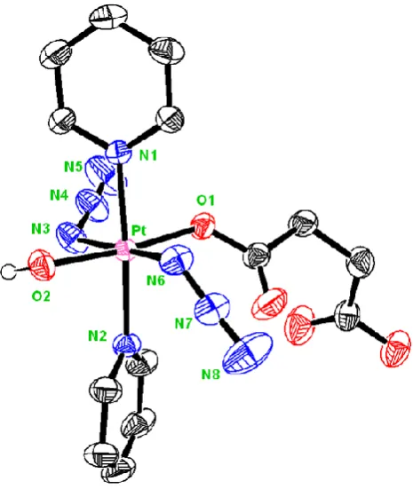
![Figure 3.5: ORTEP diagrams of the complexes reported in this chapter: A) trans, trans, trans-[Pt(N3)2(OH)(Succ)(pyr)2] 4; B) trans, trans, trans-[Pt(N3)2(OH)(Succ-Pr)(pyr)2] 5; and C) trans, trans, trans-[Pt(N3)2(OH)(N-MI)(pyr)2] 6](https://thumb-us.123doks.com/thumbv2/123dok_us/9612360.464060/94.595.173.498.84.630/figure-ortep-diagrams-complexes-reported-chapter-trans-trans.webp)