A Thesis Submitted for the Degree of PhD at the University of Warwick
Permanent WRAP URL:
http://wrap.warwick.ac.uk/88753
Copyright and reuse:
This thesis is made available online and is protected by original copyright. Please scroll down to view the document itself.
Please refer to the repository record for this item for information to help you to cite it. Our policy information is available from the repository home page.
Biosynthesis and discovery of
compounds with activity against
Acinetobacter baumannii
by
Daniel Gri
ffi
ths
Thesis
Submitted to the University of Warwick
for the degree of
Doctor of Philosophy
Supervisors: Dr. J´ozef R. Lewandowski, Prof. Gregory L. Challis
Department of Chemistry
Contents
List of Figures vii
List of Tables viii
Acknowledgements ix
Declaration x
Abstract xi
Abbreviations xii
1 Introduction 1
1.1 Natural products . . . 1
1.2 Biosynthesis using megasynthases . . . 1
1.3 Domain organisation and function . . . 2
1.3.1 PKS domains . . . 2
1.3.2 NRPS domains . . . 3
1.3.3 PKS/NRPS product prediction . . . 3
1.4 Docking and communication-mediating domains . . . 5
1.4.1 Structures of docking and COM domains . . . 5
1.5 NMR characterisation of protein domains . . . 7
1.6 Enacyloxin PKS-NRPS . . . 8
1.7 Objectives . . . 11
1.7.1 Objective I . . . 11
1.7.2 Objective II . . . 11
1.8 Synopsis . . . 12
2 Enacyloxin PKS-NRPS chain release 13 2.1 Chain release process . . . 13
2.2 Protein NMR spectroscopy . . . 14
2.2.3 Residue assignment of PCP . . . 17
2.2.4 C-terminal interaction site . . . 18
2.3 In vitro bioassays . . . 19
2.3.1 Construction of mutants . . . 19
2.3.2 Condensation assay . . . 19
2.4 Crystal structure of the condensing enzyme . . . 21
2.4.1 N-terminal appendage . . . 21
2.4.2 Loop above the active site . . . 22
2.5 Protein-protein complex . . . 23
2.6 Conclusion and future work . . . 26
3 Intermolecular docking domains and communication-mediating domains 29 3.1 Identification of docking and COM domains . . . 29
3.1.1 Profile hidden Markov models for dd1 and COM1 . . . 29
3.1.2 Novel HMMs for docking domains . . . 31
3.1.3 Identifying unknown types of docking domain . . . 31
3.1.4 (2E,4Z)-configuring dehydratase HMM . . . 34
3.1.5 Identification of Type 2 COM domains . . . 38
3.2 Bioactivity of natural products utilising type 2 COM domains . . 41
3.2.1 COM2N domain located away from a terminus . . . 46
3.3 Crosstalk at COM2 junctions . . . 47
3.3.1 Watasemycin NRPS . . . 48
3.3.2 Cross-talk assay . . . 49
3.3.3 Other carrier protein-condensation domain boundaries . . 50
3.4 Conclusion and further work . . . 52
4 Genomics-driven discovery of an enacyloxin analogue, vibroxin 55 4.1 Genomics-driven drug discovery . . . 55
4.2 Rhizosphere-associated Vibrio . . . 55
4.3 Natural products from Vibrio . . . 56
4.3.1 Genome-mining of V. rhizosphaerae . . . 58
4.4 Vibroxin structure elucidation . . . 59
4.4.1 LC-HR/MS . . . 60
4.4.2 NMR spectroscopic analysis . . . 60
4.4.3 Base hydrolysis of vibroxin . . . 61
4.5 Vibroxin gene cluster . . . 63
4.5.1 Gene annotations . . . 63
4.5.3 Proposed biosynthesis of vibroxin . . . 67
4.6 Vibroxin loading module . . . 70
4.6.1 HMM for starter MT . . . 70
4.6.2 Feeding of (13C-Me)-L-methionine . . . . 75
4.7 Antimicrobial activity . . . 76
4.7.1 Whole cell activity . . . 77
4.7.2 Vibroxin MIC . . . 78
4.8 Conclusion and further work . . . 78
5 Materials and Methods 80 5.1 Materials . . . 80
5.1.1 Plasmids . . . 80
5.1.2 Microbial Strains . . . 80
5.1.3 Culture media . . . 81
5.2 Molecular Biology . . . 82
5.2.1 Site-directed mutagenesis . . . 82
5.2.2 Sequencing . . . 83
5.2.3 Preparation of chemically competent E. coli . . . 83
5.2.4 Transformation of chemically competent E. coli . . . 84
5.3 Protein production . . . 84
5.3.1 Recombinant protein production . . . 84
5.3.2 Recombinant protein production - isotope labelling . . . . 84
5.3.3 Purification of recombinant histidine-tagged proteins . . . 85
5.4 Protein NMR spectroscopy . . . 86
5.4.1 Sample preparation . . . 86
5.4.2 PCP assignment . . . 86
5.4.3 Protein interactions by NMR . . . 86
5.5 Biochemical assays . . . 87
5.5.1 Carrier protein acetylation . . . 87
5.5.2 Condensation assay . . . 87
5.6 Vibroxin from V. rhizosphaerae . . . 88
5.6.1 Natural product extraction . . . 88
5.6.2 Bioactivity using whole cell . . . 88
5.6.3 Vibroxin isolation and purification . . . 89
5.6.4 Vibroxin characterisation by NMR . . . 89
5.6.5 Feeding experiments . . . 90
5.6.6 M.I.C measurement . . . 90
5.7.1 Hidden Markov models . . . 91
5.7.2 PKS/NRPS sequence databases . . . 91
5.7.3 Peptide binding simulation . . . 91
6 Conclusions 92 6.1 Summary and outcomes . . . 92
6.1.1 Objective I review . . . 92
6.1.2 Objective II review . . . 93
6.2 Context and outlook . . . 94
7 Appendix 110 7.1 Sequences of PKS and NRPS domains . . . 110
7.2 Protein NMR spectroscopy bu↵er screening . . . 114
7.3 dd-containing biosynthetic pathways . . . 126
7.3.1 Natural products from biosynthetic pathways using dd2 do-mains . . . 126
7.3.2 Natural products from biosynthetic pathways using dd3 do-mains . . . 126
7.4 COM2N example biosynthetic genes . . . 128
7.4.1 COM2N proteins from Atlas PKS/NRPS database . . . 130
7.5 Vibroxin biosynthetic genes . . . 131
List of Figures
1.1 Conversion toholo-carrier proteins, and PKS domain mechanisms 4
1.2 Structures of docking domains and COM domains . . . 6
1.3 Proposed biosynthetic pathway for enacyloxin . . . 10
2.1 Stages preceding chain release in enacyloxin PKS-NRPS . . . 14
2.2 SDS-PAGE of purified domains from enacyloxin PKS. . . 15
2.3 1H-15N HSQC spectra of carrier proteins with partner domains . . 16
2.4 HSQC of PCP . . . 17
2.5 Random coil deviations for PCP . . . 18
2.6 NMR signal intensities for PCP interacting with the C domain . . 19
2.7 Truncated mutant PCP . . . 20
2.8 Condensation assay investigating COM2C . . . 21
2.9 Crystal structure of COM-C didomain from enacyloxin PKS-NRPS 23 2.10 Overlay of PCP-C domains and PCP-E domain complex . . . 23
2.11 Models of COM domain interactions, and proposed interaction sites 25 2.12 Representation of the domain organisation of PCP-COM2C and COM2N-C from enacyloxin PKS-NRPS . . . 26
3.1 HMM logos for COM1C and COM1N . . . 30
3.2 HMM logo for type 1 docking domains . . . 30
3.3 Motifs from N-terminal PKS sequences identified using MEME . . 32
3.4 Examples of docking domains from nostophycin PKS and bacil-laene PKS-NRPS . . . 33
3.5 Domain organisation of curacin PKS-NRPS with tandem d4 domains 34 3.6 Biosynthetic pathways containing (2E,4Z)-configuring DH domains 37 3.7 Sequence logo of putative COM2C domains, and HMM logo for a COM2N . . . 39
3.8 Examples of natural products biosynthesised utilising COM2domains 43 3.9 Examples of domains found adjacent to COM2N domains . . . 45
3.10 COM2-boundary within aeruginosin NRPS . . . 47
3.13 Condensation assay across a non-native COM2 domain boundary . 50
3.14 Representation of C domain structures with N-terminal appendages 51
4.1 Examples of bioactive compounds isolated from Vibrio sp. . . 57
4.2 NRPS from sca↵old KL543967 of V. rhizosphaerae. . . 58
4.3 LC-MS analysis of ethyl acetate extracts fromV. rhizosphaerae . 59 4.4 MS isotopic pattern for ion 645.2803 and predicted mass spectrum for C33H47ClNaO9+ . . . 60
4.5 The structure of vibroxin from NMR correlations . . . 61
4.6 Base hydrolysis reaction for vibroxin and enacyloxin . . . 63
4.7 Vibroxin biosynthetic gene cluster . . . 64
4.8 Comparison of the vibroxin biosynthetic gene cluster and the ena-cyloxin biosynthetic gene cluster . . . 65
4.9 Comparison of domain annotations for enacyloxin and vibroxin PKS proteins . . . 68
4.10 Proposed vibroxin biosynthetic pathway . . . 71
4.11 Examples of biosynthetic pathways with sMT domains or fragments 73 4.12 Natural products utilising sMT domains . . . 74
4.13 Isotopic incorporation for vibroxin with feeding experiments . . . 76
4.14 Overlay antimicrobial assays with V. rhizosphaerae . . . 77
7.1 HSQC of PCP screening bu↵er pH from 7.0 to 5.5. . . 114
7.2 Chemical shift perturbations of PCP from pH 6 to 7 . . . 114
7.3 HSQC of PCP screening temperature from 10 C to 25 C. . . 115
7.4 CSP of PCP with temperatures from 10 C to 25 C . . . 115
7.5 Word cloud of genera from Atlas PKS/NRPS database that con-tain COM2N domains . . . 127
7.6 Example COM2-boundary: Cyanothece . . . 128
7.7 Example COM2-boundary: Calothrix . . . 128
7.8 Example COM2-boundary: Hyphomonas . . . 128
7.9 Example COM2-boundary: Finegoldia . . . 128
7.10 Example COM2-boundary: Microcystis . . . 129
7.11 Example COM2-boundary: Gamma proteobacterium HdN1 . . . . 129
7.12 Example COM2-boundary: Nostoc . . . 129
7.13 Example COM2-boundary: Crocosphaera . . . 130
7.14 Example COM2-boundary: Bradyrhizobium . . . 130
7.15 Vibroxin: 1H spectrum . . . 132
7.16 Vibroxin: COSY . . . 132
7.17 Vibroxin: 1H-13C HSQC . . . 133
List of Tables
3.1 Examples of characterised COM2N-containing biosynthetic pathways 39
4.1 NMR assignments for vibroxin . . . 62
4.2 KR stereochemistry prediction for vibroxin PKS-NRPS . . . 70
4.3 Examples of domain organisation within sMT-containing proteins 72 4.4 Resistance rates ofA. baumannii to antibiotics . . . 76
4.5 MIC and MBC for vibroxin against the ESKAPE panel . . . 78
5.1 Plasmids for protein production . . . 80
5.2 LC-MS elution profile for condensation assay . . . 88
5.3 LC-MS elution profile for natural product extracts . . . 89
5.4 HPLC elution profile for vibroxin purification . . . 90
7.1 Chemical shift assignments for PCP-COM2C. . . 115
Acknowledgements
I would like to thank my supervisors, J´ozef Lewandowski and Greg Challis, for providing the opportunity to work on engaging multidisciplinary projects, and for introducing me to the field of natural product biosynthesis. Together, you have provided sound guidance, enthusiasm, encouragement, an encyclopedic knowl-edge of natural products, humour, relentless optimism, patience and tolerance. I would like to thank all Challis group and Lewandowski group members (past and present) for creating a positive and cooperative working environment, including Lona Alkhalaf, Daniel Zabala-Alvarez, Matthew Jenner, Douglas Roberts, Si-mone Kosol, Joleen Masschelein, Chuan Huang, Yousef Dashti, Emzo De Los Santos, Angelo Gallo, Slawomir Potocki, Ruby Awodi, Shanshan Zhou, Rebin Salih, Gideon Idowu, Jade Ronan, Joshua Cartwright, Rakesh Saroay, Richard Gibson, Marrianne Costa, Xinyun Jian, Christian Hobson, Chris Perry, Alma Svatoˇs, Matt Beech, Panward Prasongpholchai.
Thanks to Joleen Masschelein for training and collaboration during LC-MS analysis of condensation assays, and vibroxin production, extraction and purifi-cation. To Douglas Roberts and Matthew Jenner for their persistent enthusiasm, and much of my chemistry and natural product biosynthesis education, through informative and outlandish conversations. To Paulina Sydor for providing the bio-chemical basis of enacyloxin chain release. To Vilmos F¨ul¨op, Dean Rea, Sheryl-Tsai, Tim Valentic for the crystal structure of the condensation domain. To Lona Alkhalaf for proof-reading and feedback on this thesis. To MOAC/MAS DTC and the EPSRC for funding. To the members of my DTC cohort. To Lijiang Song and Ivan Prokes for dutifully facilitating MS and NMR experiments, respectively. To the members of the Chemical Biology Facility.
Declaration
Abstract
Enacyloxin is a natural product from Burkholderia ambifaria that exhibits an-timicrobial activity against the multi-drug resistant pathogen Acinetobacter
bau-mannii. The biosynthesis of enacyloxin involves a hybrid modular polyketide
synthase (PKS)-nonribosomal peptide synthetase, performing sequential thiol-template catalysis. The process of chain release encompasses an unusual domain architecture that includes an acyl carrier protein domain, a non-elongating ke-tosynthase domain, a peptidyl carrier protein (PCP) domain and a standalone condensation (C) domain. Specific protein-protein interactions have been shown to facilitate chain release, by the identification, and characterisation, of a pair of communication-mediating domains across a PCP-C protein boundary. This class of communication-mediating domains was also identified within other biosyn-thetic pathways using a hidden Markov model, which demonstrates the widespread occurrence of this type of intermolecular interaction. A hybrid intermolecular in-teraction was formed using non-native communication-mediating domains from watasemycin and enacyloxin biosynthetic pathways, which demonstrate their in-herent compatibility.
Abbreviations
↵-KG ↵-ketoglutarate
A Adenylation
ACP Acyl carrier protein
AT Acyl transferase
Atd Trans-AT docking
ATP Adenosine triphosphate
Bcc Burkholderia cepecia complex
bL -lactamase-like
BME Basal medium Eagle
BSM Basal salt medium
BSM2S Basal salt medium with 2% NaCl
C Condensation (or Carbon)
CAL Coenzyme A ligase
Cd Dual condensing and epimerising
Clcl Condensation domain that condenses two l-amino acids
CLSI Clinical Laboratory and Standards Institute
COM Communication-mediating
COM2C C-terminal type 2 COM
COM2N N-terminal type 2 COM
COSY Correlation spectroscopy
CP Carrier protein
Cy Heterocyclisation
Chemical shift (or !)
DC Glutamate decarboxylase
DH Dehydratase
DHCCA Dihydroxycyclohexane carboxylic acid
dMT Dimerisation subdomain of MT (‘acyl-recruitment’)
DNA Deoxyribonucleic acid
DSS 4,4-dimethyl-4-silapentane-1-sulfonic acid
E Epimerisation
E.I.C Extracted ion chromatogram
EF-Tu Elongation-Factor Thermo-unstable
ER Enoyl reductase
ESKAPE Enterococcus faecium, Staphylococcus aureus, Klebsiella
pneu-moniae, Acinetobacter baumannii, Pseudomonas aeruginosa, Enterobacter cloacae
FADH2 Flavin adenine dinucleotide (hydroquinone form)
FAS Fatty acid synthase
GE General electric
GNAT Malonyl transferase/decarboxylase (Gcn5-N-aceyltransferase fam-ily)
H Halogenase (or Hydrogen)
HMBC Heteronuclear multiple-bond correlation spectroscopy
HMM Hidden Markov model
HPLC High-performance liquid chromatography
HSQC Heteronuclear single quantum coherence
IPTG isopropyl -d-1-thiogalactopyranoside
KAc C-terminal KS-AT linker motif
KAn N-terminal KS-AT linker motif
KLD Kinase-Ligase-DpnI
KR Ketoreductase
KS Ketosynthase
KS0 ACPtrans-acylase (non-elongating KS)
LB Lysogeny broth
LC-HR/MS Liquid-chromatography high-resolution/mass-spectrometry
MALDI-ToF Matrix-assisted laser desorption/ionization-time of flight
MBC Minimal bactericidal concentration
MH M¨uller-Hinton
MIC Minimum inhibitory concentration
MRSA Methicillin-resistant Staphylococcus aureus
MT Methyltransferase
MS Mass spectrometry
NADPH Reduced nicotinamide adenine dinucleotide phosphate
NEB New England Biolabs
NMR Nuclear magnetic resonance
NOESY Nuclear Overhauser e↵ect spectroscopy
NRPS Nonribosomal peptide synthetase
OD Optical density
Ox Oxidase
PCP Peptidyl carrier protein
PCR Polymerase chain reaction
PDB Protein data bank
PKS Polyketide synthase
PMSF Phenylmethylsulfonyl fluoride
PPTase 4’-phosphopantetheinyl transferase
PQQ Pyrroloquinoline quinone
SAH S-adenosyl-homocysteine
SAM S-adenosyl-methionine
SAT Starter-unit:ACP transacylase
SDS-PAGE Sodium dodecyl sulfate-polyacrylamide gel electrophoresis
sfp Pantetheinyl transferase from surfactin NRPS
sMT Starter methyltransferase
SOC Super optimal broth with Catabolite repression
TCEP Tris(2-carboxyethyl)phosphine
TE Thioesterase
TFA Trifluoroacetic acid
THCCA Trihydroxycyclohexane carboxylic acid
TIM Triosephosphate isomerase
TOCSY Total correlation spectroscopy
TR Thioester-reductase
TSB Tryptic soy broth
TSB2S Tryptic soy broth and 2% NaCl
TTC 2,3,5-triphenyltetrazolium chloride
U-15N Uniformly labelled with 15N isotope
Chapter 1
Introduction
1.1
Natural products
The discovery of novel antibiotics over the last 50 years has declined
dramat-ically, whilst multidrug-resistant bacterial pathogens, like methicillin-resistant
Staphylococcus aureus1 (MRSA), Pseudomonas aeruginosa,2 and Acinetobacter
baumannii3 have proliferated rapidly. This constitutes an ever-growing
health-care challenge, and is recognised as a global threat.4 Without the development of
novel antibiotics, and regulated prescription, many standard medical treatments,
that we depend on, will fail. Currently, the majority of clinically important
an-timicrobial agents are derived from bacterial natural products.5 The discovery of
novel natural products will provide important drug-leads, to combat the rise of
multidrug-resistant bacteria. A detailed understanding of natural product
biosyn-thesis can provide motivation for rational mutasynthetic approaches to develop
drug-leads, or to engineer biosynthetic pathways, developing novel compounds.
1.2
Biosynthesis using megasynthases
Many natural products are biosynthesised via enzymatic assembly lines. Similar
to product assembly lines, where each worker has a specific component to
which produce an antibiotic product. These enzymes recruit chemical building
blocks (commonly malonate from malonyl CoA, or an amino acid), append them
to the product, and pass the product to the following enzyme in a defined
or-der, to manufacture an antibiotic. A thorough understanding of natural product
assembly provides opportunities to venture towards rationally engineering these
processes, to combat the challenge of multi-drug resistant microbial pathogens.
Two foundational classes of natural products are derived from the acetate
pathway: fatty acids and polyketides, produced respectively by fatty acid
syn-thases (FASs), and polyketide synsyn-thases (PKSs). Fatty acid synthesis is
funda-mental to the production of energy and cell structure, which designates it as a
member of primary metabolism, being essential for life. FAS I is a dimeric
multi-enzyme protein6 that sequentially synthesises aliphatic acids. Each enzymatic
domain from FAS is well-characterised both structurally and biochemically, from
a diverse range of organisms.6–9 Akin to FAS, PKSs can also resemble
multi-enzyme proteins10 or can be composed of independent enzymes acting in
succes-sion11 or iteratively.12 Alternatively, natural products that are synthesised from
amino acids, can be produced by a nonribosomal peptide synthetase (NRPS).
1.3
Domain organisation and function
1.3.1
PKS domains
Type 1 PKSs are large, highly modular proteins, where enzymatic domains
catal-yse sequential reactions on carrier protein-bound intermediates.10 A prerequisite
for PKS biosynthesis is the post-translational modification of apo-acyl carrier
proteins (ACPs) to theirholo form, using a phosphopantetheinyltransferase
(PP-Tase) as shown in Fig. 1.1a. Archetypical PKSs load malonyl or methylmalonyl
extender units, based on the substrate specificity of acyl transferase (AT)
densation of (methyl) malonyl-ACP and the upstream thioester (Fig. 1.1bii). In
the case of an extender unit resulting in malonyl-ACP, methyltransferase (MT)
domains can methylate the substrate after condensation (Fig. 1.1biii).13
Addi-tional domains can act on the growing natural product, resulting in changes at the
position; reduction by a ketoreductase (KR) domain (Fig. 1.1biv), subsequent
dehydration by a dehydratase (DH) domain (Fig. 1.1bv), and enoyl reduction by
an enoyl reductase (ER) domain (Fig. 1.1bvi). Chain release, from the PKS, is
typically accomplished by a thioesterase (TE) domain, which can also facilitate
macrocyclisation.
1.3.2
NRPS domains
The typical architecture of an NRPS module includes an adenylation (A) domain,
which determines the amino acid specificity, and facilitates amino acid loading
onto a peptidyl carrier protein (PCP) domain, through an ATP-dependent
pro-cess. A condensation (C) domain condenses amino acids, to form an amide bond
between the thioester of the growing chain from the previous module, and the
amino group of the amino acid-PCP from its own module. Additional enzymes
may act within an NRPS, modifying loaded amino acids. The most common
of which is the epimerization (E) domain, that epimerizes at the ↵ position of
the proximal PCP-bound amino acid. NRPSs can also form hybrid biosynthetic
pathways with PKSs, either as distinct NRPS and PKS proteins, or as chimeric
proteins.
1.3.3
PKS/NRPS product prediction
The sequential and combinatorial nature of PKSs and NRPSs allows the
collec-tion of domains into modules, corresponding to one round of chain elongacollec-tion
of a carrier protein-tethered intermediate. The nature of the elongation unit is
determined by the specificity of AT domains in PKSs, and A domains in NRPS.
O S -KS DH MT KR ACP AT S O O -O CoA
His H O S
-KS DH MT KR ACP AT O O -O H
B HO S
KS DH MT KR ACP AT O O -O
S- S
KS DH MT KR ACP AT O O -O S ACP R O H B S KS DH MT KR ACP AT O O -O SH ACP S R O B H S KS DH MT KR ACP AT O R O SH ACP SH S KS DH MT KR ACP AT O R O N H H NH2 O H B S KS DH MT KR ACP AT O R HO S KS DH MT KR ACP AT O R
HO H B
S KS DH MT KR ACP AT O R ER N H H NH2 O S KS DH MT KR ACP AT -O R ER B H S KS DH MT KR ACP AT O R ER S KS DH MT KR ACP AT O R O NADPH NADPH -O 2C S+ NH2 H3C SAM H S KS DH MT KR ACP AT O R O
i
ii
iii
iv
vi
v
SH ACP O ACP HS HN HN OH O P O P O
O P -O
-O OOH
[image:21.595.142.495.85.702.2]N N N N NH2 O O -O O -O O O B H O ACP SH N H N H OH O P -O O O O
b
a
PPTase Mg2+ apo holo holo = Coenzyme A phosphopantetheine H B S KS DH MT KR ACP AT -O R O S KS DH MT KR ACP AT O R S KS DH MT KR ACP AT -O R HO H BFigure 1.1: a) Conversion of apo-ACP to holo-ACP, b) mechanisms of PKS
amino acids within their active site.14,15 The auxiliary KR domain, governs the
stereochemistry of the -OH (and ↵-Me if present) at a thioester. The
stere-ospecificity of any given KR domain can also be predicted based on active site
amino acids.16 For PKS modules containing a trio of KR, DH, and ER domains,
the resulting stereochemistry of an ↵-Me (if present) can be putatively assigned,
based on the presence of a conserved tyrosine amino acid17within the ER domain.
Together, the predictions of enzyme specificity, classification, and their
sequen-tial occurrence within modules, provides a useful prediction of the core molecular
structure of a PKS, or NRPS, derived natural product.
1.4
Docking and communication-mediating
do-mains
The genetic organisation of PKS and NRPS genes typically adhere to a
‘colin-earity rule’, whereby genes are arranged in agreement with the direction of the
thiol-template stages of a biosynthetic pathway. This biosynthetic order needs to
be maintained after translation, to ensure that a particular biosynthetic route is
followed. Each multi-enzymatic protein must selectively interact with an enzyme
corresponding to the subsequent stage of biosynthesis, passing a chemical
inter-mediate, whilst avoiding other enzymes within any given biosynthetic pathway. A
justification for thiol-template biosynthetic sequential order has been identified,
in part, in the form of N- and C-terminal docking domains (dd) in PKSs, and
communication-mediating (COM) domains in NRPSs.
1.4.1
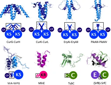
Structures of docking and COM domains
To date, docking domains and COM domains have been structurally characterised
from the biosynthetic pathways of curacin,18 erythromycin,19 pikromycin,20
vir-giniamycin,21 macrolactin,22 tubulysin,23 surfactin24 (Fig. 1.2), and enacyloxin
bound-aries, typically form a coiled-coil structure. The C-terminal binding partner also
consists of one, or two, helices that bind across the dimeric interface.
Creat-ing hybrid pairs of dockCreat-ing domains, and mutants have revealed indications of
docking domain selectivity, via key electrostatic interactions.18 The selectivity of
docking domains, is most apparent in a study that measures binding between
each docking domain from erythromycin PKS and pikromycin PKS. It shows
that the binding affinity of native docking domain pairs interact with binding affinities near 100µm, whereas no binding was observed with non-native pairings of docking domains.20 Interestingly, the addition of an adjacent KS domain has
been reported to provide a twofold increase in the affinity of the intermolecular interaction, and addition of a whole module provides a ninefold increase.20
KS KS
CP CP
EryAI-EryAII
KS
CPKS
CPCurK-CurL
KS
CP
KS
CP
CurG-CurH
(SrfB)-SrfC TubC
MlnE
C
E
KRKS
CP
KS
CP
PikAIII-PikAIV
C
KS
CP
[image:23.595.128.501.350.635.2]VirA-VirFG
Figure 1.2: Structures of docking domains and COM domains from the following
boundaries, possesses a helix-turn-helix structure (e.g. VirA-VirFG and MlnE),
which forms a four-helical bundle in its heterodimeric complex. This class of
docking domain appears across split modules, with di↵erent types of PKS en-zymes found adjacently. The low micromolar dissociation constant measured for
this class of docking domain, support the capability of mediating the association
of neighbouring domains.22Truncation and swapping docking domains show their
importance for forming protein complexes, and their pairwise portability.22
A structure of an N-terminal COM domain is reported from SrfC within the
surfactin NRPS, which forms a -sheet structure with the adjacent condensation
domain. A mimic of the corresponding C-terminal interaction partner, within
SrfB, forms an ↵-helix that makes contacts with the N-terminal COM domain,
as well as the condensation domain. At a PCP-C protein boundary
(TubB-TubC from tubulysin NRPS), an isolated N-terminal interaction domain was
structurally characterised, which revealed a distinctively di↵erent class of COM domain (herein referred to as ‘type 2’) exhibiting a ↵ ↵↵ fold.
1.5
NMR characterisation of protein domains
Small protein domains from PKSs and NRPSs are ideal candidates for structural
characterisation by protein NMR spectroscopy. Docking domains,19,21 COM
do-mains,23 and carrier proteins,25 represent types of small biosynthetic protein
do-mains that have been structurally characterised by NMR. Carrier proteins display
inherent conformational flexibility, making them less desirable for other common
biophysical methods, such as X-ray crystallography. Protein NMR spectroscopy
can uncover atom specific details of protein domains, describing protein
interac-tions in solution, by monitoring changes in the electromagnetic environment of
atoms during a domain-domain interaction. This can provide a molecular-level
description of significant interactions, during the sequential stages of enacyloxin
chain release.
nuclei. To improve sensitivity and enable the use of multidimensional
experi-ments, proteins are often isotopically labelled with 15N and 13C isotopes. One
of the most frequently used solution NMR experiments is the two-dimensional
1H-15N heteronuclear single quantum coherence spectroscopy (HSQC) sequence,
where each 1H-15N correlation signal is assigned to a specific amino acid residue
in the primary sequence of the protein. The HSQC spectrum provides a snapshot
of the electromagnetic environment for each 1H-15N bond within a protein, in
the form of chemical shift values. Since the chemical shifts depend on their local
environment, perturbations, or changes in cross peak intensity, in the presence of
a binding partner can be used to map interaction sites with other proteins.
1.6
Enacyloxin PKS-NRPS
PKSs and NRPSs have been identified in bacteria, fungi, plants and some
ani-mal genomes.26,27 Recently, the proteobacteria Burkholderia has been identified
as a promising candidate for the discovery of bioactive compounds, based on
the genome-guided discovery of natural product biosynthetic gene clusters.28 The
Burkholderia genus consists of over forty Gram-negative species that occupy
re-markably diverse ecological niches, through the production of secondary
metabo-lites.29 A group of seventeen closely related Gram-negative bacteria, so-called
Burkholderia cepacia complex (Bcc), are known to demonstrate biopesticidal
in-teractions against fungal diseases in plants,30 biodegrade pollutants,31,32and have
the ability to cause devastating lung infections in individuals with cystic
fibro-sis.33 Screening a large collection of Bcc strains has identified a strong activity
against Acinetobacter baumannii, which exhibits resistance to most antibiotics
and is one of the most problematic pathogens in healthcare institutions globally.
This antimicrobial activity has been identified from Bukholderia ambifaria, and
the anti-Gram-negative activity was mapped to a biosynthetic gene cluster using
The proposed biosynthetic pathway for enacyloxin IIa is outlined in Fig. 1.3.
It consists of a large modular PKS, NRPS domains, a pathway for a
shiki-mate derived compound, and tailoring enzymes. The loading module in protein
Bamb 5925 consists of a MT domain, a GNAT domain (malonyl
transferase/de-carboxylase from the Gcn5 histone acyltransferase family) and an ACP domain,
which initiates polyketide assembly with a propionyl starter moiety. Modular
ex-tension continues in a largely sequential manner for ten rounds of chain exex-tension
from PKS proteins belonging to thecis-AT PKS phylogeny, despite many of these
modules lacking an AT domain within their modules. Polyketide chain release is
preceded by a PKS module within Bamb 5919, belonging to the trans-AT PKS
phylogeny, which does not perform chain elongation. The natural product
inter-mediate is proposed to be passed from the ACP of Bamb 5919, to the adjacent
non-elongating ketosynthase (KS0), that lacks the necessary catalytic histidine
for chain elongation. The chain transfer continues from the KS0 domain to a
PCP domain within Bamb 5917, where specific protein-protein interactions are
postulated to contribute to the directionality of this process. Chain release is
ac-complished by the condensation of the 3’-hydroxyl group of a shikimate derived
dihydroxy-cyclohexane carboxylic acid (DHCCA), with the PCP-bound
polyke-tide. The biosynthetic pathway for the DHCCA compound includes the proteins
Bamb 5912 Bamb 5913, Bamb 5914, Bamb 5916 and Bamb 5918. The
conden-sation reaction is catalysed by an NRPS-like C domain within Bamb 5915. The
released intermediate then becomes the substrate for a series of tailoring enzymes
S O GNA T KR S O S O S O HO S O HO HO S O
HO HO HO
S
O
HO HO HO
S
O
HO HO HO
S
O
S
O
HO HO HO
HO HO HO
S
O
HO HO HO
S
O
HO HO HO
O
HO HO HO
1.7
Objectives
1.7.1
Objective I
The primary objective is to elucidate the biophysical basis for the unusual chain
release mechanism from the enacyloxin biosynthetic pathway. This process
in-volves two surplus enzymes, in terms of biochemistry, so it is proposed that a
se-ries of compatible interactions take place between ACP, KS0, PCP and C protein
domains. This could explain the necessity for this unusual domain architecture.
Elucidating the molecular basis for product release from the enacyloxin pathway,
will facilitate the application of this unusual enzymology to synthetic biology
approaches, for novel antibiotic production. A major challenge for engineering
biosynthetic pathways is developing a molecular-level understanding of
domain-domain and protein-protein interactions to faciliate efficient catalysis. The de-scription of interactions during enacyloxin chain release could describe rules of
interactions that could facilitate bioengineering novel biosynthetic pathways, and
describe unidentified features from characterised biosynthetic pathways.
1.7.2
Objective II
The secondary objective is to search for enacyloxin analogues, exploit any
bioac-tive properties and to investigate the biosynthesis of enacyloxin analogues. A
genomics-driven approach will be used to extensively search databases of
se-quenced organisms as experimental leads. Resulting organisms that contain
com-ponents related to the enacyloxin biosynthetic gene cluster, will be acquired,
cultured and their metabolic profile will be surveyed for the production of
enacy-loxin analogues. The structure, spectrum of activity, and biosynthetic pathway
of any enacyloxin-analogues will be investigated. Any beneficial bioactivity of
enacyloxin analogues could provide promising drug-leads, for chemical synthesis
1.8
Synopsis
This body of work encompasses three result chapters on the theme of the
biosyn-thesis of enacyloxins. Chapter 2 probes an unusual chain release process during
the biosynthesis of enacyloxin, where specific protein-protein interactions are
pro-posed to facilitate biosynthesis. Investigating protein interaction by NMR leads
to the discovery of an interaction site at the C-terminus of a PCP domain. A
crystal structure of the condensing enzyme revealed an additional N-terminal
communication-mediating (COM) domain. Together, this provided a full
ac-count of the intermolecular interaction during chain release between PCP and
C domains. Following the discovery of the COM interaction within the
enacy-loxin biosynthetic pathway, an HMM was developed to search for similar motifs
within other biosynthetic pathways (chapter 3). Many biosynthetic pathways
were identified using this HMM, and a hybrid COM interaction was investigated
at a non-native COM boundary from watasemycin NRPS and enacyloxin
PKS-NRPS. Other types of docking domains were also investigated using HMMs, which
led to the identification of a novel KS-DH protein boundary feature.
The discovery of an enacyloxin analogue is described in chapter 4. A
genome-mining approach revealed an enacyloxin-like biosynthetic gene cluster within
Vib-rio rhizosphaerae. The production of an enacyloxin analogue, referred to as
vi-broxin, was purified from extracts of solid cultures, and characterised. Some
of the di↵erences between the molecular structures of vibroxin and enacyloxin could be predicted from the di↵erences in biosynthetic gene clusters. One sig-nificant di↵erence between enacyloxin and vibroxin is a dimethylation of the starter unit during vibroxin biosynthesis, which was investigated by isotope
feed-ing experiments. Rigorous inspection of the PKS proteins, revealed fragments of
AT domains, suggesting intermediate stages between cis-acting AT domains and
trans-acting AT domains. The spectrum of activity of vibroxin is tested against
Chapter 2
Enacyloxin PKS-NRPS chain
release
2.1
Chain release process
During the biosynthesis of polyketides and non-ribosomal peptides, chain release
is ordinarily accomplished by a TE domain. This domain is dedicated to the
hydrolysis of the final thiol-bound intermediate, and often mediates
macrocycli-sation. In contrast, within the enacyloxin biosynthetic pathway, four enzymes
are involved in the formation of an ester bond during chain release. This unusual
system includes an ACP domain, a non-elongating KS domain (KS0), a PCP
domain and a stand-alone C domain. The KS0 domain lacks a catalytic histidine
residue involved in the mechanism for chain elongation. Thus, the KS0 domain
is proposed to act as an adapter between ACP and PCP domains, acting as a
trans-acylase (TA), passing the polyketide intermediate from the ACP domain
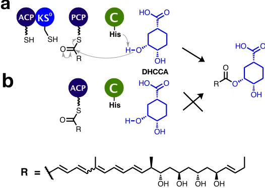
to the PCP domain. Subsequently, the catalytic C domain is then proposed to
selectively attack the 3’-OH of dihydroxy-cyclohexane carboxylic acid (DHCCA),
which then forms an ester with the PCP-bound polyketide (Fig. 2.1a). However,
the C domain has also been shown to accept a number of other intermediates
)-DHCCA does not appear to be a substrate for the C domain, but (10S,30R,40S
)-DHCCA is accepted by the C domain.35. For the remainder of this thesis,
the substrate of the C domain will be referred to as (10S,30R,40S)-DHCCA, or
shikimate, where stated. One alternative pathway describes the condensation
of (10S,30R,40S)-DHCCA with the ACP-bound polyketide, instead of the
PCP-bound polyketide (Fig. 2.1b). However, previous work has shown that in vitro
assays with acetyl-ACP, (10S,30R,40S)-DHCCA and the C domain do not perform
catalysis.36It is proposed that specific domain interactions play an important role
in this unusual chain release process, requiring four enzymes to produce one ester
bond. The biophysical basis of such specific interactions was investigated, using
a range of biophysical techniques.
ACP PCP
SH SH S
KS
O
R OH
O O HO
His
H C 0
OH O
O HO
R O
OH OH OH OH
R =
ACP
S O
R OH
O O HO
His
H C
a
b
DHCCA [image:31.595.190.453.350.536.2]b
Figure 2.1: Stages preceding chain release in enacyloxin PKS-NRPS. a) The
proposed chain release pathway, b) an equivalent biosynthetic pathway without KS0 and PCP domains.
2.2
Protein NMR spectroscopy
2.2.1
Sample preparation
in-Bamb 5919), KS0(encoded by gene Bamb 5919), PCP (encoded by gene Bamb 5917)
and C (encoded by gene Bamb 5915) were reconstituted in vitro with N-terminal
His6-tags to facilitate purification. For the reminder of Chapter 2 and for the
sake of simplicity the designations ACP, PCP, C and KS0 will refer to the above
mentioned constructs unless specified otherwise.
In order to facilitate NMR studies and whenever required,15N and13C were
in-corporated in proteins by using isotopically labeled precursors as the sole sources
of carbon and nitrogen during recombinant overproduction of proteins in E. coli.
Proteins were purified using nickel affinity column. The SDS-PAGE of the pu-rified domains are presented in Fig. 2.2. Because KS0 and C domains are very
large, and thus challenging for solution NMR (75 and 60 kDa respectively), only
smaller and amenable to solution NMR ACP and PCP domain where isotopically
labelled and observed in 15N and 13C-filtered experiments. For protein-protein
interaction experiments, carrier proteins were uniformly isotopically labelled with
15N (i.e. [U-15N]PCP and [U-15N]ACP), and the larger domains, KS0 and C, were
not isotopically enriched.
75 kDa
60 kDa C
KS0
15 kDa
PCP
22 kDa
ACP
14 kDa
Figure 2.2: SDS-PAGE of purified domains from enacyloxin PKS.
2.2.2
Carrier protein interactions with partner domains
Protein-protein interactions, or the formation of protein-protein complexes, can
be characterised by following changes in 2D 1H-15N HSQC spectra of uniformly
15N-labelled carrier protein upon addition of a larger domain, e.g. natural
changes of positions (or chemical shifts) of the sites involved in binding inform on
the interaction. In the case of slow chemical exchange the interaction manifests
as a decrease of the intensity of the peak for free site and increase of intensity of
the peak for the bound site as a function of interacting partner concentration. As
the size of the complex increases the decrease in the overall rotational di↵usion correlation time leads to improvement of transverse relaxation and associated
line broadening. For very large complexes, as it is in our case, the signals of the
carrier protein in the complex may become broadened beyond detection.
pH 6-7.5 Temp. 15-25oC
a
b
[image:33.595.124.516.269.532.2]c
d
Figure 2.3: 1H-15N HSQC spectra of carrier proteins in isolation (blue), and
with the addition of a partner domain at a 1:1 ratio (red). a) ACP-KS0, b)
PCP-KS0, c) PCP-C, d) ACP-C.
Within the HSQC spectra of protein complexes ACP-KS0, PCP-KS0 and
PCP-C (Fig 2.3a-c), there are significant changes in the HSQC spectra of carrier
proteins following the addition of a larger partner domain. This indicates an
in-teraction between them. On the contrary, the ACP-C complex HSQC spectrum
shows no significant changes when compared with those observed for the other
The residue assignment of PCP was then undertaken, to uncover the specific
details of the interaction site on PCP.
2.2.3
Residue assignment of PCP
The bu↵er conditions for NMR samples were screened for stability of PCP, includ-ing changes to pH (Appendix Fig. 7.1, 7.2) and temperature (Appendix Fig. 7.3,
7.4). A suite of three-dimensional NMR experiments could then be performed
(Section 5.4.2), to sequentially deduce the assignment of [U-13C,15N]PCP. The
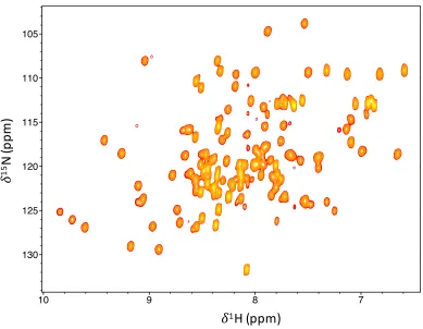
backbone assignment of PCP was achieved for 96% of backbone amides (Fig. 2.4,
Appendix Table 7.1), excluding the N-terminal purification tag. The dispersion
of the hydrogen chemical shifts are characteristic of a well-folded protein, but the
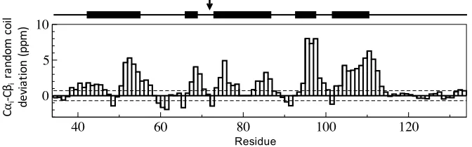
random coil deviations, proximal to the C-terminus, are indicative of an
unstruc-tured peptide, with a high degree of disorder (Fig. 2.5).
10 10
9 9
8 8
7 7
2 - 1H (ppm)
130 130
125 125
120 120
115 115
110 110
105 105
1
-
15
N (ppm)
Title: PCP17-288 -- hsqc_1000_2014_11_26-13C_15N_PCP17:1 User: daniel Date: Thu Nov 10 21:23:46 2016
Positive contours: low 8.00e+05 levels 30 factor 1.20
!
1H (ppm)
!
15
N
(p
pm
[image:34.595.128.517.388.690.2])
Figure 2.4: HSQC of PCP where the amide assignment is listed in Appendix
δobse
rve
d
-δrand
om
co
il
(p
p
m)
Residue
C
⍺i
-C
βi
ra
nd
om
c
oi
l
de
vi
at
io
n
(ppm
[image:35.595.125.461.108.213.2])
Figure 2.5: Random coil chemical shift deviations for PCP based on the di↵
er-ence in C↵iand C ichemical shift values for each residue,i. Predicted secondary
structure is represented above the bar chart, where bold lines indicate helices and the active site serine residue is identified with an arrow.
2.2.4
C-terminal interaction site
The aforementioned HSQC spectra, that monitor interactions between PCP and
C domains, display peak broadening, where the C-terminal region displays the
most intense signals, even with a relatively high concentration of the C domain.
The high intensities of these correlations correspond to a region of the protein
that does not conform to the core helical bundle, observed in other carrier protein
structures. In fact, the chemical shifts of this C-terminal extension are consistent
with an unstructured, or disordered, peptide. This lack of secondary structure
is congruous with the observed high signal intensities. The decay in each amide
correlation signal, during interaction of the PCP and C domain, can be observed
across the PCP protein to identify interaction sites during complex formation. As
well as the core PCP domain, significant changes were observed at the disordered
C-terminus of PCP (Fig. 2.6a-e) from about residue 125 to residue 134, with
in-creased amounts of C domain. This suggests that the C-terminal region is
impor-tant for the complex formation of the PCP and C domains, and consequentially
facilitating ester formation, during chain release. This C-terminal domain,
down-stream of PCP, shall be referred to as a C-terminal communication-mediating
Figure 2.6: Normalised signal intensities for PCP-COM2Camide HSQC
correla-tions upon titration with natural abundance C domain, for the following C:PCP ratios: a) 1:16, b) 1:8, c) 1:4, d) 1:2, e) 1:1.
2.3
In vitro
bioassays
2.3.1
Construction of mutants
The role of the COM2C domain can be investigated using assays that depend
upon the COM interaction. To evaluate the biochemical importance of the COM
domains, nine amino acids were removed from the C-terminus of PCP-COM2C
to form PCP. This was achieved by site-directed mutagenesis, where a premature
stop codon was substituted on the PCP-COM2C expression plasmid. The
trun-cated protein, PCP, was recombinantly overproduced and purified (Fig. 2.7a).
Mass spectrometry of PCP (Fig. 2.7bi) and PCP-COM2C (Fig. 2.7bii) provide
ions consistent with predicted molecular weights, 14466.3 Da and 13403.1 Da
respectively.
2.3.2
Condensation assay
A condensation assay was developed using: acetyl-PCPs (Bamb 5917), the C
0 200 400 600 800
In
te
ns
ity
[a.
u.]
0 100 200 300
12500 13000 13500 14000 14500 15000 m/z 0
200 400 600 800
In
te
ns
ity
[a.
u.]
0 100 200 300
12500 13000 13500 14000 14500 15000 m/z
PCP PCP
PCP
a
b
i
ii
Figure 2.7: Truncated mutant PCP. a) SDS-PAGE of PCP, b) MALDI-ToF
spectra of i) PCP truncated mutant, ii) PCP-COM2C.
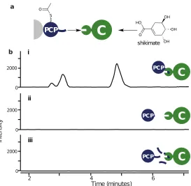
could be used to verify the role of the COM2 boundary during biosynthesis, in
particular the e↵ect of COM2C. A mimic of the wild-type system,
acetyl-PCP-COM2C with the C domain and shikimate, yielded a product consistent with
acetylated shikimate, monitored by LC-MS. The extracted ion chromatogram
cor-responding to the sodiated adduct of acetylated shikimate displays three species
with di↵erent retention times (Fig.2.8b). This could be due to acetylation of each of the three hydroxyls on the cyclohexene during condensation, or acetyl
rearrangement thereafter. The truncated mutant PCP was used to replace the
PCP-COM2C protein in the assay, to investigate the biochemical significance of
the COM2Cdomain. No product formation was observed for assays containing the
COM2C truncated PCP (Fig.2.8bii). The interpretation of this result is that the
breakdown of the COM2C-dependent intermolecular interaction prevents efficient
complex formation, a prerequisite for catalysis in this case. The assay with the
full length PCP-COM2Cwas then repeated with the addition of a tenfold excess of
COM2C peptide (11 amino acid length chemically synthesised by Insight
Biotech-nologies). From this assay, no product was detected (Fig.2.8biii). Competitive
inhibition of the protein-protein interaction is proposed to account for
signifi-cantly decreasing catalysis upon the addition of supplementary COM2Cpeptides.
Together these results support the biochemical significance of the COM2Cdomain
i
ii
[image:38.595.184.457.80.347.2]iii
Figure 2.8: a) Schematic representation of the condensation assay. b) E.I.C
corresponding to the sodiated adduct of acetylated shikimate for the assay with the C domain, shikimate and either, i) acetyl-PCP-COM2C, ii) truncated
acetyl-PCP, iii) acetyl-PCP-COM2C with a tenfold excess of COM2C peptide.
2.4
Crystal structure of the condensing enzyme
In order to determine the possible binding site of COM2C, the structure of the
C domain from enacyloxin PKS-NRPS was solved. This was part of a
collabora-tion between the University of Warwick and UC Irvine, which included Timothy
Valentic, Sheryl Tsai, Paulina Sydor, Dean Rea and Vilmos F¨ul¨op. This structure
deviates from other NRPS-like C domain structures, by a loop above the active
site, and the addition of an N-terminal domain.
2.4.1
N-terminal appendage
The additional N-terminal domain consists of an ↵ ↵↵ structure, and appears
recurrently within NRPS proteins, typically identified upstream of condensation
or heterocyclisation domains using homology searches and motif finders. An
(Fig. 2.9a), and its function was proposed to perform an intermolecular interaction
across a protein boundary. Subsequently, this type of domain will be herein
referred to as a ‘type 2’ N-terminal communication mediating (COM2N) domain.
This will distinguish it from ‘type 1’ COM domains, regularly identified between
condensation domain and epimerisation domain boundaries (see Chapter 3 for
further details).37
The crystal structure of COM2N-C from enacyloxin PKS-NRPS, provides the
first structural account of the relative positioning of a COM2N domain, to its
catalytic neighbouring domain (Fig. 2.9b). A short linker allows the COM2N
domain to situate near the C-terminal subdomain of the condensing enzyme,
positioned adjacently to the entrance of the downstream substrate. Amino acids
Thr2 and Tyr51 from the COM2N domain engage in polar contacts with Glu380
and Ser383 from the C domain respectively, which stabilise the localisation of
COM2N.
2.4.2
Loop above the active site
The structure of the C domain contains some variations from other NRPS C
do-main structures, namely between Ser418 and Ser453. Within this region, a loop
extends above the C-terminal subdomain, forming little secondary structure. In
other structures this region typically forms a -sheet with the N-terminal
subdo-main with additional helical components, displaying an overall more structured
fold. These di↵erences could arise from the isolated nature of this particular con-densation domain, as its placement di↵ers from the archetypal location within an NRPS module. Whilst the upstream PCP delivers a polyketide to the accepting
face of the condensing enzyme, access to the donating face is proposed only to
facilitate a relatively small substrate (e.g. DHCCA), not a typical PCP-bound
a
b
T2
Y51 S383 E380
H454
linker loop
Figure 2.9: a) COM2N structure from tubulysin NRPS.23 b) COM2N-C
dido-main structure from enacyloxin PKS-NRPS. Linker between COM2N domain and
C domain is shown in blue. The atypical loop within the C domain is shown in red.
2.5
Protein-protein complex
A crystal structure of a PCP-E didomain38 (Fig. 2.10a) from gramicidin S NRPS
provides a reference for modelling the PCP-C complex from enacyloxin
PKS-NRPS, as E domains and C domains possess the same overall fold. The PCP
domain from enacyloxin PKS-NRPS was overlayed on the PCP-E structure, and
the COM2N-C domains were aligned to the relative position of the E domain
(Fig. 2.10b).
PCP
E
PCPC
a
b
Figure 2.10: a) PCP-E didomain structure from gramacidin NRPS.38 b)
Align-ment of the COM2N-C domains and PCP from enacyloxin PKS-NRPS to the
Models of the interaction between COM2N and COM2C domains have been
proposed from tubulysin NRPS23(Fig. 2.11ai) and myxothiazol NRPS39(Fig. 2.11aii).
Using the structure of COM2N, and the partner interaction site COM2C, a
protein-peptide molecular dynamics simulation was performed to objectively investigate
the COM2C-COM2N domain interaction during the biosynthesis of enacyloxin.
Small amounts of helical propensity were observed for the COM2Cpeptide during
simulations, but it remained largely devoid of any secondary structure generally.
Four clusters of solutions of the COM2N and COM2C complex are represented
in Fig. 2.11b. Each of the protein-peptide complex models represent clusters
of hundreds of states, where the COM2C peptide samples proximal regions of
the COM2N surface. These solutions may represent a ‘fuzzy’ complex, whereby
degenerate multiplicity exists, as is commonly observed for interaction sites
in-volving disordered peptides.40,41 It is unlikely that the proposed interaction sites
on the COM2N can be simultaneously occupied by the COM2C domain, due to
their relatively large expanse. The COM domain complex solutions provide an
insight into the role of the hydrophobic amino acids on COM2C. There are two
pockets, adjacent to the -hairpin, that locate the hydrophobic residues (namely
i
ii
iii
iv
b
a i
ii
Figure 2.11: a) COM2N domain interaction sites (sticks) proposed from i) a
COM2N from tubulysin NRPS,23 ii) a COM2N domain homology model from
myxothiazol NRPS.39 bi-iv) Solutions from simulations of the COM
2C domain
(purple, C-terminus represented as sphere) with the COM2N domain from
ena-cyloxin PKS-NRPS.
Coupling the PCP-C model, and the COM2N-COM2C models, provides a
rep-resentation of the entire complex (Fig. 2.12). It describes intermolecular
com-munication, and the interactions of adjacent enzymes responsible for catalytic
activity during enacyloxin chain release. A model solution for the COM
domain-complex was linked to the C-terminus of the core PCP domain by building in
a loop, which was subsequently subjected to energy minimisation. The
PCP-COM2C and COM2N-C complex model describes the proposed interactions, and
a schematic of the states it must accommodate during biosynthesis. After
trans-lation, the intermolecular interactions that are required between the PCP and C
domain, are proposed to be facilitated by a pair of compatible COM domains.
This ensures a particular biosynthetic pathway is followed, and acts to increase
the e↵ective concentration of the PCP domain with respect to the C domain. Following COM domain interactions, the PCP is proposed to deliver a
polyke-tide intermediate to the C domain, for condensation with a shikimate derived
terminal thiol of holo-PCP can be made acessible, to load another molecule of
the polyketide from the KS0 domain, before redelivery to the C domain for ester
formation with DHCCA.
PCP
accept
substrate
substrate
donate
C
[image:43.595.204.437.165.548.2]COM
interaction
Figure 2.12: Representation of the domain oragnisation of PCP-COM2C and
COM2N-C from enacyloxin PKS-NRPS. COM domains are propsed to direct
intermolecular interactions, where the PCP domain accepts polyketides from the previous module to donate to the C domain in a recursive manner.
2.6
Conclusion and future work
domains have been observed by protein NMR spectroscopy, and are consistent
with the proposed biosynthetic pathway. Biophysical investigation of the chain
release process has uncovered a pair of COM domains, which form specific
inter-molecular interactions across the PCP and C protein boundaries. The residue
assignment of PCP, and PCP-C mixing experiments, led to the identification of a
C-terminal COM domain, exhibiting high levels of disorder. This COM domain
has been shown to be necessary for efficient catalysis, performing a fundamental intermolecular interaction across a protein-protein boundary.
The structure of the C domain revealed an additional N-terminal COM
do-main, and its position relative to the catalytic C domain. The nature of the COM
domain interaction was simulated, which provided clusters of statistical solutions.
These states were subsequently evaluated based upon the NMR interaction data
with the C-terminal binding partner. However, an experimentally determined
structure of the protein complex, or an experimentally verified description of
the interactions sites on the N-terminal COM domain interface, would provide
additional insight to this particular intermolecular interaction.
The COM domain interaction has led to an updated description of chain
re-lease within enacyloxin biosynthesis, and is proposed to drive complex formation
to facilitate catalysis. This COM domain interaction site allows the quaternary
structure to be maintained, whilst permitting accessibility for the PCP domain
to locate the active sites of both the C domain, and the KS0 domain. Given
the significance of the COM domain interaction within enacyloxin biosynthesis,
further experiments could investigate the exploitation of this interaction to
ra-tionally engineer intermolecular interactions to develop hybrid biosynthetic
path-ways. Furthermore, to test the promiscuity of the enzymes across COM domain
boundaries, creating a chimeric protein of ACP-COM2C (instead of PCP-COM2C)
would present valuable insight to the tolerance of ACP and C domains, and
ven-tures for engineering COM domain intermolecular interactions to biosynthesise
domain interaction, new tools could be developed for their annotation within
Chapter 3
Intermolecular docking domains
and communication-mediating
domains
3.1
Identification of docking and COM domains
Given the large number of known PKS and NRPS proteins, docking domains and
COM domains are scarcely identified within them. The development of new tools
to identify these terminal domains will provide a more detailed understanding of
intermolecular interactions within PKSs and NRPSs.
3.1.1
Profile hidden Markov models for dd
1and COM
1Docking domains and COM domains can be identified using profile hidden Markov
models (pHMMs), which consist of a statistical description of a protein family.
Currently, antiSMASH42 utilises two pHMMs that describe N-terminal and
C-terminal docking domains (denoted dd1N and dd1C respectively) and two pHMMs
for COM domains (COM1N and COM1C). The pHMM for COM1N contains a
region that can be considered part of a condensation or cyclisation domain from a
num-ber of results that could be considered potential false positive annotations. As a
consequence, it is commonplace to identify COM1N domains upstream of
conden-sation domains that do not lie at a terminus. The pHMM for the corresponding
binding partner, COM1C, contains a small region that can be attributed to an
epimerisation (E) domain. Despite this, COM1C domains can also be identified
following PCP domains, at PCP and condensation domain protein boundaries
(e.g. VpsA-VpsB from vancomycin NRPS).
C
a
b
E
Figure 3.1: HMM logos for a) COM1C and b) COM1N HMMs from antiSMASH.
Analogous to COM domains, docking domains have been identified within a
number of PKSs. These have also been identified using pHMMs for dd1N and
dd1C, corresponding to the N-terminal and C-terminal interaction sites,
respec-tively (Fig. 3.2). The pHMM that includes the dd1Cdomain, also includes a region
describing an additional dimerisation domain.19This region has been structurally
characterised from eryAI, and is regularly unidentified within PKS proteins. This
is a limitation of this relatively long pHMM. In contrast, the N-terminal
bind-ing partner, dd1N, more accurately represents the profile of this domain, and is
consistent with a structurally characterised dd1N domain.
a
b
dimerisation
dd1N
dd1C
Figure 3.2: HMM logo for type 1 docking domains used by antiSMASH, a)
3.1.2
Novel HMMs for docking domains
A second class of docking domains (dd2) are based on protein termini identified
at ACP and KS boundaries within curacin PKS (CurG-CurH and CurK-CurL).18
These dd2 domains were also reported within 22 other biosynthetic pathways at
ACP and KS protein boundaries.18This set of N-terminal and C-terminal docking
domains (dd2N and dd2Crespectively) was used to build two HMMs (dd2N, dd2C)
to facilitate the identification of these domains.
The dd2N and dd2C HMMs were used to search the MIBiG database43 for
biosynthetic pathways that contain dd2 domains. This revealed a large
num-ber of dd2-containing biosynthetic pathways, which were previously
unidenti-fied. The results include biosynthetic pathways for: anabaenopeptin, ambruticin,
burkholdac, chondrochloren, crocacin, ebelactone, enacyloxin, gephyronic acid,
gulmirecin, leupyrrin, microsclerodermins, nannocystin, nostophycin, pellasoren,
polyoxypeptin, puwainaphycin and spiruchostatin. Each of these examples
ex-hibit class 2 docking domains, which are proposed to facilitate biosynthesis across
ACP and KS boundaries. One exception is nostophycin PKS, where dd2 domains
are not located at protein boundaries of NpnA. Within this protein, dd2N and
dd2C domains are found in between the ACP from module 1 and the KS from
module 2 (Fig. 3.4a). It is clear that docking domains between modules 1 and 2
are not required as they are part of the same protein. These modules may have
originated from separate genes where the dd2 domains would have facilitated
biosynthesis across a protein boundary, but mutations in the stop codon of the
upstream gene could have led to a genetic merger, if the downstream gene was
situated in the same reading frame.
3.1.3
Identifying unknown types of docking domain
Whilst HMMs for docking domains have revealed many biosynthetic pathways
utilising these terminal features, there are many PKS and NRPS proteins where
unanno-tated terminal gaps provide motivation to investigate patterns in these terminal
regions, which could uncover novel types of docking domains. To address this,
a database of PKS and NRPS amino acid sequences was curated from
charac-terised biosynthetic pathways, and the termini were extracted to provide a list of
N-terminal and C-terminal amino acid sequences for inspection. These sequences
were analysed for novel motifs (recurring, fixed-length patterns) using MEME.44
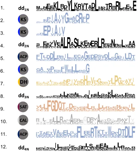
The resulting motifs for N-terminal PKS sequences are listed in Fig. 3.3.
KS
KS
KS
DH
dd1N
dd2N
dd3N
SAT
CAL
dd4N
ACP ACP
1.
2.
3.
4.
5.
6.
7.
8.
9.
10.
11.
[image:49.595.204.435.253.508.2]12.
Figure 3.3: Motifs from N-terminal PKS sequences identified using MEME,
listed by statistical significance. Each motif is labelled corresponding to its cat-alytic domain, or type of docking domain.
The N-terminal motifs could be attributed to docking domains as well as
enzymatic domains that are situated directly at the N-terminus. KS and ACP
domains from type II PKSs were identified by MEME, but did not contain
termi-nal docking domains, as well as KSQdomains involved in loading modules. Other
N-terminal domains include starter-unit:ACP transacylase (SAT) domains from
fungal biosynthetic pathways, and coenzyme A ligase (CAL) domains, conferring
con-ACP and KS domains at protein boundaries.
The fourth most significant motif was used to develop a HMM to describe a
docking domain with some similarity to type 2 docking domains (coined dd2N ↵
and dd2C ). During instances where multiple HMMs identify a docking
do-main within the same amino acid sequence, the highest scoring HMM is chosen.
The dd2N ↵ and dd2C HMMs identified docking domains from 66 biosynthetic
pathways (Appendix 7.3.1) at protein boundaries between ACP domains and KS
domains. The eighth most significant motif described another pair of putative
docking domains, which were converted into paired HMMs (dd3N and dd3C).
These HMMs were used to identify 22 biosynthetic pathways (Appendix 7.3.2)
from the MIBiG database, which putatively utilise dd3 domains. Most examples
represent boundaries between at ACP and KS domains, but dd3 domains were
also identified at PCP-KS and ACP-C protein boundaries. An example of an
ACP-C boundary is BaeM-BaeN, from bacillaene PKS-NRPS, where a dd3N
do-main was identified at the N-terminus of BaeN (or PksN), illustrated in Fig. 3.4b.
Within the gobichelin biosynthetic pathway, a dd3C domain was identified
adja-cent to a PCP at a PCP-C protein boundary, but no corresponding N-terminal
binding partner was identified next to the condensation domain.
KS DH
ER KR KR
PCP AT ACP KS AT ACP
KR
npnA
dd2C-dd2N
KR
KS A PCP
A
dd3N
ACP C
b baeM baeN
... ...
[image:50.595.232.405.500.650.2]a
Figure 3.4: Examples of docking domains in unusual locations, a) nostophycin
PKS with dd2 domains incisb) bacillaene PKS-NRPS showing a PKS-like dd3N
domain adjacent to a condensation domain.
The twelfth motif was identified both at the N- and C-terminus of biosynthetic