ISSN 0015–5659 www.fm.viamedica.pl
Address for correspondence: B. Ercakmak, MD, Hacettepe University Faculty of Medicine, Department of Anatomy, Ankara, Turkey, tel: +90-312 3052117, e-mail: [email protected]
Association between frontal sinus development
and persistent metopic suture
S. Bilgin
1, U.H. Kantarcı
2, M. Duymus
2, C.H. Yildirim
3, B. Ercakmak
4, G. Orman
2,
C. Gunenc Beser
4, M. Kaya
3, M. Gok
2, A. Akbasak
31Department of Anatomy, Kafkas University Faculty of Medicine, Kars, Turkey 2Department of Radiology, Kafkas University Faculty of Medicine, Kars, Turkey 3Department of Neurosurgery, Kafkas University Faculty of Medicine, Kars, Turkey 4Department of Anatomy, Hacettepe University Faculty of Medicine, Ankara, Turkey
[Received 10 June 2013; Accepted 14 July 2013]
Background: Frontal sinuses are 2 irregular cavities, placed between 2 lamina of frontal bone. Expansion continues during childhood and reaches full size after puberty. Persistent metopic suture is one of the factors that are related to abnormal frontal sinus development. In this study, we want to discuss about the coexistence of persistent metopic suture and abnormal frontal sinus development using radiological techniques.
Materials and methods: In this retrospectively planned study, images of 631 pa- tients were examined, 217 (34.4%) of them were men and 414 (65.6%) of them were women. Brain computed tomography and magnetic resonance images were retrieved from the electronic archive for analysis.
Results: In this study, frontal sinus development is categorised as right side atrophy, left side atrophy, bilateral atrophy and bilaterally developed sinuses. The presence of metopic suture was accepted as persistent metopic suture. Frontal sinus atrophy was found in 22.7% and persistent metopic sutures were found in 9.7% of overall.
Conclusions: In this study, no significant results were detected that were related to the frontal sinus agenesis or dismorphism associated with persistent metopic suture. We conclude that, although publications propounding metopism that leads to abnormal frontal sinus development are present in the literature, no reasonable explanation has been mentioned in these articles; and we believe that these findings are all incidental. (Folia Morphol 2013; 72, 4: 306–310)
Key words: frontal sinus, atrophy, metopic suture, computed tomography imaging, magnetic resonance imaging
INTRODUCTION
Frontal sinuses are 2 irregular cavities placed be-tween 2 lamina of frontal bone [29]. They are the last paranasal sinuses to develop [23]. They are rudi-mentary at birth and can be radiologically visible by 6 years. Expansion continues during childhood and reaches the full size after puberty. Size and shape of the frontal sinuses differ by age and sex. Also
2 sinuses of the same person differ in shape and size be- cause of the septum deviated from the median plane [29]. Several septa may be seen besides the median septum [9, 10]. As the radiological morphology is individualised, shape of the sinuses can be valuable
in identification of a person [29].
or ethnic groups [6, 8]. Persistent metopic suture is one of these factors [7, 20, 28]. Metopic suture is a kind of dentate suture [4, 12, 13]. It lies between 2 halves of the frontal bone and extends from nasion to bregma [1, 19, 28]. This suture normally closes
be-tween first and second year of life, however fusion can
be completed until the age of 7 [27]. If the metopic suture persists after the 7th year of life, it is known
as ‘metopism’ [28], and it is said that frontal sinus development may be affected by this entity [20, 21]. Metopic suture persistence can be visualised particu-larly or completely [12, 20]. Some authors think that abnormal frontal sinus development can be grounded on particular or complete persistent metopic suture that extends to the inferior part of the frontal bone [20, 28]. Frontal sinuses of a person with persistent metopic suture develop separately on either side of the suture and this entity can be used to differentiate persistent metopic suture from fracture [29].
MATERIALS AND METHODS
In this retrospectively planned study, the images that were taken from the patients who applied to our center between September 2011 and April 2012 were
analy-sed. We identified brain computed tomography (CT) and brain magnetic resonance (MR) images that could
be retrieved from the electronic archive for the analysis. All CT scans were obtained with 64-slice
spi-ral scanner (Toshiba Aqulion). As a standard, slice
thickness and interval were set at 5 and 3 mm. Scan-ning parameters included 550 mA, 135 kV and tube rotation time of 1.5 s. When the imaging was per-formed, the head was in neutral position.
MR scans were performed with 1.5 tesla MR
scan-ner (Siemens, Essenza). T1 and T2 axial, coronal and
sagittal images were examined.
Then both MR and CT images were forwarded
into the viewer program (Aquarius intuition viewer version 4.4.6).
The study parameters were age, sex, frontal sinus development, and metopic suture presence. Frontal sinus development was noted as right side atrophy, left side atrophy, bilateral atrophy, and bilaterally developed. Unfusion of metopic suture was accepted as persistent metopic suture.
Statistical analysis
SPSS (Statistical Package for Social Sciences) ver -sion 17 was used to make statistical analyses. In-dependent Sample T test was used to detect the
correlation between metopic suture and frontal sinus atrophy. A p value of less than 0.5 was indicative of
statistical significance.
RESULTS
219 cranial CT (34.7%) and 412 cranial MR (65.3%)
images retrieved from the electronic archive were
identified. 217 (34.4%) of the study patients were men, 414 (65.6%) were women. The mean age of the
patients was 48.37 ± SD (between 30–93). Frontal
sinus development and the presence of metopic su-tures were investigated.
In this study, frontal sinus development is catego-rised as right-side atrophy, left-side atrophy, bilateral atrophy and bilaterally developed sinuses. The pres-ence of metopic suture was accepted as persistent metopic suture.
Frontal sinus atrophy was found in 22.7% of over
-all, including 9.4% right sided, 2.9% left sided and 10.5% bilaterally.
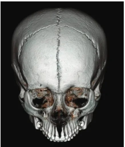
Persistent metopic sutures were found in 9.7% of overall (Fig. 1).
Frontal sinus atrophy was found in 24.6% of the patients with persistent metopic suture (Table 1). Out of
No correlation between persistent metopic suture
and frontal sinus development was detected (p > 0.05) (Table 3).
Table 1. The correlation between persistent metopic suture and frontal sinus atrophy
Atrophy Total
+ –
Metopic suture + 15 (24.6%) 46 (75.4%) 61 – 128 (22.5%) 442 (77.5%) 570
Total 143 488 631
Table 2. Side distributions of the patients with metopic suture and frontal sinus atrophy together
Metopic suture Atrophy Frequency Per cent Valid per cent Cumulative per cent
Exist Absent Missing System 46 100,0
Exist Valid Right 7 46.7 46.7 46.7
Left 2 13.3 13.3 60.0
Bilateral 6 40.0 40.0 100.0
Total 15 100.0 100.0
Absent Absent Missing System 442 100.0
Exist Valid Right 52 40.6 40.6 40.6
Left 16 12.5 12.5 53.1
Bilateral 60 46.9 46.9 100.0
Total 128 100.0 100.0
Table 3. P value of the c2 tests is 0.828 and showed no significant result related to frontal sinus agenesis or dismorphism associated
with persistent metopic suture
Value df Asymp. Sig. (2-sided) Exact Sig. (2-sided) Exact Sig. (1-sided)
Pearson c2 0.143a 1 0.705
Continuity correctionb 0.047 1 0.828
Likelihood ratio 0.141 1 0.708
Fisher’s exact test 0.748 0.405
Linear-by-linear association 0.143 1 0.705
No. of valid cases 631
a0 cells (0%) have expected count less than 5. The minimum expected count is 13.82; bComputed only for a 2 × 2 table
atrophy was detected as 46.7% right-sided (Fig. 2), 13.3% left-sided and 40% bilaterally. Bilateral deve
-lopment was found in 75.4% of the patients with persistent metopic suture (Table 2).
Frontal sinus atrophy was found in 22.5% of the patients without persistent metopic suture (Table 1).
Out of the patients without persistent metopic suture,
frontal sinus atrophy was detected as 40.6% right
--sided, 12,5% left-sided and 46.9% bilaterally (Table 2). Bilateral development was found in 77.5% of the
patients without persistent metopic suture.
their major development, evolution of the frontal sinuses begin according to the continuous growth of the outer table in response of the stimulus of the growing nasomaxillary facial complex [27].
Many authors have performed descriptive or experimental studies regarding cranial sutures and their evolution either in man [9, 16, 19, 23], or in animals [3, 18, 23]. In these studies, histological and microradiographic aspect of the metopic suture was
well defined before, during, and after its closure.
Although there is abundant data about the closure of the metopic suture, little or no information is encoun-tered on persistent form of it. Neither embryologic, nor histological information can be obtained from the investigations performed until today regarding the persistence of metopic suture and its relation to frontal sinus agenesis. Pneumatisation of the frontal sinuses occurs after the 5th or 6th postnatal year, while
frontal bones and metopic suture are developing in early foetal life; thus there should be no embryologic, and temporal relation concerning the development of these 2 anatomic structures. Adverse effect of a persistent metopic suture on the development of the frontal sinuses seems unlikely. Conversely, it is well known that premature closing of the cranial sutures in craniosynostosis results in atretic frontal sinuses due to increased intracranial pressure hindering pneuma-tisation of the sinuses. In a study conducted by Locher et al. [17], frontal sinus pneumatisation following bilateral fronto-orbital advancement was seen in 24 of 33 patients with craniosynostosis [14]. On the other hand, individuals with persistent metopic suture are otherwise neurologically and physically normal.
There are publications suggesting the persistent metopic suture was associated with the absence of frontal sinus; however, none of them are based on
a scientific work or explanation [2, 6]. Since the plain
X-ray exams are not as sensitive as the CT and MRI studies, correct results from the investigations related to skull anatomy can be obtained from the CT and MRI scans, or from the autopsy material and cadavers
only. Results acquired from the material consisting of
X-rays are prone to misinterpretation arising either from the erroneous evaluation of the radiograms or
their insufficient resolution. Baaten et al. [2] declared
that the absence of frontal sinus was found in 7 of the 8 cases of metopism in a study they carried out on 968 skull X-rays. In that study they also admit that some irrelevant results to the literature they obtained could be due to the use of X-rays rather than cadavers.
DISCUSSION
There are 2 types of cranial bones in the head;
tho-se formed through the ossification of a cartilaginous
intermediate are known as ‘endochondral bones’,
and those formed through the direct ossification in
the mesenchyme are known as ‘membrane bones’ or ‘dermal bones’.
The cranium is composed of 3 layers. The en-dochranium, which encloses the brain and helps to form the sensory capsules that support and protect the olfactory organs, eyes, and inner ears; an
exter-nal protective layer of membrane (dermal) bones;
and the viscerocranium that supports the jaws. The cranial bones can be divided into the neurocranium and viscerocranium. The neurocranium encompasses the bones surrounding and protecting the brain and sensory organs — the endochondral bones of the base of cranium and sensory organs, and the dermal bones of the skull vault. The viscerocranium encompasses the bones of face and pharyngeal arches [5, 26].
In humans, the chondrocranium is the portion of
the skull formed by endochondral ossification. It is
developed from 3 pairs of precursors. These cartilages contribute to the cranial base and, together with cartilaginous capsules that develop around the otic and nasal pits help to protect the brain and sensory
organs. This portion of the skull is the first to form in the embryo. The flat bones of the cranial vault
calvaria are consisted of membrane-bone armor that covers the skull.
The bones of the cranial vault do not complete
their growth during foetal life. The soft, fibrous su -tures allow them to continue growing throughout infancy and childhood. During foetal life, the so-called sutural space — metopic suture, separates 2 frontal
bones; it consists of fibrous tissue and mesenchymal
cells responsible for the growth of the frontal bones [18]. The metopic suture closes normally before the second year of life, while the closure of the rest of the calvarial sutures occurs between 26th and 36th month.
The frontal sinuses do not form until the 5th or
6th postnatal year and they expand throughout
ad-olescence. Each frontal sinus actually consists of 2 independent spaces that develop from different so- urces. One is developed from the expansion of the ethmoid sinus into the frontal bone, and other is developed from an independent invagination of the middle meatus of the nasal passage; they never coa-lesce [25]. As the growth of the inner table ceases at
6. Cakur B, Sumbullu MA, Bayindir Durna N (2011) Aplasia
and agenesis of the frontal sinus in Turkish individuals: a retrospective study using dental volumetric tomography.
Int J Med Sci, 8: 278–282.
7. Das S, Suri R, Kapur V (2005) Anatomical observations on
os inca and associated cranial deformities. Folia Morphol,
64: 118–121.
8. Del Sol M, Binvignat O, Bolini PD, Prates JC (1989) Meto
-pism in Brazilians. Rev Paul Med, 107: 105–107. 9. Dhem A, Dambrain R, Thauvoy CH (1983) Contribution
to the histological and microradiographical study of the
craniostenosis. Acta Neurochir, 69: 259–272.
10. Dwivedi AND, Sıngh KK (2010) CT of the paranasal sinuses:
normal anatomy, variants and pathology. J Optoelectronics
Biomedical Materials, 4: 281–289.
11. Eisenberg RL (1994) Skull and spine imaging: an atlas of
differential diagnosis. Raven Press, New York.
12. Gulisano M, Pacini P, Orlandini GE (1978) Frontal sinus
dimentions in reletion to the cranial index. Boll Soc Ital
Bio Sper, 54: 66–69.
13. Hussain Saheb S, Mavishetter GF, Thomas ST, Prasanna LC
(2010) Incidence of metopic suture in adult South Indian skulls. J Biomed Sci and Res, 2: 223–226.
14. Jane JA, Edgerton MT, Futrell JW (1978) Immediate correction of sagittal synostosis. J Neurosurg, 49: 705–710. 15. Krogman WM (1962) The human skeleton in forensic
medicine. Spring field, Thomas.
16. Latham RA, Burston WR (1966) The postnatal pattern of
growth at the sutures of the human skull. Dental
Practi-cioner, 17: 61–679.
17. Locher MC, Sailer HF, Haers P (1998) Development of fron -tal sinus following bilateral fronto-orbi-tal osteotomies.
J Cran-Max Surg, 26: 129–135.
18. Manzaranes MC, Goret-Nicaise M, Dhem A (2002) Metopic sutural closure in human skull. J Anat, 161: 203–215. 19. Moss ML (1958) Fusion of the frontal suture in the rat.
Am J Anat, 102: 141–165.
20. Murlimanju BV, Prabhu LV, Pai MM, Goveas AA,
Dhanan-jaya KVN, Somesh MS (2011) Median frontal sutures-
incidence, morphology and their surgical, radiological
importance. Turkish Neurosurgery, 21: 489–493. 21. Newton T, Potts GD (1971) Radiology of the skull and brain.
Vol. 1. The Skull The C. V. Mosby Company, Saint Louis.
22. Ponde JM, Andrade RN, Via JM (2008) Anatomical variations of the frontal sinus. Int J Morphol, 26: 803– –808.
23. Pritchard JJ, Scott JH, Girgis FG (1956) The structure and
development of cranial and facial sutures. J Anat, 90:
73–86.
24. Reeder MM, Bradley WG (1993) Reeder and Felson’gamuts
in radiology. 3rd Ed. Springer-Verlag, New York.
25. Schoenwolf GC, Bleyl SB, Brauer PR, Francis-West PH (2009)
Development of the pharyngeal apparatus and face. In: Schoenwolf GC ed. Larsen’s human embryology. Churchill
Livingstone Elsevier, Philadelphia, pp. 543–584. 26. Scuderi AJ, Harnsberger GR, Boyer RS (1993) Pneumatiza
-tion of the paranasal sinuses: Normal features of impor-tance to the accurate interpretation of CT scans and MR
images. AJR, 160: 1101–1104.
27. Shapiro R, Shorr SA (1980) A consideration of systemic factors that influence frontal sinus pneumatization. Invest Radiol, 15: 191–202.
28. Skrzat J, Walocha J, Zawilinski J (2004) A note on the
morphology of the metopic suture in the human skull.
Folia Morphol, 63: 481–484.
29. Standring S (2008) Gray’s anatomy. 40th Ed. Churghill Livingstone Elseiver, Spain.
30. Swischuk L (1984) Differential diagnosis in pediatric ra -diology. Williams & Wilkins, Baltimore.
Moreover, in many studies the absence of frontal
si-nuses was found at rates changing from 5% to 27.9%
regardless of persistence of the metopic suture [12, 15, 22].
Normal skull anatomy such as vascular grooves,
developmental fissures, and straighter-appearing su -ture lines of the lamina interna can mimic, and be mistaken for fractures. Persistent metopic suture is one of these markings and may cause misdiagnosis as a linear fracture. Misdiagnosed metopic suture may also cause therapeutic mistakes and unnecessary interventions. Neurosurgeons desire to be informed
about all anatomical configurations on the skull be -fore a cranial surgery. A persistent metopic suture should be revealed prior to a frontal craniotomy. It should be kept in mind that routine radiographs of the skull can be more sensitive than CT in the detec-tion of linear fractures and other linear formadetec-tions. Therefore, meticulous radiographic examinations, including 3-dimensional CT, should be performed for the correct diagnosis. On the other hand, prominent suture areas can also be noted in hydrocephalus, cerebritis, brain neoplasms, metastasis, leukaemia, lymphoma, and other causes of increased intracranial pressure [11, 24, 30].
No data were found correlating the persisten-ce metopic suture to the frontal sinusitis and other pathologies of the frontal sinuses in the literature.
CONCLUSIONS
In the present study, no significant result was detec -ted relating to frontal sinus agenesis or dismorphism that is associated with persistent metopic suture. We conclude that, although publications propounding metopism that leads to abnormal frontal sinus deve-lopment are present in the literature, no reasonable explanation has been mentioned in these articles; and
we believe that these findings are all incidental.
REfERENCES
1. Ajmani ML, Mittal RK, Jain SP (1983) Incidence of the me
-topic suture in adult Nigerian skulls. J Anat, 137: 177–183. 2. Baaten PJJ, Haddad M, Abi-Nader K (2003) Incidence of me
-topism in the Lebanese population. Clin Anat, 16: 148–152. 3. Babler WJ, Persing JA, Nagorsky MJ (1987) Restricted gro -wth at the frontonasal suture: alterations in craniofacial
growth in rabbits. Am J Anat, 178: 90–98.
4. Bademci G, Kendi T, Agalar F (2007) Persistent metopic
suture can mimic the skull fractures in emergency setting?
Neurocirugia, 18: 238–240.
5. Bryce TH (1915) Osteology and arthrology. In: Quain’s