www.fm.viamedica.pl
Address for correspondence: Dr Samar Al-saggaf, Department of Anatomy, Faculty of Medicine, King Abdulaziz University, P.O. Box. 42806 Jeddah 21551, Kingdom of Saudi Arabia, tel: 6716065/ext. 222, 6400000/ext. 24159, fax: 6404064,
e-mail: [email protected]
Variations in the insertion of the extensor
hallucis longus muscle
Samar Al-saggaf
Department of Anatomy. Faculty of Medicine, King Abdulaziz University, Jeddah, Kingdom of Saudi Arabia
[Received 30 January 2003; Revised 6 May 2003; Accepted 6 May 2003]
Reports that describe abnormalities of the muscles and tendons of the leg and foot are important with regard to understanding the function of the lower limb and correlating these abnormalities with the clinical aspects. Variations in the insertion of the extensor hallucis longus muscle were studied in 60 adult human cadavers. Three different patterns of insertion were noticed. Pattern I (65%): the extensor hallucis longus muscle had a single tendinous insertion on the dorsal aspect of the base of the distal phalanx of the big toe. Pattern II (26.67%): the muscle terminated in two tendons. Pattern III (8.33%): the muscle terminat-ed in three tendinous slips. Various sites of insertion of the extensor hallucis longus muscle were recorded other than the dorsal aspect of the base of the distal phalanx of the big toe. These were the dorsal aspect of the base of the proximal phalanx of the big toe and the capsule of the first metatarso-pha-langeal joint or a connection with the tendon of the extensor hallucis brevis. The frequency of occurrence of lateral deviation of the big toe at the metatars-phalangeal joint (hallux valgus) was recorded. It is suggested that the presence of these variations in the insertion of the extensor hallucis longus muscle may have a role in explaining its association with hallux valgus.
key words: anomalous muscle, extensor hallucis longus, variation, hallux valgus
INTRODUCTION
The extensor hallucis longus muscle is classically described as originating from the middle half of the anterior surface of the fibula and from the in-terosseous membrane. It is inserted into the base of the distal phalanx of the big toe. Its action is to ex-tend the big toe and to exex-tend the foot at the ankle joint. It also assists in inversion of the foot at the subtalar and transverse tarsal joints [9, 16].
Hallux valgus is a common condition in which there is lateral deviation of the big toe, at the meta-tarso-phalangeal joint. Once the deformity is estab-lished, it is progressively worsened by the pull of the flexor hallucis longus and the extensor hallucis
lon-gus muscles. Later osteoarthritic changes occur in the metatarso-phalangeal joint, which then becomes stiff and painful; the condition is then known as hallux rigidus [12].
Numerous operations have been devised to cure the condition of hallux valgus. The extensor hallucis longus tendon is displaced medially and maintained in position by suturing, so that by its contraction the big toe is kept in alignment [13].
Folia Morphol., 2003, Vol. 62, No. 2
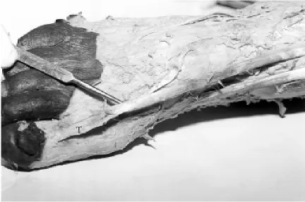
Figure 1. A photograph showing single tendinous insertion (T) of the extensor hallucis longus muscle on the dorsal aspect of the base of the distal phalanx of the big toe.
MATERIAL AND METHODS
The study was performed on 60 lower extremities of adult human cadavers that were used in our labo-ratory for educational gross anatomy at the Faculty of Medicine, King Abdulaziz University, Saudi Arabia. The insertion of the extensor hallucis longus muscle was dissected in order to study accurately the different forms of its attachment. The number and the sites of attachment of the tendinous termi-nation on which the extensor hallucis longus muscle gained insertion, as well as the incidence of each type of insertion, was recorded. The frequency of occurrence of hallux valgus with each type of inser-tion was also recorded.
RESULTS
The dissection of 60 lower extremities of human cadavers showed that the extensor hallucis longus insertion was found as a single tendon in 39 cases (65%) (Fig. 1, 2), or two tendons in 16 cases (26.67%) (Fig. 3–7), while in five cases it was three tendinous slips (8.33%) (Fig. 8, 9). Three different patterns of variation in the insertion of the extensor hallucis lon-gus were noticed (Fig. 10, Table 1).
Pattern I: was found in 39 cases (65%), in which the extensor hallucis longus muscle had a single
ten-dinous insertion on the dorsal aspect of the base of the distal phalanx of the big toe (Fig. 1). Association with lateral deviation of the big toe at the metatar-so-phalangeal joint (hallux valgus) was seen in three cases (5%) (Fig. 2).
Pattern II: was observed in 16 cases (26.67%), in which extensor hallucis longus terminated in two tendons (main and accessory). The distribution of the variations in this pattern is as follows:
— In nine cases (15%) the main tendon was in-serted into the dorsal aspect of the base of the distal phalanx of the big toe. The accessory tendon was smaller and passed medial to the main tendon and was inserted separately into the dorsal aspect of the base of the proximal phalanx of the big toe, just dis-tal to the insertion of extensor hallucis brevis (Fig. 3). — In three cases (5%) the main tendon was in-serted into the dorsal aspect of the base of the dis-tal phalanx of the big toe. The accessory tendon was smaller and passed medial to the main tendon to join the termination of the extensor hallucis brevis into the dorsal aspect of the base of the proximal phalanx of the big toe (Fig. 4).
Figure 2. A photograph showing a single tendinous insertion (T) of the extensor hallucis longus muscle on the dorsal aspect of the base of the distal phalanx of the big toe. Note the lateral deviation of the big toe at the metatarso-phalangeal joint. The head (H) of the first metatarsal bone is seen to be deviated medially.
Folia Morphol., 2003, Vol. 62, No. 2
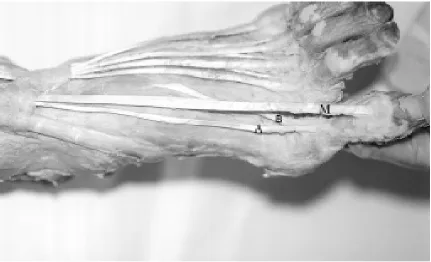
Figure 4. A photograph showing the two tendons of extensor hallucis longus muscle. The main tendon (M) is inserted into the dorsal aspect of the base of the distal phalanx of the big toe. The accessory tendon (A) is smaller and passes medial to the main tendon and joins the termination of extensor hallucis brevis (B) into the dorsal aspect of the base of the proximal phalanx of the big toe. Note the slight lateral deviation of the big toe at the metatarso-phalangeal joint.
Figure 6. A photograph showing the two tendons of extensor hallucis longus muscle. The main tendon (M) is inserted into the dorsal aspect of the base of the distal phalanx of the big toe. The accessory tendon (A) is smaller and passes lateral to the main tendon and joins the middle of extensor hallucis brevis tendon (B) forming a common tendon (C). The common tendon is inserted into the dorsal aspect of the base of the proximal phalanx of the big toe. Note the slight lateral deviation of the big toe.
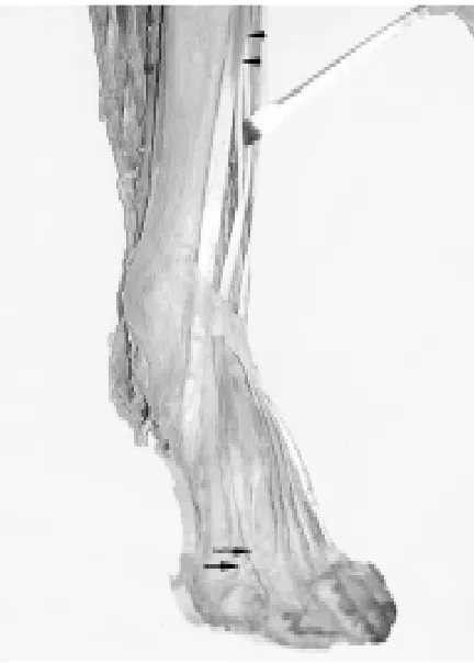
Figure 7. A photograph of the same specimen as in Figure 3 showing the 2 tendons (arrows) of the extensor hallucis longus muscle arising from separate fleshy bellies (arrowheads).
was smaller and passed medial to the main tendon and was inserted separately into the dorsal aspect of the proximal phalanx of the big toe medial to the attachment of the extensor hallucis brevis tendon (Fig. 5).
— In two cases (3.33%) the main tendon was inserted into the dorsal aspect of the base of the distal phalanx of the big toe. The accessory tendon was smaller and passed lateral to the main tendon and joined the middle of extensor hallucis brevis ten-don forming a common tenten-don which was inserted into the dorsal aspect of the base of the proximal phalanx of the big toe (Fig. 6).
In Pattern II an association with various grades of lateral deviation of the big toe at the metatarso-phalangeal joint was seen in 10 cases (16.67%) (Fig. 3, 4, 6). In one case (1.67%), the same case as in Figure 3, the two tendons of the extensor hallucis longus were seen arising from separate fleshy bel-lies (Fig. 7). In the remaining 59 cases (98.33%) the muscle was formed of one fleshy belly (unipennate muscle).
Folia Morphol., 2003, Vol. 62, No. 2
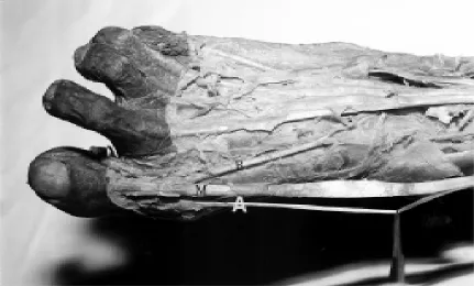
Figure 8. A photograph showing the three tendinous slips of extensor hallucis longus. The main tendon (M) is inserted into the dorsal aspect of the base of the distal phalanx of the big toe. Two accessory tendons (A) arise from the medial side of the main tendon proximal to the first metatarso-phalangeal joint. The accessory tendons are attached to the capsule (P) of the first meta-tarso-phalangeal joint. Note the marked lateral deviation of the big toe at the metameta-tarso-phalangeal joint and the medial deviation of the head (H) of the first metatarsal bone. The extensor hallucis brevis tendon (B) is seen.
Figure 10. Shows the three different patterns of the variations in the insertion of the extensor hallucis longus muscle.
Table 1. Patterns of insertion of the extensor hallucis longus
Pattern No. of tendinous slips No. of cases (%)
I One 39 65
II Two 16 26.67
III Three 5 8.33
Total 60 100
Table 2. Incidence of accessory tendons in patterns II and III
Pattern Accessory tendons (AT)
Medial AT Lateral AT Medial and lateral AT No. of (%) No. of (%) No. of (%)
cases cases cases
II 14 23.33 2 3.33 – –
III 3 5 – – 2 3.33
Total 17 28.33 2 3.33 2 3.33
Table 3. Incidence of hallux valgus and normal big toe in different patterns
Pattern Hallux valgus Normal big toe
No. of cases (%) No. of cases (%)
I 3 5 36 60
II 10 16.67 6 10
III 5 8.33 0 0
Total 18 30 42 70
— In three cases (5%) the main tendon was in-serted into the dorsal aspect of the base of the dis-tal phalanx of the big toe. The two accessory dons arose from the medial side of the main ten-don proximal to the first metatarso-phalangeal joint and gained insertion into the capsule of the joint. (Fig. 8).
— In two cases (3.33%) the main tendon was inserted into the dorsal aspect of the base of the distal phalanx of the big toe. The two accessory ten-dons (medial and lateral) arose from the medial and lateral sides of the main tendon respectively and gained insertion into the capsule of the first meta-tarso-phalangeal joint (Fig. 9).
— Hallux valgus was seen in the five cases (8.33%) of pattern III, with marked lateral deviation of the big toe at the metatarso-phalangeal joint and medi-al deviation of the head of the first metatarsmedi-al bone (Fig. 8, 9).
The accessory tendons were, seen in 21 cases (35%). The distribution is as follows: Pattern I has no accesso-ry tendons. Medial accessoaccesso-ry tendons were seen in 17 cases (28.33%), 14 cases (23.33%) appeared in Pat-tern II and 3 cases (5%) in PatPat-tern III. A lateral accesso-ry tendon was seen in Pattern II in two cases (3.33%). Both lateral and medial accessory tendons were seen in Pattern III in two cases (3.33%) (Table 2).
Hallux valgus was seen in 18 cases (30%). The cases are distributed as follows: 3 cases (5%) in Pat-tern I, 10 cases (16.67%) in PatPat-tern II, 5 cases (8.33%) in Pattern III.
A normal big toe was seen in the remaining 42 cases (70%). The cases are distributed as follows: 36 cases (60%) in Pattern I, 6 cases (10%) in Pat-tern II. No case of PatPat-tern III showed a normal big toe (Table 3).
DISCUSSION
Most of the available anatomical texts have de-scribed the insertion of the extensor hallucis lon-gus muscle into the dorsal aspect of the base of the distal phalanx of the big toe [9, 12, 16].
In the present work the extensor hallucis longus muscle was seen in 65% of cases with a single tendi-nous insertion on the dorsal aspect of the base of the distal phalanx of the big toe. The same observa-tion was made by Kaneff [4] in 51.12% of the cases examined by him.
con-Folia Morphol., 2003, Vol. 62, No. 2
nection to the tendon of extensor hallucis brevis . In 8.33% of cases extensor hallucis longus was seen to terminate in 3 tendinous slips (one main and two accessory). The main tendon is inserted into the dorsal aspect of the distal phalanx of the big toe, the accessory tendons into the capsule of the first metatarso-phalangeal joint.
Only in one case (1.67%) in this work were the two tendons of the extensor hallucis longus seen arising from separate fleshy bellies, while the mus-cle was unipennate in the remaining cases. The pres-ence of accessory tendons for the extensor hallucis longus muscle described in the present study is in agreement with the work of Kaneff [4], who de-scribed additional tendons of the musculus exten-sor hallucis longus in 48.88% of the cases examined by him. Williams et al. [14] stated that at the meta-tarso-phalangeal articulation a slip from each side of the extensor hallucis longus tendon covers the joint’s dorsal aspect. An expansion from its medial side to the proximal phalangeal base is also usually present and it may send a slip to the second toe.
Wood [15] mentioned that doubling of the mus-cle may occur anywhere along its length with the ex-tramuscle (extensor primi internodii hallucis longus) running parallel and lateral to the main muscle. It may be joined to the tendon of extensor hallucis brevis distally or inserted independently onto the first meta-tarsal, the proximal part of the first phalanx, or both phalanges of the big toe. It arises rarely below the usual extensor, or it may arise from the tendon of the extensor at the ankle, or three inches above it.
Macalister [7] found that the muscle may be dou-bled, may have a common muscular slip between it and extensor digitorum; or a slip to the extensor hal-lucis brevis at the junction of its muscle and tendon. Gruber [2] mentioned that occasionally this mus-cle, which is usually unipennate, has three bellies and three tendons and is named extensor hallucis longus tricaudatua. When one of these three bellies is repre-sented as a separate (and usually smaller) muscle, it is named extensor hallucis longus minor.
In the present study, the accessory tendon was seen arising from the medial side of the main tendon in 28.33% of the examined cases, while in only 3.33% it arose from the lateral side. In addition, in 3.33% of cases both the medial and lateral accessory tendons were found. This finding is in agreement with that described by Kaneff [4], who said that most of these supplements (additional tendons and muscles) are positioned on the medial side of the main tendons with only a few on the lateral side.
The presence of accessory tendons for the exten-sor hallucis longus muscle as described in the present study is in agreement with previous reports in the literature. However, the coexistence of an additional insertion of the extensor hallucis longus muscle asso-ciated with hallux valgus has not been reported in the available literature. While Gunal et al. [3] described the anomalous tibialis posterior muscle as an etiolog-ical factor of hallux valgus, Brenner [1] stated that this deformity (hallux valgus) developed as a result of a specific type of insertion of the tendon of the tibia-lis anterior muscle.
In the present study, hallux valgus was seen in 30% of cases; in Pattern II, in which the muscle has two tendinous insertions, various degrees of hallux valgus were seen in 16.67% of the cases, while in Pattern III, in which the muscle has three tendinous insertions, hallux valgus was seen with marked lateral deviation of the big toe and medial deviation of the head of the metatarsal in all cases (8.33%). In Pattern I, in con-trast, in which the muscle has a single tendinous inser-tion, hallux valgus is seen in only 5% of cases.
The presence of an additional insertion of the extensor hallucis longus muscle, described in this work has functional and clinical implications. From a functional viewpoint, the accessory tendon of ex-tensor hallucis longus with its insertion into the base of the proximal phalanx of the big toe either di-rectly or through its connection to the extensor hallucis brevis tendon and also to the capsule of the first metatarso-phalangeal joint would help to extend the joint irrespective of the position of the interphalangeal joint. This would add flexibility to the movement of extension of the big toe that usu-ally accompanies the mechanism of walking and running. The clinical significance of the accessory tendons of extensor hallucis longus muscle is its possible association with deformity of the big toe, such as hallux valgus. It is expected that the pres-ence of accessory tendons, particularly those on the medial side of the main tendon, may exert a medial pull on the base of the proximal phalanx, in addi-tion to extending it. This would counteract the lat-eral pull exerted on the distal phalanx of the big toe caused by the pull of the main tendon of the muscle, which increases the predisposition to the development of hallux valgus.
depends on a number of factors, such as the type of sport practised and its degree of rigour.
It is known that different sports put varied pres-sures on the foot. Walking generates a force in the forefoot equal to 80% of the body’s weight; running increases it to 250% of body weight [5]. The severity and progression of the hallux valgus deformity de-pend on the particular sport involved. For instance, dancers develop hallux valgus at a younger age than is seen in the general population [10, 11]. A deformi-ty may affect one patient differently from another because various sports make different demands on the foot. For example, sprinters require an extended range of motion in both dorsiflexion and plantarflex-ion of the first metatarso-phalangeal joint, while mid-dle- and long-distance runners do not [6]. The pres-ence of multiple tendons of the extensor hallucis lon-gus muscle, as reported in the present work, could be added as a predisposing factor for the develop-ment of hallux valgus. This is probably due to the imbalance created by the accessory tendons, with dif-ferent attachments along the axis of pull of the joints of the big toe.
It is concluded that this study emphasises the importance of realising the functional and clinical significance of detecting variations in the muscles of the lower limb. These variations may have a sig-nificant correlation with the clinical aspects.
REFERENCES
1. Brenner E (2002) Insertion of the tendon of the tibialis anterior muscle in feet with and without hallux val-gus. Clin Anat, 15 (3): 217–223.
2. Gruber W (1876) Ein neuer Fall von Musculus extensor hallucis longus tricaudatus. Arch Anat Physiol Wissen Med, 750–752.
3. Gunal I, Sahinoglu K, Bergman RA (1994) Anomalous tibialis posterior muscle as an etiologic factor of hal-lux valgus. Clin Anat, 7 (1): 21–25.
4. Kaneff A (1986) The upright posture of man and the morphological evolution of the musculi extensors dig-itorum pedis with reference to evolutionary mycolo-gy. III. Gegenbaurs Morphol Jahrb, 132 (5): 681–722 (Article in German).
5. Lam SF, Hodgson AR (1958) A comparison of foot forms among the non-shoe and shoe wearing Chinese population. J Bone Joint Surg (Am), 40: 1058–1062. 6. Lillich J.S, Baxter D.E (1986) Bunionectomies and
related surgery in the elite female middle – distance and marathon runner. Am J Sports Med, 14 (6): 491–493.
7. Macalister A (1875) Additional observations on mus-cular anomalies in human anatomy (third series) with a catalogue of the principal muscular variations hith-erto published. Trans Roy Irish Acad Sci, 25: 1–134. 8. Mann RA, Coughlin MJ (1986) Hallux valgus and com-plications of hallux valgus. In: Mann RA (ed.). Surgery of the foot. 5th ed. St Louis, CV Mosby, pp. 65–130.
9. Moore KL, Dalley AF (1999) Clinically oriented anato-my. 4th ed. Lippincott Williams & Wilkins, p. 577.
10. Sammarco GJ, Miller E.H (1982a) Forefoot conditions in dancers: part I. Foot Ankle, 3 (2): 85– 92.
11. Sammarco GJ, Miller EH (1982b) Forefoot conditions in dancers: part 2. Foot Ankle, 3 (2): 93–98.
12. Snell RS (2000) Clinical anatomy for medical students, 6th ed. Lippincott Williams & Wilkins, p. 561.
13. Thorek Ph (1962) Anatomy in surgery, 2nd ed. J.B.
Lip-pincott Company, p. 845.
14. Williams PL, Warwick R, Dyson M, Bannister L.H (1989) Gray’s anatomy. 37th ed. Churchill Livingstone,
Edin-burgh–London, p. 646.
15. Wood J (1867) Variations in human mycology observed during the winter session of 1866–67 at King’s Col-lege. Proceedings of the Royal Society. Lond. B Lon-don(???). 15: 518–546.
16. Woodburne RT, Burkel WE (1994) Essentials of human anatomy, 9th ed. Oxford University Press, New York