Original article:
EFFECT OF
FICUS GLOMERATA
LEAF EXTRACT IN
STREPTOZOTOCIN-INDUCED EARLY DIABETIC
COMPLICATIONS AND ITS CHARACTERIZATION BY LC-MS
Abusufyan Shaikh1, 2*, Mohammed Ibrahim3
, Mohib Khan4
1 School of Pharmacy, Anjuman-I-Islam’s Kalsekar Technical Campus, New Panvel,
Maharashtra, affiliated to Mumbai University, Mumbai, India 2 Research Scholar, JNTUH, Kukatpally, Telangana, India 3 PNR College of Pharmacy, Telangana, India
4 Oriental College of Pharmacy, Navi Mumbai, India
* Corresponding author: Abusufyan Shaikh, Phone:(+91-022) 2748 1247; E-mail: [email protected]
http://dx.doi.org/10.17179/excli2019-1441
This is an Open Access article distributed under the terms of the Creative Commons Attribution License (http://creativecommons.org/licenses/by/4.0/).
ABSTRACT
Diabetes mellitus is a complex metabolic disorder that leads to various micro-vascular complications. The present study elucidated the effect of chloroform extract of leaves of Ficus glomerata (CHFG) in streptozotocin-induced
early diabetic renal and neural complications. Wistar rats were injected with STZ (55 mg/kg, i.p.) to produce experimental diabetes. Two weeks after the stabilization of diabetes, CHFG extract at the dose of 200 and 400 mg/kg (CHFG 200 and CHFG 400) and metformin at the dose of 250 mg/kg (Met 250) was administered to the diabetic rats for further two weeks. Diabetic rats showed an increase in blood glucose, plasma urea, uric acid, creatinine, triglyceride, and total cholesterol level. The change in behavioral parameters such as thermal hyperal-gesia and cold allodynia with compromised sciatic nerve and kidney antioxidant status were seen in diabetic rats. Diabetic rats treated with CHFG 200, CHFG 400, and Met 250 showed a decrease in blood glucose, plasma urea, uric acid, creatinine, triglyceride, and total cholesterol level. Also, it improved altered behavioral parameters such as thermal hyperalgesia and cold allodynia. It also restored the sciatic nerve and kidney antioxidant status. The results of kidney and sciatic nerves histopathological study were in line with the results of biochemical parameters that confirmed the favorable role of CHFG. Characterization of CHFG by LC-MS revealed the presence of diverse phytoconstituents, which might be responsible for its protective effect.
Keywords: Antidiabetic activity, antioxidant activity, Ficus glomerata, phytoconstituents, streptozotocin
INTRODUCTION
Diabetes mellitus (DM) is a complicated disorder caused by genetic or environmental factors (Bronwyn et al., 1995; Fatemeh et al., 2009; Todd et al., 1987). It is one of the most common endocrine diseases with its global prevalence of 9.3 % of the world population and is predicted to rise to 10.2 % by 2030 and
Chronic hyperglycemia stimulates the polyol pathway, causing osmotic stress, in-creases the generation of reactive oxygen spe-cies, which leads to oxidative stress in the tis-sues. An increasing body of data supports the role of oxidative stress in the pathogenesis of diabetic microvascular complications (Ah-mad et al., 2015; Bashan et al., 2009; Mahmood and Neaimy, 2008; Nishikawa et al., 2000). There are many drugs to treat dia-betes, but none have been successful for the treatments of its complications completely. Therefore its treatment remained as one of the challenge faced by modern medicine. Hyper-glycemic control remains a primary therapeu-tic target when dealing with diabetherapeu-tic compli-cations in both types of diabetes (Mahmood and Neaimy, 2008; Sima and Kamiya, 2006). A significant benefit of the use of phytocon-stituents, minerals, and vitamins is thought to be due to its capability to scavenge free radi-cals and thus lowering the incidence of dia-betic complications (Pirat, 1977; Dal and Si-grist, 2016). Plants are always a good source of active pharmacological agents, and many of the currently available therapeutic agents have been derived from them (Patel et al., 2012). Many biologically active compounds from plant sources such as phenolics, flavo-noids, lycopene, saponins, triterpeflavo-noids, and many others have the antidiabetic effect (Grover et al., 2002). Traditional, comple-mentary, and alternative medicines are in-creasingly used for the treatment of diabetes and its complications (Dodda and Ciddi, 2014; Snehal et al., 2017; Dal and Sigrist, 2016).
Various medicinal plants, especially those belonging to the genus Ficus and its phyto-constituents, have been used for the treatment of diabetes and related chronic complications since ancient times (Deepa et al., 2018). Ficus glomerata (FG), family Moraceae is well known as a Cluster fig tree, Goolar, Indian fig tree, Umber, Udumbara, Rumadi, Dimri, Blue lotus found in the Indian subcontinent, Indo-China, Australia and Malaysia (Joy et al., 2001; Kunwar and Bussmann, 2006). All parts of FG are medicinally important and
used widely in the treatment of jaundice, wound healing, dysentery, diabetes, biliary disorders and inflammatory conditions (Nad-karni, 1976; Patil et al., 2009; Satish et al., 2014). Our previous study revealed the in vitro antidiabetic and antioxidant activity of CHFG (Abusufyan et al., 2018). However, there is no in vivo data about the efficacy of this extract available in diabetic renal and neural complications. Based on this, CHFG was selected to investigate its effect on early diabetic renal and neural complications.
METHODS AND MATERIALS
Chemicals reagents
A free sample of metformin procured from USV Pvt Ltd, Mumbai. Streptozotocin was purchased from Sisco Research Labora-tories Pvt. Ltd, Mumbai. All other chemicals were of analytical grade and obtained from commercial sources.
Preparation and selection of FG leaf extract
The leaves of FG were collected from the Sindhudurg district of Maharashtra, India, and identified with authentication no. 15-023 by botanist Dr. A. S. Upadhye of Agharkar Research Institute, Pune, India. The powdered leaf material was extracted by successive sol-vent extraction with the solsol-vents of increasing order of polarity and concentrated by using a rotary evaporator. Based on our previous study, chloroform extract (CHFG) was se-lected for the final investigation as it showed maximum antidiabetic and considerable anti-oxidant activity (Abusufyan et al., 2018).
CHFG extract analysis
the MS system. Solvents used were 100 % of water (A) and 100 % of acetonitrile (B). The elution started with a linear gradient; within 30 minutes of acquisition time, the initial 2 minutes, the flow of solvent composition A: B was 95:5. The injection volume was 3 µl, and the flow rate was 0.2 ml min-1. The MS signals were used for qualitative analysis based on specific mass spectra. The chroma-tographic data were analyzed by using the software from Agilent, USA. For the detec-tion of compounds, the MS spectra of the an-alyzed samples were compared with the spec-tra from the library.
Acute oral toxicity test
Acute oral toxicity test was conducted as per the OECD guideline No. 423. A single oral dose of 2000 mg/kg of CHFG was admin-istered to overnight fasting Wistar albino rats. The sign and symptoms of toxicity were ob-served in the animals for 24 hours. The mor-tality, if any, was noted for two weeks.
Oral glucose tolerance test (OGTT) and selection of dose
Doses equivalent to 200, 400, and 600 mg/kg of CHFG extract were calculated and suspended in 1 % v/v of tween 80 solutions. OGTT was carried out on overnight fasting normal rats. 1 % v/v of tween 80, CHFG (200, 400, and 600 mg/kg) and metformin (250 mg/kg) were administered to five groups of rats, respectively. Glucose (2 g/kg) was fed 30 min after pre-treatment with 1 % v/v of tween 80, CHFG, and metformin. Blood glucose levels were observed at 0, 30, 60, 120, and 240 min after glucose load to assess the effect of the extract. Two doses with optimum dose-dependent anti-hyperglycemic activity against glucose tolerance in normal rats were selected for the final study in STZ induced di-abetic rats.
Animal’s treatments and experimental design
Healthy male Wistar albino rats weighing 300-350 g were procured from the National Institute of Biosciences, Pune, Maharashtra,
India, and housed in polypropylene cages at the temperature of 22 ± 1 °C and relative hu-midity of 50 to 60 % with a 12 h light/dark cycle. The animal experiments were approved by the Institutional Animal Ethics Committee (IAEC), School of Pharmacy-AIKTC, New Panvel (Approval No. AIKTC/SoP/IAEC/ Approval/2016/1). Standard pellet diet and water ad libitum were provided to the experi-mental animals throughout the experiment. Diabetes was induced by a single injection of streptozotocin (STZ, 55 mg/kg, i.p.) freshly prepared in 0.1 M citrate buffer pH 4.5) in rats (Azahar et al., 2012). Blood glucose level was measured, and rats with blood glucose levels above 250 mg/dl were considered diabetic. Two weeks post-STZ injection diabetic rats with stable blood glucose level were divided into different groups as follow:
GroupI: Normal control (n = 6), normal rats treated with 10 ml/kg, p.o. of distilled water. Group II: Diabetic control (n = 6), STZ in-duced diabetic rats treated with vehicle (1 % v/v of tween 80 in distilled water 10 ml/kg, p.o.).
Group III: STZ + Met 250 (n = 6), STZ in-duced diabetic rats treated with Metformin 250 mg/kg.
Group IV: STZ + CHFG 200 (n = 6), STZ in-duced diabetic rats treated with CHFG 200 mg/kg.
Group V: STZ + CHFG 400 (n = 6), STZ in-duced diabetic rats treated with CHFG 400 mg/kg.
of tissue antioxidant status and histopatholog-ical study.
Measurements of fasting blood glucose
The fasting blood glucose was measured to study the effect of pre-treatment, one and two-week post-treatment on lateral tail vein blood samples using a OneTouch glucometer (Lifescan Scotland Ltd) throughout the treat-ment period.
Behavioral assessment of early diabetic neuropathy
Both thermal hyperalgesia and cold allo-dynia were studied after one and two-week post-treatment. Thermal hyperalgesia was studied by measuring paw and tail withdrawal latency (sec) by hot plate and tail immersion test at 49 ± 0.5 oC. Shortening of the paw and tail withdrawal latency indicates thermal hy-peralgesia. Cold allodynia was studied by measuring tail withdrawal latency (sec) by dipping the tail in the water at a cold temp (10 ± 0.5 °C), which is usually innocuous.
Determination of the serum biochemical parameters
Serum urea, uric acid, creatinine, triglyc-erides, and total cholesterol levels were meas-ured by using the standard commercially available kits and as per the instructions of manufacturer’s.
Determination of tissue antioxidant status
At the end of the experiment, the sciatic nerve and kidney were isolated and washed with ice-cold saline. Then homogenate was made and used for determination of MDA, an end product of lipid peroxidation (Mihara and Uchiyama, 1978), reduced glutathione (GSH) (Ellman, 1959), and superoxide dismutase (SOD) (Misra and Fridovich, 1972).
Histopathological examinations
The part of the isolated kidney and sciatic nerves were fixed in 10 % formalin. The par-affin embedded sections of 5 μm thickness were cut and stained with hematoxylin and
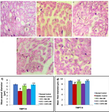
eosin (H&E). The kidney histological sec-tions were examined by light microscopy at 20x, whereas the sciatic nerve sections were examined by oil immersion lens (100x). Quantitative estimation of urinary space, glo-merular tuft area (GA) of kidney sections, mean axonal, and fiber diameter in sciatic nerve sections was determined by using Im-ageJ software. The glomerular volume (GV) was calculated by converting GA to GV by applying a spherical approximation formula (GV = 1.2545 (GA)1.5) (Gopala and Greg, 2007). For each parameter, six images from six different samples of each study group were analyzed. Five non-overlapping fields from each image were captured. Five differ-ent readings from every captured photo were counted, and the mean was calculated for each image.
Data analysis and statistics
One-way analysis of variance (ANOVA) followed by post-hoc Tukey’s HSD test was performed to conduct statistical analysis. All values were expressed as means ± SEM. Dif-ferences were considered significant when *P < 0.05 and highly significant when **P < 0.01.
RESULTS
Preliminary phytochemical analysis and LC-MS characterization of CHFG
Preliminary phytochemical estimation re-vealed the presence of alkaloids, flavonoids, glycosides, phenolics, and saponins. CHFG was characterized by LC-MS, and phytocon-stituents in the extract were identified by the compound library. The list of the compounds detected is given in Table 1.
Acute oral toxicity study
Table 1: The list of the compounds detected from
the CHFGby LC-MS
Effect on oral glucose tolerance test and selection of dose
Hypoglycemic activity of CHFG 200, CHFG 400, and CHFG 600 of extract and Met 250 on glucose tolerance was examined in glucose loaded rats (Figure 1). Rats treated with CHFG 200, CHFG 400, and CHFG 600 and Met 250 mg/kg significantly (P < 0.01) lower the FBG level in comparison to the nor-mal control group. The dose-dependent de-crease in FBG level was observed in CHFG 200 and CHFG 400. However, CHFG 600 fails to show a significant dose-dependent ac-tivity as compared to its lower dose of CHFG 400. Hence, for the final study in STZ induced diabetic rats, two doses, CHFG 200 and CHFG 400, were selected.
Figure 1: Effect of single-dose treatment on oral glu-cose tolerance in normoglycemic rats. All values were expressed as Mean + SEM, (n = 6); *P < 0.05, **P < 0.01 as compared to normal control group.
Effect on fasting blood glucose level
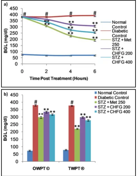
The fasting blood glucose levels for both single and repeated dose treatment with Met 250, CHFG 200, and CHFG 400 were meas-ured in STZ-induced diabetic rats (Figure 2a and 2b). Fasting blood glucose level of single-dose treatment was fall significantly (P < 0.01) after 4 and 6 hours post-treatment in Met 250, CHFG 200, and CHFG 400 group as compared to the diabetic control group (Fig-ure 2a). There were 41.27 %, 17.88 %, and 29.13 % fall in blood glucose level after 4 hours post-treatment in Met 250, CHFG 200, and CHFG 400 groups, respectively, as com-pared to the diabetic control group. There was 50.53 %, 21.64 %, and 34.98 % fall in blood glucose level after 6 hours post-treatment in Met 250, CHFG 200, and CHFG 400 groups, respectively, as compared to the diabetic con-trol group.
Figure 2:a) Effect of single-dose treatment on blood
glucose level in STZ-induced diabetic rats. b) Effect of repeated-dose treatment on blood glucose level in STZ-induced diabetic rats. OWPT: One Week Post Treat-ment, © = observation recorded three weeks after in-duction of diabetes, TWPT: Two Weeks Post Treat-ment, ® = observation recorded four weeks after induc-tion of diabetes, All values were expressed as Mean + SEM, (n = 6); #P < 0.01, as compared to normal control group; *P < 0.05, **P < 0.01 as compared to diabetic control group.
40 90 140
0 30 60 90 120
FBG
(mg
/d
l
)
Time (min)
Normal Co ntrol Met 250 CHFG 200 CHFG 400 CHFG 600 **
** **
** **
** ** **
** ** **
Repeated dose treatment with Met 250, CHFG 200, and CHFG 400 showed signifi-cant (P < 0.01) fall in blood glucose level compared to the diabetic control group (Fig-ure 2b). There was 21.64 %, 11.12 %, and 16.64 % fall in fasting blood glucose level af-ter one-week post-treatment (OWPT) in Met 250, CHFG 200, and CHFG 400 groups re-spectively as compared to the diabetic control group. There were 41.29 %, 20.58 %, and 26.21 % fall in fasting blood glucose level af-ter two weeks post-treatment (TWPT) in Met 250, CHFG 200, and CHFG 400 groups re-spectively as compared to the diabetic control group.
Effect on thermal hyperalgesia
The pain threshold was significantly re-duced in diabetic rats as compared with the normal control group (Figure 3a and 3b). Treatment of diabetic rats with Met 250, CHFG 200, and CHFG 400 revealed an in-crease in pain threshold after OWPT and TWPT as compared to the diabetic control group.
Effect on cold allodynia
Tail flick latency at 10 °C was signifi-cantly (P < 0.01) lower in diabetic rats after two weeks of diabetes as compared with the normal control group (Figure 4). Treatment of diabetic rats with CHFG 400 showed statisti-cally significant (P < 0.01) increase in pain threshold after OWPT and TWPT as com-pared to the diabetic control group. Although Met 250 and CHFG 200 fails to show a statis-tically significant difference in OWPT but showed a statistically significant increase in pain threshold in Met 250 (P < 0.05), CHFG 200 (P < 0.01) after TWPT as compared to the diabetic control group.
Figure 3: a) Effect of repeated-dose treatment on paw
withdrawal latency (PWL) using a hot plate test at 49 °C in STZ-induced diabetic rats. b) Effect of repeated-dose
treatment on tail-flick latency (TFL) using water immer-sion test at 49 °C in STZ-induced diabetic rats. OWPT: One Week Post Treatment, © = observation recorded three weeks after induction of diabetes, TWPT: Two Weeks Post Treatment, ® = observation recorded four weeks after induction of diabetes, All values were ex-pressed as Mean + SEM, (n = 6); # P < 0.01, as com-pared to normal control group; *P < 0.05, **P < 0.01 as compared to diabetic control group.
Effect on body weight, kidney weight and kidney weight to body weight ratio
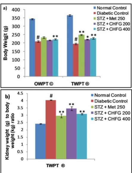
A statistically significant (P < 0.01) de-crease in body weight in the diabetic control group was observed as compared to the nor-mal control group. OWPT treatment with CHFG 400 showed a statistically significant (P < 0.01) increase in the body weight as com-pared to the diabetic control group. Consider-able increase in the body weight after TWPT with Met 250 (P < 0.01), CHFG 200 (P < 0.05), and CHFG 400 (P < 0.01) was observed against the diabetic control group (Figure 5a). Moreover, there was a remarkable elevation in kidney weights to body weight ratio in the diabetic control group. In contrast, kidney weight to body weight ratio was significantly (P < 0.01) lower in the TWPT with Met 250, CHFG 200 and CHFG 400 treatment group as compared to the diabetic control group (Fig-ure 5b).
Effect on biochemical parameters
Table 2 presents the study on serum urea, uric acid, creatinine, triglycerides, and total cholesterol level. In comparison to the normal control group, STZ treated diabetic rats showed a significant (P < 0.01) increase in se-rum urea, uric acid, creatinine, triglyceride, and total cholesterol level. Met 250, CHFG 200, and CHFG 400 showed a significant (P < 0.01) decrease in serum urea level as com-pared to the diabetic control group. TWPT with Met 250 and CHFG 400 significantly (P < 0.01) reduced serum uric acid, creatinine, triglyceride, and total cholesterol level as compared to the diabetic control group. TWPT with CHFG 200 significantly (P < 0.05) decreased serum triglyceride, but it fails to show a significant change in serum creati-nine and total cholesterol level as compared to the diabetic control group.
Figure 5: a) Effect of repeated-dose treatment on body
weight in STZ-induced diabetic rats. b) Effect of re-peated-dose treatment on kidney weight to body weight ratio in STZ-induced diabetic rats. OWPT: One Week Post Treatment, © = observation recorded three weeks after induction of diabetes, TWPT: Two Weeks Post Treatment, ® = observation recorded four weeks after induction of diabetes, All values were expressed as Mean + SEM, (n = 6); #P < 0.01, as compared to normal control group; *P < 0.05, **P < 0.01 as compared to di-abetic control group.
Effect on serum antioxidant status
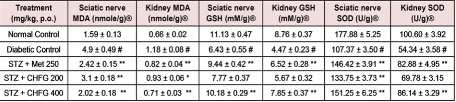
Significant (P < 0.01) increase in MDA level (3.08 fold in sciatic nerve and 1.79 fold in kidney tissue), decrease in GSH (1.73 fold in sciatic nerve and 1.96 fold in kidney tissue) and decrease in SOD (1.66 fold in sciatic nerve and 1.85 fold in kidney tissue) were ob-served in diabetic control rats as compared to normal control rats (Table 3).
Table 2: Effect of repeated-dose treatment on serum biochemical parameters in STZ-induced diabetic
rats
® = observation recorded two-week post-treatment and four weeks after induction of diabetes. All values were expressed as Mean + SEM, (n = 6); #P < 0.01, as compared to normal control group; *P < 0.05, **P < 0.01 as compared to diabetic control group.
Table 3: Effect of repeated-dose treatment on antioxidant activity of sciatic nerve and kidney tissue in
STZ-induced diabetic rats
® = observation recorded two-week post-treatment and four weeks after induction of diabetes. All values were expressed as Mean + SEM, (n = 6); #P < 0.01, as compared to normal control group; *P < 0.05, **P < 0.01 as compared to diabetic control group.
TWPT with CHFG 200 showed a signifi-cant decrease in MDA level (1.58 fold in the sciatic nerve and 1.27 fold in kidney tissue) as compared to their level in respective tissues in the diabetic control group. Although TWPT with CHFG 200 increases GSH level by 1.21 fold in the sciatic nerve and 1.27 fold in kid-ney tissue but this improvement was found to be non-significant as compared to their level in respective tissues in the diabetic control group. Also, CHFG 200 showed a significant increase in SOD by 1.25 fold in sciatic nerve (P < 0.01) and 1.28 fold in kidney tissue (P-ns) as compared to their level in respective tis-sues in the diabetic control group (Table 3). Whereas treatment with CHFG 400 for two weeks significantly (P < 0.01) decreases MDA (2.43 fold in sciatic nerve and 1.66 fold in kidney tissue), increases (P < 0.01) GSH (1.58 fold in sciatic nerve and 1.76 fold in kid-ney tissue) and increases (P < 0.01) SOD
(1.41 fold in sciatic nerve and 1.59 fold in kid-ney tissue) as compared to their level in re-spective tissues in diabetic control group (Ta-ble 3).
Effect of CHFG on histopathological analysis of kidney and sciatic nerve
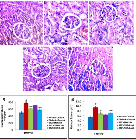
CHFG 400) and absence of capillaries con-gestion without hemorrhage (Figure 6c, e, f, and g). Whereas, the TWPT with CHFG 200 showed statistically insignificant changes in
glomerular volume and urinary space as com-pared to the diabetic control group. Also, it showed the evidence of hemorrhage with the minor blood-filled area (Figure 6d, f, and g).
Figure 6: Light microscopic representative images of kidney tissues (HE stained sections, 20X, Scale bar 50 μm).
a) Normal control, showing normal urinary space, and glomerular volume (arrowhead). b) Diabetic control, showing
an increase in urinary space (arrowhead), increased in glomerular tuft area (arrowhead) and the presence of capil-laries congestion with hemorrhage (arrow) as compared to normal control. c) STZ + Met 250 showed a decrease
in urinary space (arrowhead), decreased in glomerular tuft area (arrowhead), and the absence of capillaries con-gestion without hemorrhage. d) STZ + CHFG 200 showed an insignificant decrease in urinary space (arrowhead),
decreased in glomerular tuft area (arrowhead), and evidence of hemorrhage (arrow). e) STZ + CHFG 400 showed a decrease in urinary space (arrowhead), decreased in glomerular tuft area (arrowhead), and absence of capillaries congestion with no evidence of hemorrhage. f) Effect of repeated-dose treatment on the glomerular volume of
kidney tissue. g) Effect of repeated-dose treatment on urinary space of kidney tissue. TWPT: Two Weeks Post
Histopathological study of the normal control group showed normal sciatic nerve ar-chitecture (Figure 7a). Axonal atrophy with significant (P < 0.01) decrease in mean ax-onal, and fiber diameter was seen in the sci-atic nerve section of the diabetic control group as compared to the normal control group (Figure 7b, f, and g). The TWPT with Met 250 and CHFG 400 showed statistically
significant (P < 0.01) preservation of mean axonal and fiber diameter in the sciatic nerve section of the treatment group as compared to the diabetic control group (Figure 7c, e, f, and g). Whereas the TWPT with CHFG 200 showed the statistically insignificant change in mean axonal and fiber diameter in the sci-atic nerve section as compared to the diabetic control group (Figure 7d, 7f, and 7g).
Figure 7: Light microscopic representative images of sciatic nerve tissue (HE stained sections, 100X, immersion oil, Scale bar 5 μm). a) Normal control showing normal histological features of axonal fiber (arrow). b) Diabetic
control showing axonal atrophy (arrow). c) STZ + Met 250 showing less axonal atrophy as compared to the diabetic control group. d) STZ + CHFG 200 showed axonal atrophy (arrow). e) STZ + CHFG 400, showing less axonal atrophy as compared to the diabetic control group. f) Effect of repeated-dose treatment on the mean axonal
DISCUSSION
Diabetes is one of the major endocrine disorders that leads to impairment of renal and neural microvasculature (UK Prospective Diabetes Study (UKPDS), 1991; Yagihashi et al., 1992). Hyperglycemia-induced oxidative stress is a crucial element initiating diabetic microvascular complications (Jha et al., 2016). Oxidative stress in diabetes is the re-sult of a series of pathological events that leads to an imbalance between the pro-oxi-dant levels and counteracting antioxipro-oxi-dant de-fense mechanisms, consequently both kidney and nerves which are an essential part of the body are susceptible to hyperglycemia-in-duced oxidative damage (Asieh and Moham-mad, 2013; Jha et al., 2016).
The antioxidant effects on the diabetic kidney (Lee et al., 2008) and nerves (Salma et al., 2017) disorders were positively correlated with medicinal herbs supplementation. Alt-hough our previous in vitro study showed sig-nificant antidiabetic and antioxidant activity (Abusufyan et al., 2018) to CHFG, no study was conducted to observe the effects of CHFG in diabetic renal and neural complica-tions. We have planned our investigation to evaluate the influence of CHFG against dia-betes-induced renal and neural complications.
In the current study, diabetic rats were af-fected by their behavioral and biochemical parameters. Diabetic rats showed severe hy-perglycemia, an increase in plasma urea, uric acid, creatinine, triglyceride, and total choles-terol level. In addition, diabetic rats revealed altered behavioral parameters such as thermal hyperalgesia and cold allodynia, decreased body weight, and increased kidney weight to body weight ratio beside compromised sciatic nerve and kidney antioxidant status. Previous studies have shown that hyperglycemia can induce similar early renal and neural compli-cations in diabetic rats (Bhaskar et al., 2014; Morsy et al., 2014). The administration of CHFG to the STZ induced diabetic rats im-proved the parameters discussed above. This protective effect of CHFG has not been re-ported earlier.
The CHFG showed a significant decrease in blood glucose levels at 200 mg/kg and 400 mg/kg body weight in STZ induced diabetic rats both in a single dose and in one and two weeks repeated dose treatment. Our previous study showed in vitro inhibition of α -gluco-sidase and α-amylase enzymes by CHFG ex-tract (Abusufyan et al., 2018). α-glucosidase and α-amylase are two important enzymes that convert starch to more simple sugars. Therefore, the inhibition of these enzymes re-duces the rate of absorption of glucose that helps in controlling the postprandial rise in the blood glucose levels.
Neuropathic pain associated with diabetes is characterized by two important sensory symptoms such as hyperalgesia i.e. an in-creased response to painful stimuli, and allo-dynia, i.e. pain in response to a non-painful stimulus (Bianchi et al., 2012). In the present study, diabetic rats showed a significant in-crease in paw and tail withdrawal latency at 49 °C (Thermal hyperalgesia) and tail with-drawal latency at 10 °C (Cold allodynia) as compared to the normal control group. Simi-lar observations were also reported in earlier findings (Arun et al., 2008). Administration of Met 250 and CHFG 400 resulted in a con-sequent decrease in both thermal hyperalgesia and cold allodynia, compared to diabetic rats. Free radical-induced oxidative stress has been implicated to play an important role in the pathogenesis of diabetic neuropathy (Nasiry et al., 2017). In the present study, there was a significant increase in sciatic nerve MDA level and decrease in SOD and GSH level in diabetic rats, which is indicative of impaired oxidative balance in the sciatic nerve of dia-betic rats. Treatment with Met 250 and CHFG 200 and CHFG 400 reduces oxidative stress parameters of the sciatic nerve. The anti-noci-ceptive effect of CHFG may be attributed to its neuroprotective effect, which might be due to its antioxidant activity.
treated four weeks of diabetic rats at a dose of 55 mg/kg, i.p. The significant decline of body weight could be due to hyperglycemia, hypo-insulinemia, loss of tissue proteins, and in-creased muscle wasting (Cheng and Liang, 2013; Zafar and Naqvi, 2010). In our experi-mental study, the administration of CHFG to diabetic rats significantly improved the body weight that indicates the inhibition of loss of protein and muscle tissue damage caused by hyperglycemia. CHFG also improved kidney weight to body weight ratio, which could be attributed to its protective effect against oxi-dative stress induced cell injury in the renal tissues.
Changes in kidney weights to body weight ratio in diabetic rats were also associated with an increase in serum urea, uric acid, and cre-atinine, which is considered as the marker of kidney impairment (Karahan et al., 2005; Soussi et al., 2018). Catabolism of protein and nucleic acid due to hyperglycemia result in the formation of non-protein nitrogenous compounds such as urea, uric acid, and creat-inine in diabetic rats (Yassin and Mwafy, 2007). In the present study, the diabetic rats showed an increase in the serum levels of urea, uric acid, and creatinine. The treatment with Met 250, CHFG 400, significantly atten-uated the hyperglycemia-induced kidney im-pairment indicated above. Kidney function impairment could be attributed to oxidative damage caused by hyperglycemia (Jha et al., 2016; Soussi et al., 2018). Our experimental results revealed a state of oxidative stress in diabetic rats by the significant elevation in kidney MDA, decreased in SOD and GSH. Treatment with Met 250 and CHFG 400 re-duces kidney MDA levels and increases the level of SOD and MDA.
The previous study revealed that diabetic patients with high cholesterol and triglyceride levels are at high risk of nephropathy and pe-ripheral neuropathy (Aguiar et al., 2015; Wheeler and Bernard, 1994). In the present study, there was a significant increase in se-rum triglyceride and total cholesterol levels in diabetic rats as compared to normal control rats. Treatment with Met 250 and CHFG 400
reduces serum triglyceride and total choles-terol levels. This hypolipidemic activity of CHFG might also improve blood supply to the kidney and peripheral nerve, and in turn, it reduces its ischemic degeneration. The bio-chemical parameters were correlated with re-nal and neural histological studies. The histo-pathological observation in the kidney tissue of diabetic control rats showed an increase in glomerular volume, urinary space, and capil-lary congestion with hemorrhage. The previ-ous study conducted by Malatiali et al. (2008), Kartiawati et al. (2017), and Krishan et al. (2017) showed a similar change in typi-cal architecture of kidney tissue of diabetic rats. Our study also reports that CHFG 400 showed marked improvement in the histolog-ical sections with a decrease in urinary space and glomeruli volume. Morphological assess-ments of sciatic nerve showed a decline in the mean nerve fiber and axonal fiber diameter in the sciatic nerve tissue in diabetic control rats. The study conducted by Sameni et al. (1994) showed similar changes in the sciatic nerve of diabetic rats. The administration of CHFG 400 markedly reduced these changes in the sciatic nerve tissues. The findings of kidney and sciatic nerves histopathological study were in line with the biochemical parameters that revealed the beneficial role of CHFG in the treatment of diabetic renal and neural complications.
de Oliveira et al., 2015) and Anethole was re-ported for its hypoglycemic, antioxidant and neuroprotective activities (Sheikh et al., 2015; Bing et al., 2018). Dual antidiabetic and anti-oxidant activity of CHFG phytoconstituents might be responsible for their protective ef-fect against early diabetic renal and neural damage in STZ rats.
CONCLUSION
Our findings revealed a protective effect of CHFG against STZ-induced oxidative damage, alteration of biochemical parame-ters, and histopathology. Favorable health ef-fects of FG leaf extracts have been docu-mented and can partially explain the observed reno-protective and neuroprotective effects of CHFG. The current investigation, therefore, provides evidences that support the beneficial effect of CHFG against STZ-induced oxida-tive damage on the renal and neural system.
Acknowledgments
The present work was partially supported by the University of Mumbai, Minor Re-search Grant. The authors are grateful to Dr. Abdul Razak Honnutagi, Director, Anjuman-I-Islam’s Kalsekar Technical Campus, and Dr. Shariq Syed, Dean, School of Pharmacy, Anjuman-I-Islam’s Kalsekar Technical Cam-pus for providing facilities to carry out this work.
Conflict of interest statement
The authors report no conflicts of interest associated with this manuscript.
REFERENCES
Abusufyan S, Ibrahim M, Mohib K. Comparative in vitro antidiabetic and antioxidant activity of various
extracts of Ficus species. Pharmacog J.
2018;10:349-54.
Aguiar PCM, della Coletta MV, de Souza JJS. The as-sociation of dyslipidemia and peripheral diabetic neu-ropathy: The influence of urea. Diabetol Metab Syndr. 2015;7(1):A30.
Ahmad NK, Rahmat AK, Mushtaq A, Nadia M. Role of antioxidant in oxidative stress and diabetes mellitus. J Pharmacogn Phytochem. 2015;3:217-20.
Arun VK, Cindy SYL, Matthew CK. Activity-depend-ent excitability changes suggest Na+/K+ pump
dysfunc-tion in diabetic neuropathy. Brain. 2008;131:1209-16.
Asieh H, Mohammad A. Diabetic neuropathy and oxi-dative stress: therapeutic perspectives. Oxid Med Cell Longev. 2013;6:1-15.
Azahar MA, Al-Naqeb G, Hasan M, Adam A. Hypo-glycemic effect of Octomeles sumatrana aqueous
ex-tract in streptozotocin-induced diabetic rats and its mo-lecular mechanisms. Asian Pac J Trop Med. 2012;5: 875-81.
Bashan N, Kovsan J, Kachko I, Ovadia H, Rudich A. Positive and negative regulation of insulin signaling by reactive oxygen and nitrogen species. Physiol Rev. 2009;89:27-71.
Bhaskar N, Pratap RK. Protective effect of Coccinia grandis leaf extract: behavioral, electrophysiological,
biochemical and histological features of diabetic neu-ropathy. Int J Pharm Sci Res. 2014;5:4302-9.
Bianchi R, Cervellini I, Porretta S, Oggioni N, Burkey B, Ghezzi P, et al. Beneficial effects of PKF275-055, a novel, selective, orally bioavailable, long-acting dipep-tidyl peptidase IV inhibitor in streptozotocin-induced diabetic peripheral neuropathy. J Pharmacol Exp Ther. 2012;340:64-72.
Bing W, Guoxin Z, Mei Y, Ning L. Neuroprotective effect of anethole against neuropathic pain induced by chronic constriction injury of the sciatic nerve in mice. Neurochem Res. 2018;43:2404-22.
Bronwyn AO, Brian VH, Donald PC, David JA. Beta-cell apoptosis is responsible for the development of IDDM in the multiple Low-dose streptozotocin model. J Pathol.1995;178:176–81.
Cheng D, Liang B, Li Y. Antihyperglycemic effect of
Ginkgo biloba extracts in streptozotocin-induced
dia-betes in rats. Biomed Res Int. 2013;2013:162724
Dal S, Sigrist S. Protective effect of antioxidant con-sumption on diabetes and vascular complications. Dis-eases. 2016;4(3):E24.
Deepa P, Sowndhararajan K, Kim S, Park SJ. A role of
Ficus species in the management of diabetes mellitus:
A review. J Ethnopharmacol. 2018;6(215):210-32.
De Oliveira TM, De Carvalho RB, Da Costa IH, De Oliveira GA, De Souza AA, De Lima SG, et al. Evalu-ation of p-cymene, a natural antioxidant. Pharm Biol. 2015;53:423-8.
Ellman GL. Tissue sulfhydryl groups. Arch Biochem Biophys.1959;82:70-7.
Fatemeh NR, Soheila A, Zakieh VA, Jalal VA, Payam G. Effects of diabetes mellitus on gastric motility in rats. Pak J Physiol. 2009;5(1):20-4.
Gopala KR, Greg HT. Methods in renal research quan-tification of renal pathology by image analysis. Neph-rology. 2007;12:553-8.
Grover JK, Yadav S, Vats V. Medicinal plants of India with anti-diabetic potential. J Ethnopharmacol. 2002; 81(1):81-100.
International Diabetes Federation (IDF). IDF diabetes atlas, 19th ed. IDF, 2019. https://www.diabetesat- las.org/upload/resources/2019/IDF_Atlas_9th_Edi-tion_2019.pdf
Jha JC, Banal C, Chow BSM, Cooper ME, Jandeleit-Dahm K. Diabetes and kidney disease: Role of oxida-tive stress. Antioxid Redox Signal. 2016;25:657–84.
Joy PP, Thomas J, Mathew S, Skaria BP. Medicinal plants. In: Bose TK, Kabir J, Das P, Joy PP (eds): Trop-ical horticulture, Vol. 2 (pp 449–632). Calcutta: Naya Prokash, 2001.
Karahan I, Ateşşahin A, Yilmaz S, Ceribaşi AO, Sakin F. Protective effect of lycopene on gentamicin-induced oxidative stress and nephrotoxicity in rats. Toxicology. 2005;215:198–204.
Kartiawati A, Ema PS, Madihah, Tia S, Nining R, De-sak MM. Kidney histology in streptozotocin-induced diabetic male Wistar rats treated with combined extract of Temulawak rhizome and Belimbing wuluh fruit.
Nusantara Biosci. 2017;9:312-7.
Khandelwal KR. Preliminary phytochemical screen-ing. 22nd ed. In: Vrunda KS (ed): Practical
pharmacog-nosy techniques and experiments (pp 25.1-25.6). Pune: Nirali Prakashan, 2013.
Krishan P, Singh G, Bedi O. Carbohydrate restriction ameliorates nephropathy by reducing oxidative stress and upregulating HIF-1α levels in type-1 diabetic rats. J Diabetes Metab Disord. 2017;19:16-47.
Kunwar RM, Bussmann RW. Ficus (Fig) species in
Nepal: a review of diversity and indigenous uses. Lyo-nia. 2006;11:85-97.
Lee HS, Ku SK. Effect of Picrorrhiza rhizoma extracts on early diabetic nephropathy in streptozotocin-in-duced diabetic rats. J Med Food. 2008;11:294-301.
Loo AY, Jain K, Darah I. Antioxidant activity of com-pounds isolated from the pyroligneous acid, Rhi-zophora apiculata. Food Chem. 2008;107:1151–60.
Mahmood IH, Neaimy KSAA. Antioxidant status in type 2 diabetic neuropathy. Bahrain Med Bull. 2008; 30(1):1-7.
Malatiali S, Francis I, Barac-Nieto M. Phlorizin pre-vents glomerular hyperfiltration but not hypertrophy in diabetic rats. Exp Diabetes Res. 2008;2008:305403.
Manikanta V, Rema R. Ellagic acid ameliorates the progression of nephropathy in streptozotocin induced diabetic rats. Int Res J Pharm. 2017;8(1):55-60.
Mihara M, Uchiyama M. Determination of malonalde-hyde precursor in tissues by thiobarbituric acid test. Anal Biochem. 1978;86:271-8.
Misra C, Fridovich I. Superoxide dismutase activity was measured in dialyzed extracts with a modified ep-inephrine assay. J Biol Chem. 1972;247:6960-7.
Morsy MA, Ibrahim SA, Amin EF, Kamel MY, Abdel-wahab SA, Hassan MK. Carvedilol ameliorates early diabetic nephropathy in streptozotocin-induced dia-betic rats. Bio Med Res Int. 2014;2014:105214.
Nadkarni KM (ed.). Indian materia medica.Bombay: Popular Prakashan, 1976.
Nasiry D, Khalatbary AR, Ahmadvand H, Amiri FT, Akbari E. Protective effects of methanolic extract of
Juglans regia L. leaf on streptozotocin-induced
dia-betic peripheral neuropathy in rat. BMC Comple-ment Altern Med. 2017;17:(476):1-11.
Nishikawa T, Edelstein D, DU XL. Normalizing mito-chondrial superoxide production blocks three path-ways of hyperglycemic damage. Nature. 2000;404: 787-90.
Ohtsuji M, Yagi K, Shintaku-Kubota M, Kojima-Koba Y, Ito N, Sugihara M, et al. Decreased ADP-ribosyl cyclase activity in peripheral blood mononuclear cells from diabetic patients with nephropathy. Exp Diabetes Res. 2008;2008:897508.
Patel DK, Prasad SK, Kumar R, Hemalatha S. An over-view on antidiabetic medicinal plants having insulin mimetic property. Asian Pac J Trop Biomed. 2012; 2(4):320-30.
Patil VV, Pimprikar RB, Sutar NG, Barhate AL, Patil LS, Patil AP, et al. Anti- hyperglycemic activity of Fi-cus racemosa Linn leaves. J Pharm Res. 2009;2:54-7.
Salma AEM, Eman RZ, Heba MIA, Mahmoud SA. Therapeutic effects of aqueous, methanol and ethanol extracts of Egyptian Artemisia herba-alba in
STZ-in-duced diabetic neuropathy in rats. J Appl Pharm Sci. 2017;7:180-7.
Satish AB, Deepa RV, Nikhil CT, Vinodkumar SD, Saurabh ST. Ficus racemosa Linn.: a comprehensive
review. J Applicable Chem. 2014;3:1423-31.
Sameni HR, Panahi M, Sarkaki A, Saki GH, Makvandi M. The neuroprotective effects of progesterone on ex-perimental diabetic neuropathy in rats. Pakistan J Biol Sci. 1994;11:2000-8.
Sheikh BA, Leelavinothan P, Ayyasamy R, Ramasamy C. Trans-anethole, a terpenoid ameliorates hyperglyce-mia by regulating key enzymes of carbohydrate metab-olism in streptozotocin induced diabetic rats. Bio-chimie. 2015;112:57-65.
Sima AA, Kamiya H. Diabetic neuropathy differs in type 1 and type 2 diabetes. Ann N Y Acad Sci. 2006; 1084:235-49.
Snehal NM, Jayesh BD, Sangita BK, Archana RJ. At-tenuation of diabetic nephropathy in streptozotocin-in-duced diabetic rats by Punica granatum Linn. leaves
extract. J Tradit Complement Med. 2017;7:273-80.
Soussi A, Gargouri M, Akrouti A, Feki AEI. Antioxi-dant and nephro-protective effect of Juglans regia
veg-etable oil against lead-induced nephrotoxicity in rats and its characterization by GC-MS. EXCLI J. 2018; 17:492-504.
Todd JA, Bell JI, McDevitt HO, HLA DQ. Beta gene contributes to susceptibility and resistance to insulin dependent diabetes mellitus. Nature. 1987;329:599– 604.
UK Prospective Diabetes Study (UKPDS). VIII. Study design, progress and performance. Diabetologia. 1991; 34:877–90.
Wheeler DC, Bernard DB. Lipid abnormalities in the nephrotic syndrom: causes, consequences, and treat-ment. Am J Kidney Dis. 1994;23:331-46.
Yagihashi S, Kamijo M, Baba M, Yagihashi N, Nagai K. Effect of aminoguanidine on functional and struc-tural abnormalities in peripheral nerve of STZ-induced diabetic rats. Diabetes. 1992;41:47–52.
Yassin M, Mwafy N. Protective potential of glime-piride and nerium oleander extract on lipid profile, body growth rate, and renal function in streptozotocin-induced diabetic rats. Turk J Biol. 2007;31:95-102.