Huseyin Aktug
A–C, E, Aysegul Uysal
C–F, Altug Yavasoglu
A–D, Fatih Oltulu
B–D,
Saadet O. Akarca
B–D, Ozlem Yilmaz-Dilsiz
D–F, Gulperi Oktem
A–C, EThe Detrimental Effects of Diabetes on Pluripotency
Determined by KLF4, SOX2, C-MYC and OCT4
Immunoreactivity in Rat Testes*
Szkodliwy wpływ cukrzycy na omnipotencjalność określoną
przez immunoreaktywność Klf4, Sox2, c-myc i Oct4 w jądrze szczura
Ege University Faculty of Medicine, Department of Histology and Embryology, Bornova, Izmir, Turkey
A – research concept and design; B – collection and/or assembly of data; C – data analysis and interpretation;
D – writing the article; E – critical revision of the article; F – final approval of article; G – other
Abstract
Background. Diabetes mellitus (DM) is a multisystem disorder. Type 1 DM can be experimentally induced in rats with streptozotocin (STZ). Diabetic conditions result in testicular oxidative stress and suppressed male reproduc-tive activity as well as decreases in both testicular organ weights and subject weights.
Objectives. The purpose of this study was to investigate immunohistochemical differences in testicular tissue due to STZ induced diabetes regarding pluripotency via transcription factors like Klf4, Sox2, c-Myc and Oct4, and to determine weight changes in both the subjects and the testes during the experiment.
Material and Methods. Diabetes was induced in male adult ratsfor this study. A healthy control group and a dia-betic group were observed for one month. Blood glucose levels over 250 mg/dL were considered diadia-betic.
Results. On days 0, 3, 15 and 30, the subjects’ weights and testicular organ weights were determined and analy-zed. The results revealed statistically significant decreases (p < 0.05 and p < 0.001, respectively). Semiquantitative immunohistochemical analyses of Klf4, Sox2, c-Myc and Oct4 were studied in testes paraffin sections via light microscopy. Decreased immunoreactivity of Klf4 was observed in the diabetic group in comparison to the controls. Spermatogonial cells and Sertoli cells showed increased immunostaining for Sox2 and c-Myc,while decreased immunoreactivity of Oct4was noted for both spermatogenic and Sertoli cellscompared to the control group.
Conclusions. This study clearly demonstrated that Klf4, Sox2 and Oct4 immunopositive cells in adult male rat testes manifested sustainable pluripotency and that diabetes has dramatically detrimental effects on this trait (Adv Clin Exp Med 2013, 22, 3, 327–335).
Key words: Klf4 protein, SOX2, c-Myc transcription factor, Oct4 protein, rat, testes, diabetes mellitus, experimental.
Streszczenie
Wprowadzenie. Cukrzyca (DM) jest wielonarządową chorobą. Typ 1 DM można doświadczalnie wywołać u szczu- rów za pomocą streptozotocyny (STZ). Warunki, jakie wywołuje cukrzyca, powodują stres oksydacyjny jąder i hamują aktywność reprodukcyjną osobników płci męskiej, a także zmniejsza się masa jąder i samych osob-ników.
Cel pracy. Zbadanie immunohistochemicznych różnic tkanki jąder na skutek cukrzycy wywołanej przez STZ dotyczących omnipotencjalności za pomocą czynników transkrypcyjnych, takich jak Klf4, Sox2, c-myc i Oct4, oraz ustalenie zmian masy ciała u badanych osobników i ich jąder podczas badania doświadczalnego.
Materiał i metody. W tym badaniu cukrzycę wywołano u dorosłych samców szczurów. Grupę kontrolną i badaną
Adv Clin Exp Med 2013, 22, 3, 327–335 ISSN 1899–5276
OrIGINAL pApErS
© Copyright by Wroclaw Medical University
obserwowano przez miesiąc. Jeśli stężenie glukozy we krwi było powyżej 250 mg/dl, osobnika uznawano za chorego na cukrzycę.
Wyniki. W dniach 0, 3, 15 i 30 zmierzono masę ciała osobników i ich jąder. Wyniki wykazały jej statystycznie istotne zmniejszenie (p < 0,05 i p < 0,001). Wykonano półilościową immunohistochemiczną analizę Klf4, Sox2, c-myc i Oct4 parafinowych wycinków tkanki jąder za pomocą mikroskopu optycznego. Wykryto zmniejszoną immunoreaktywność Klf4 w grupie chorych na cukrzycę, w porównaniu do grupy kontrolnej. Spermatogonia i komórki Sertoliego wykazały zwiększone barwienie immunologiczne dla Sox2 i c-myc, a zmniejszoną immunoreaktywność Oct4 zaobserwowano zarówno dla spermatogoniów, jak i komórek Sertoliego w porównaniu z grupą kontrolną.
Wnioski. Badanie to wyraźnie pokazało, że immunopozytywne komórki Klf4, Sox2 i Oct4 w jądrach dorosłych szczurów wykazują zrównoważoną omnipotencjalność i że cukrzyca ma znacząco niekorzystny wpływ na tę cechę (Adv Clin Exp Med 2013, 22, 3, 327–335).
Słowa kluczowe: białko Klf4, Sox2, czynnik transkrypcyjny c-Myc, białko Oct4, szczur, jądra, cukrzyca, badanie doświadczalne.
Diabetes mellitus (DM) is a multisystemic dis-order which, in the end, results with serious micro and macrovascular diseases [1].Type 1 DM can be induced with streptozotocin (STZ) in rats by de-creasing insulin receptors on target cells, thereby inhibiting pancreatic insulin release and tyrosine kinase activity [2]. Diabetic conditions result in tes-ticular oxidative stress and suppressed male repro-ductive activity in experimental animals, which also exhibit testicular dysfunction as well as decreased testicular weight, sperm count and sperm motility. previous studies about morphological evaluations of the testes of diabetic rats revealed a decrease in seminiferous tubule diameter as well as increased numbers of hollow testicular tubules and apoptotic germ cell death [1, 3, 4].Other studies, in which adult tissues were investigated, reported early de-velopmental markers such as embryonic antigen and transcription factors being expressed via a pop-ulation of testicular stem cells [5]. Sun and Liu [6] have already pointed out: “Induced pluripotent stem (ipS) cells are a group of pluripotent stem cells artificially derived from non-pluripotent cells typi-cally by a forced expression of specific transcription factors.”This technology offers novel opportunities for the development of stem cell-based therapies.
reprogramming mouse fibroblasts to a pluripotent-like state by viral transfection of four transcription factors – Oct3/4, Sox2, Klf4 and c-Myc – has been reported previously [7]. These transcription fac-tors and their expressions were adopted in order to generate ipS cells from many types of differentiated somatic cells [8]. Klf4 is highly expressed both in terminally differentiated and undifferentiated pro-liferating cells. Klf4 contributes to the generation of induced pluripotent stem cells, and increased expression of Klf4 has been reported for adult tis-sues such as skin, colon and testes [9]. Klf4 interacts directly with Oct4 and Sox2 in ipS cells and embry-onic stem cells (ESC). Direct interaction of Oct4, Sox2, and Klf4 indicates that they function through down-regulation and re-programming [10]. Oct4 is
critical to maintaining the pluripotency of the inner cell mass (ICM) and the survival of germ cells [11, 12]. Oct4 is also expressed in primordial germ cells (pGCs) and adult gonads [11, 12]. Sox2 is a High Mobility Group (HMG) box transcription factor; it was first identified while screening for genes related to the sex-determining gene, SrY. Oct4 and Sox2 act together as pluripotency-promoting transcrip-tion factors and induce differentiatranscrip-tion [13]. Addi-tionally, Myc transcription factor is known to be re-quired for the successful growth and replication of all the cell types examined; therefore it is associated with apoptosis as well [14]. c-Myc has molecular interactions with Oct4, Sox2 and Klf4, but recently the role of c-Myc in the identification of ipS cells and reprogramming is being discussed [15].
In this study, immunostaining of ipS cell mark-ers Klf4, Sox2, c-Myc and Oct4 and the potency of ipS capacity were evaluated in adult controls and in an experimental diabetic group in this context. The authors hypothesized that stem cells in the testes and the pluripotency capacity of these cells probably play critical roles in diabetes related changes and in the repair of the injured testes tissue.
Material and Methods
The Animals and the
Experimental Design
a dosage of 55 mg/kg body weight, dissolved in 0.1 mol/L sodium citrate buffer (pH:4.7). At 24 and 48 hours after administration of STZ, the tail vein blood glucose level was measured in all ani-mals. Blood glucose levels of 250 mg/dL and above were considered diabetic and measured at the end of the first month. All the rats were weighed be-fore the commencement of the study (day 0) and also after the diabetes induction with STZ on day 3, day 15 and day 30. Two rats in Group 2 died during the experiment. At the end of the observa-tional and experimental period, the animals were sacrificed and the testes were removed in order to determine organ weight alterations in comparison to the controls.
Histologic
and Immunohistochemical
Procedures
The removed testes were fixed by overnight immersion in 4% paraformaldehyde (Merck); then dehydrated, embedded in paraffin and sectioned via microtome (Leica rM 2145). For the immuno-histochemical analyses, 5 µm-thick sections were used. In brief, the deparaffinization procedure was conducted in xylene for one hour. rehydration was done in sequential 100%, 95%, 80%, and 70% alcohol series for two minutes each. After leaving the sections in distilled water for five minutes, en-dogenous peroxidase activity was blocked by in-cubation with 3% H2O2 in methanol for 20 minu-tes. Then the tissues samples were delineated on object slides with a marker pen (Dakopen, Glos-trup, Denmark), washed in phosphate buffered saline (pBS) for ten minutes, and then left in 2% trypsin (Sigma Chemical Co., St. Louis, Missouri, USA) in 50 mM Tris buffer (pH 7.5), at 37°C for 15 minutes and washed again with pBS. To reduce non-specific background staining, the slides were incubated at room temperature for 30 minutes in 0.3% bovine serum albumin/1× Tris-buffered sali-ne. The primary antibodies – Klf4 and Oct4 (Ab-cam), Sox2 (Abnova) and c-Myc (Santa Cruz), all diluted at 1/300 – were then applied in a humidity chamber in an incubator at 57°C for one hour. The sections were washed three times with pBS for fi-ve minutes each time, followed by incubation with biotinylated secondary antibody (Histostain plus peroxidase kit, Zymed Laboratories Inc., South San Francisco, CA, USA) and then with strepta-vidin conjugated to horseradish peroxidase in pBS for 30 minutes each (Santa Cruz Biotechnology, Santa Cruz CA sc-2053) and 3,3-diaminobenzidi-ne tetrahydrochloride (DAB). After being washed with distilled water, the sections were stained with Mayer’s hematoxylin (Zymed Laboratories) and
the slides were dehydrated, cleared in xylene, and mounted with entellan. The presence of a brown precipitate indicated positive findings for the pri-mary antibodies. The negative control samples were processed in an identical manner; instead of primary antibodies, the same type IgGs were used. All the sections were examined and photographed with an Olympus C-5050 digital camera. An Olym-pus BX51 microscope connected to a computer system running the software-program (Image-pro Express, Media-Cybernetics, Inc., Bethesda, MD, USA) was used for the analysis. The immunoreac-tivity pattern was evaluated independently by two investigators. Nuclear immunostaining was scored for all the primary antibodies. Both investigators, blind to the group distinctions of the specimens, obtained at least five images from ten different se-ctions per animal for each primary antibody. The preparations were screened systematically using a random start. Sampling at magnification ×100 was started at the top right corner of the prepa-ration and intensely immune reactive cells were analyzed. The intensity of Klf4, Sox2, c-Myc and Oct4 immunohistochemical stainings was graded semiquantitatively according to the nuclear im-munoreaction of the spermatogonial series cells, Sertoli and Leydig cells in testes sections as fol-lows: (–) no immunostaining, (+) weak staining, (++) moderate staining, (+++) strong staining.
Statistical Analysis
Alterations in the weights of the subjects on day 0, day 3, day 15 and day 30 were analyzed in pairwise comparisons via the Bonferroni test used in multiple comparisons, and the generated data were displayed on a general linear model. The testicular organ weights were statistically compa-red for both the experimentally-induced diabetes group and and the control group via univariate analysis of variance (ANOVA). All hypotheses were analyzed using SpSS (Statistical package for the Social Sciences) software for Windows (versi-on 17.0), and significance was defined as a P value of < 0.05.
Results
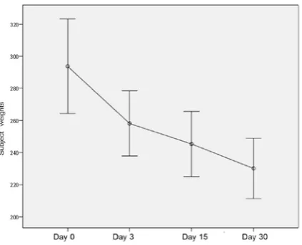
showed a significant decrease in the diabetic group weights on a daily basis (p < 0.05, see Fig. 1). Testes organ weights were also found to be significantly decreased for the diabetic group on a daily basis (p < 0.001, see Fig. 2).
Comparisons of Klf4, Sox2, c-Myc and Oct4 immunoreactivity were studied in the rat testes sec-tions from both the control and diabetic groups. The intensity of immunoreactivity was graded semi-quantitatively, and the scores are shown in Table 2.
Table 1. Themultiple comparison analysis of the weight of the subjects on day 0, day 3, day 15 and day 30 showed a significant decrease (p < 0.05)
Tabela 1. Wielokrotna analiza porównawcza masy ciała badanych osobników w dniu 0, 3, 15 i 30 wykazała jej znaczne zmniejszenie (p < 0,05)
(1) Days**
(J) Days MeanDifference (l–J) Std Error Sig.* 95% Confidence Interval for Differencea
lower bound upper bound 1 2 3 4 35.625*
48.375* 63.625*
7.874 9 0.264 9.519
0.016 0.007 0.002
6.997 14.693 29.017
64.253 82.057 98.233 2 1 3 4 –35.625*
12.750* 28.000*
7.874 2.684 3.756
0.016 0.013 0.001
–84.253 2.990 14.344
–6.997 22.510 41.656 3 1 2 4 –48.375*
–12.750* 15.250*
9.264 2.684 1.666
0.007 0.013 0.000
–82.057 –22.510 9.191
–14.693 – 2.990 21.309 4 1 2 3 –63.625* –28.000*
–15.250* 9.519 3.756 1.666
0.002 0.001 0.000
–98.233 –41.656 –21.309
–29.017 – 14.344 – 9.191 Based on estimated marginal means
*. The mean difference is significant at the 0.05 level, a. Adjustment for multiple comparisons: Bonferroni, **. The days compared to each other are as follows: 1: day 0; 2: day 3; 3: day 15; 4: day 30.
Fig. 1. Statistical analysis of the changes in the weights of the subjects for the diabetic model showed a signifi-cant decrease on a general linear model (p < 0.05)
Ryc. 1. Analiza statystyczna zmian masy ciała osob-ników na modelu cukrzycy wykazała ich znaczące zmniejszenie zgodnie z ogólnym modelem liniowym (p < 0,05)
Fig. 2. Testicular organ weights compared statistically for both the experimental induced-diabetes group and the control group were found to be significantly decreased for the induced-diabetes group on a daily basis (p < 0.001)
The normal histological structure of the testes was preserved in the cells of spermatogonial series and the Sertoli and Leydig cells in the control group. A significant loss of normal morphology for the sper-matogonial series of the diabetic testes was noted.
Klf4 Immunostaining
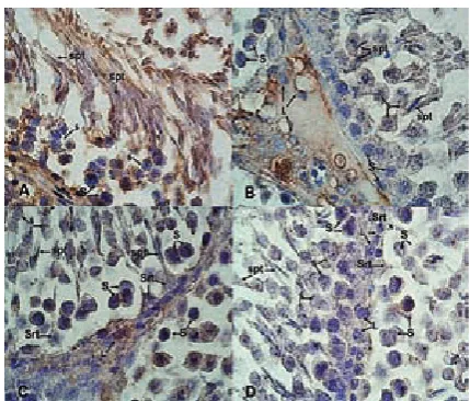
Strong (+++) immunostaining for Klf4 was observed not only in thecellsof thespermatogenic series,such as spermatogonium, spermatocyte and spermatid cells, but also in a remarkable number of Leydig and Sertoli cells in the control group (Figs. 3A and 3B). Immunostaining for Klf4 for the diabetic group was significantly decreased in spermatogenic cells and graded as weak (+) com-pared to the control group. Weak staining (+) of Klf4 was also determined in the Leydig and Sertoli cells of the diabetic group. Additionally, signifi-cant loss of early spermatids was observed in the diabetic testes (Figs. 3C and 3D).
Sox2 Immunostaining
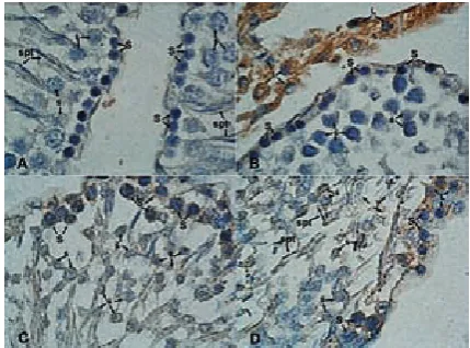
While some of the spermatogonial cells and Sertoli cells showed weak (+) immunostaining for Sox2, there was no staining (–) in spermatocytes and spermatids (Figs. 4A and 4B) in the control group. Sox2 immunostaining in the Leydig cells of the control group revealed extensive strong (+++) staining (Fig. 4B). There was no immunostaining (–) determined either for the whole
spermatogo-Table 2. The intensity of immunostaining is grouped into the following categories: – no staining; + weak staining; ++ mod-erate staining; and +++ strong staining
Tabela 2. Intensywność wybarwienia podzielono na następujące kategorie: – brak barwienia, + słabe barwienie; + + umiar-kowane barwienie, + + + silne barwienie
Immunostaining
(Wybarwienie) Spermatogonia(Spermatogonia) Spermatocytes(Spermatocyty) Spermatids(Spermatydy) Sertoli cells(Komórki Serto-liego)
Leydig cells (Komórki Ley-diga)
Klf4
Control +++ +++ +++ +++ +++
Diabetes + + + + +
Sox2
Control + + +++
Diabet +
c-Myc
Control + + + +++
Diabetes +++ +++ ++ +++ ++
Oct4
Control +++ +++ +++ ++
Diabetes + + ++
Intensity scores: – no staining; + weak staining; ++ moderate staining; +++ strong staining.
Fig. 3. Klf4 immunostaining: Klf4 expression is strongly observed in spermatogenic cells (spermatogonium – S, spermatocyte – ś, spermatid – spt) and in a remarkable number of Leydig (L) and Sertoli (Srt) cells in the control group (A, B). A significant loss of early spermatids was also observed in the diabetic testes. Klf4 expression is sig-nificantly decreased and weakly stained in the spermato-genic cellsas well as in the Leydig and Sertoli cells com-pared with the control group (C, D). Magnification x100
nial series of cells or the Sertoli cells in the diabetic group (Figs. 4C and 4D). Sox2 immunostaining in the Leydig cells of the diabetic group was weak (+) compared to the control group (+++) (Fig. 4D).
c-Myc Immunostaining
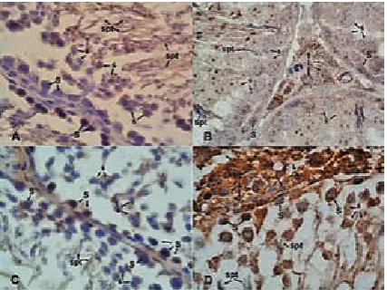
Strong (+++) immunostaining was observed for the spermatogonia, spermatocytes and Sertoli cells in the diabetic group compared to the con-trol group (+) (Fig. 5A). There was strong staining (+++) of the Leydig cells (Fig. 5B) and no stain-ing (–) in the spermatids of the control group (Fig. 5A). The spermatogonia, spermatocytes and Sertoli cells manifested increased immunostain-ing patterns, which were graded strong (+++) in the diabetic group (Fig. 5C). The spermatids and Leydig cells in the diabetic group showed moder-ate staining (++) compaired to the control group (Fig. 5D, Table 2).
Oct4 Immunostaining
In the control group, strong immunostaining (+++) for Oct4 was observed in the spermatogonia,
spermatocytes and Sertoli cells, but no immunos-taining (–) was found in the spermatids (Figs. 6A and 6B); moderate immunostaining (++) in the Leydig cells was observed (Fig. 6B). Decreased Oct4 immunostaining (+) pattern was observed for the spermatogonia and primer spermatocytes, while there was no staining (–) for the spermatids and Sertoli cells (Figs. 6C and 6D). Moderate stain-ing (++) in the Leydig cells was also determined in the diabetic group (Fig. 6D).
Discussion
In this study, in vivo expression patterns of the most common transcriptional factors (Klf4, Sox2, c-Myc and Oct4) that are in use for the generation of ipS cells from somatic cells and their possible effects on induced-diabetes adult rat testes tissue were investigated [7].The STZ model, widely used for many previous experimental observations, was used for the induction of diabetes in this study [2]. Four-month-old male rats were randomly chosen due to the fact that spermatogenesis is completed by 56 days of age, while the testes would continue
Fig. 4. Sox2 immunostaining.While some of the spermatogonial (S) and Sertoli cells (Srt) showed weak Sox2 expression, there was no staining in the spermatocytes (ś) and spermatids (spt) in the control group. Sox2 expression in the Leydig cells (L) also revealed an extensive strong staining pattern compared with the diabetic group (A, B). No Sox2 expression was determined for spermatogonial and Sertoli cells in the diabetic group (C, D). Magnification x100
Ryc. 4. Immunowybarwianie Sox2. Niektóre z sper-matogoniów (S) i komórek Sertoliego (Srt) wykazały słabą ekspresję Sox2, ale nie było barwienia w sper-matocytach (ś) i spermatydach (spt) w grupie kontrol-nej. Ekspresja Sox2 w komórki Leydiga (L) ujawniło również szeroki silny wzór barwienia w porównaniu z grupą chorych na cukrzycę (A, B). Brak ekspresji Sox2 wykazano dla spermatogoniów i komórek Sertoliego w grupie chorych na cukrzycę (C, D). powiększenie x100
Fig. 5. c-Myc immunostaining. Weak expression was observed for the spermatogonial cells (S) and spermatocytes (ś), while, the Leydig cells (L) were strongly stained (A, B). In addition, the spermatids (spt) showed no staining (5A). Nevertheless, the sper-matogonial cells and spermatocytes (ś) demonstrated increased expression (C). The spermatids and Leydig cells also showed c-Myc expression in the diabetic group (D). Magnification x100
to grow through 108 days of age [16, 17].During the one-month observation period following the induction of diabetes, the subjects not only showed lower body weight gain but also a significant de-crease in testes weights as well, which is concordant with the previous reports [17, 18].Some studies have demonstrated that Klf4 is strongly expressed in spermatids in mice [19, 20]and in humans [21], and also expressed in testicular Sertoli cells [31]. While all the cells in the control group testes – inc-luding spermatogonia, spermatocytes, spermatids, Sertoli cells and Leydig cells – manifested strong immunostaining patterns, STZ induction, probab-ly due to the negative effect of diabetes on cellular pluripotency, significantly decreased these expres-sions. It has previously been demonstrated that Klf4 possibly inhibits Myc-induced apoptosis via p53 repression [22]. On the other hand, other re-searchers have argued that Klf4 supressed cellular proliferation via activation of p21CIp1[24], while still other researchers commented that the antip-roliferative function of Klf4 was most likely inhibi-ted by c-Myc via supression of p21CIp1 expressi-on [25]. Many papers have previously emphasized
the importance of a balance between c-Myc and Klf4 for the generation of ipS cells. The dramatical-ly increased c-Myc expression levels in this study for the diabetes group demonstrates a mechanism that seems to be triggered in order to balance Klf4 accordingly. At the same time, Oct4 is known as a vital transcription factor for the maintenance of pluripotency in both embryonic and epiblast stem cells of mouse embryos [26, 27]. Moreover, a few studies related to Oct4 expression in adult gonads have appeared recently reporting post-embryonic roles for Oct4 in the mature gonads [26].
In the current study, the lack of expression in the spermatids while the control group showed strong immunoexpression of Oct4 for all the other cells of the testes, including the Sertoli and Leydig cells, can be explained by a loss of pluripotency during the second meiotic division in spermatoge-nesis. The most characteristic alterations in Oct4 expression for the diabetic rat group were noted for the Sertoli cells, where no Oct4 immunoexp-ression was detected. Considering the Sertoli cell, its connections with all the cells of the spermato-genetic series in the testes and its function of sup-port, the current authors noted that the pluripo-tency of the Sertoli cells has significantly declined. The Leydig cells in the diabetes group are the least affected cell in this study in terms of Oct4 im-munoexpression. Sox2 is known to be expressed differentially not only in female and male germi-nal stem cells, but also in the epiblast, in embr-yonic stem cells (ESCs) and subsequently in cells of the neuroepithelium and multipotent neural stem cells (NSCs). Sox2 has been designated first in a study during a screen for genes related to the sex-determining gene, SrY, by the possession of a high mobility group (HMG)-box DNA-binding domain [28]. The immunohistochemical analysis of the same study group described other modes of action for Sox2 chaperoned by Oct3/4 and Nanog under the positive regulation of c-Jun N-terminal kinases1 (JNK), and stronger expression patterns of Sox2 were reported in female germinal stem cel-ls compared to the males[28]. In the present study, immunoexpression of Sox2 was not determined for the cells of the spermatogenic series (like sper-matocytes and spermatids) in the control group, while spermatogonia and Sertoli cells revealed on-ly low levels of positivity. The marked immuno-positivity described for the Leydig cells is striking. However, considering the important roles of the Leydig cells during steroidogenesis, the authors propose probable interactions of mitogen-activa-ted protein kinase (MApK) pathways and the JNK cascade, as molecular and functional evidence for this was noted. Due to the induction of diabetes, not only did the immunoexpression of the Leydig
Fig. 6. Oct4 immunostaining. Strong expression was observed for the spermatogonial cells (S), spermato-cytes (ś) and Sertoli cells (Srt), though the spermatids (spt) are not stained (A, B). On the other hand, Oct4 expression was also observed in the Leydig cells (L) (B). Decreased Oct4 expression was observed in the spermatogonial cells and spermatocytes, although sper-matids and Sertoli cells were not stained (C, D). The Leydig cells (L) also showed Oct4 expression in the diabetic group (D). Magnification x100
cells fade, but all the other cells in the testes also lost immunoexpression. Quantification of gene expression by confocal immunomicroscopy would provide even more insight into the subject.
Another factor that was investigated in this study is the transcription factor c-Myc. Its expres-sion is generally known to be induced by mitogens and suppressed by growth-inhibitory signals in normal cells [30]. A ‘Myc’ transcription factor is crucial for the successful growth and replication cycle in the cell. For the adaptation, regeneration and apoptosis of β cell islets, c-Myc is also signi-ficantly relevant. Furthermore, recent literature describes the possible contribution of glucotoxi-city to Myc-induced apoptosis [14, 31] c-Myc pro-tein is believed to induce histone acetylation natu-rally [30], so that Oct3/4 and Sox2 would bind to their specific target loci. In contrast to the expres-sion patterns of Oct4 and Sox2 in diabetes, c-Myc demonstrated an increased expression pattern for the Sertoli cells and spermatids as a consequence of glucotoxicity.
The authors concluded that this study has demonstrated an increase in c-Myc immunoexp-resssion for the diabetic group that revealed dif-ferences from other markers. This fortifies myc-induced apoptosis as an expected outcome due to diabetes. The data that emerged from the study showed that for both control and diabetic groups
the immunoexpression of Sox2 and Oct4 was in accord with results reported in previous studies. Increased expression of Sox2 in the Leydig cells (among other cells of the testes) designated MAp kinase and JNK cascades. In addition, the effect of c-Myc (proliferative) and Klf4 (antiproliferative) have changed in terms of the balance of apoptosis and cell cycle in the diabetes group. The decrease in weight observed in this study was also concor-dant with previous studies, both for the weight of the subjects and the testicular weight. This study clearly demonstrated that Klf4, Sox2 and Oct4 im-munopositive cells in adult rat testes manifested sustainable pluripotency. Although the reported changes could be the consequence of the toxic agent streptozotocin on the testes tissue, rather than sustained hyperglycemia, there is a need for further sophisticated studies with non-toxic ani-mal models of diabetes such as NOD mice or Aki-ta mice to test this latter possibility. Furthermore, more descriptive and informative results can also be obtained by evaluating human testes specimens with these markers. The authors’ final conclusion here is that stem cells in testes and the pluripo-tency capacity of these cells are critically impaired during diabetes-related changes, and repair of the injured testes tissue is delayed. Until proven ot-herwise, it appears that diabetes has dramatically detrimental effects on pluripotency.
Acknowledgements. The authors would like to thank Dr. Timur Köse, associate professor at the Department of Biostatistics at the Ege University Faculty of Medicine, Turkey, for his support.
References
[1] Amaral S, Oliveira PJ, Ramalho-Santos J: Diabetes and the impairment of reproductive function: possible role of mitochondria and reactive oxygen species. Curr Diabetes rev 2008,4(1), 46–54.
[2] Masuzawa K, Goto K, Jesmin S, Maeda S, Miyauchi T, Kaji Y et al.: An endothelin type A receptor antagonist reverses upregulated VEGF and ICAM-1 levels in streptozotocin-induced diabetic rat retina. Curr Eye res 2006, 31(1), 79–89.
[3] Agarwal A, Saleh RA, Bedaiwy MA: role of reactive oxygen species in the pathophysiology of human reproduc-tion. Fertil Steril 2003, 79(4), 829–843.
[4] Navarro-Casado L, Juncos-Tobarra MA, Cháfer-Rudilla M, de Onzoño LÍ, Blázquez-Cabrera JA, Miralles-García JM: Effect of experimental diabetes and STZ on male fertility capacity. Study in rats. J Androl 2010, 31(6), 584–592.
[5] Ratajczak MZ, Machalinski B, Wojakowski W, Ratajczak J, Kucia M: A hypothesis for an embryonic origin of pluripotent Oct4 (+) stem cells in adult bone marrow and other tissues. Leukemia 2007, 21(5), 860–867.
[6] Sun C, Liu YK: Induced pluripotent cancer cells: progress and application. J Cancer res Clin Oncol 2011 Jan, 137(1), 1–8. doi: 10.1007/s00432-010-0955-z. Epub 2010 Nov 26.
[7] Takahashi K, Yamanaka S: Induction of pluripotent stem cells from mouse embryonic and adult fibroblast cul-tures by defined factors. Cell 2006, 126(4), 663–676.
[8] Watanabe S, Hirai H, Asakura Y, Tastad C, Verma M, Keller C et al.: MyoD gene suppression by oct4 is required for reprogramming in myoblasts to produce induced pluripotent stem cells. Stem Cells 2011, 29(3), 505–516.
[9] Godmann M, Kosan C, Behr R: Kruppel-like factor 4 is widely expressed in the mouse male and female repro-ductive tract and responds as an immediate early gene to activation of the protein kinase A in TM4 Sertoli cells. reproduction 2010, 139(4),771–782.
[10] Wei Z, Yang Y, Zhang P, Andrianakos R, Hasegawa K, Lyu J et al.: Klf4 interacts directly with Oct4 and Sox2 to promote reprogramming. Stem Cells 2009, 27(12), 2969–2978.
[12] Schöler HR, Ruppert S, Suzuki N, Chowdhury K, Gruss P: New type of pOU domain in germ line-specific pro-tein Oct-4. Nature 1990, 344(6265), 435–439.
[13] Niwa H: How is pluripotency determined and maintained? Development 2007, 134(4), 635–646.
[14] Pelengaris S, Khan M, Evan G: c-Myc: more than just a matter of life and death. Nature rev Cancer 2002, 2(10), 764–776.
[15] Lengner CJ: ipS cell technology in regenerative medicine.Ann NY Acad Sci 2010, 1192, 38–44.
[16] Ekwall H, Jansson A, Sjöberg P, Plöen L: Differentiation of the rat testis between 20 and 120 days of age. Arch Androl 1984, 13(1), 27–36.
[17] Taşkara E, Gör A, Kutlu O, Karagüzel E, Cobanoğlu U, Topbaş M et al.: Does propofol prevent testicular ischemia-reperfusion injury due to torsion in the long term? pediatr Surg Int 2011, Apr 6. [Epub ahead of print]
[18] Shalaby MA, Hamowieh AR: Safety and efficacy of Zingiber officinale roots on fertility of male diabetic rats. Food Chem Toxicol 2010, 48(10), 2920–2924.
[19] Godmann M, Kromberg I, Mayer J, Behr R: The mouse Kruppel-like factor 4 (Klf4) gene: four functional poly-adenylation sites which are used in a cell-specific manner as revealed by testicular transcript analysis and multiple processed pseudogenes. Gene 2005, 361, 149–156.
[20] Godmann M, Gashaw I, Katz JP, Nagy A, Kaestner KH, Behr R: Kruppel-like factor 4, a “pluripotency transcrip-tion factor” highly expressed in male postmeiotic germ cells, is dispensable for spermatogenesis in the mouse. Mech Dev 2009, 126(8–9), 650–664.
[21] Behr R, Deller C, Godmann M, Muller T, Bergmann M, Ivell R et al.: Kruppel-like factor 4 expression in normal and pathological human testis. Mol Hum reprod 2007, 13(11), 815–820.
[22] McLean DJ, Friel PJ, Pouchnik D, Griswold MD: Oligonucleotide microarray analysis of gene expression in follicle-stimulating hormone treated rat Sertoli cells. Mol Endocrinol 2002, 16(12), 2780–2792.
[23] Zindy F, Eischen CM, Randle DH, Kamijo T, Cleveland JL, Sherr CJ et al.: Myc signaling via the ArF tumor suppressor regulates p53-dependent apoptosis and immortalization. Genes Dev 1998, 12(15), 2424–2433.
[24] Zhang W, Geiman DE, Shields JM, Dang DT, Mahatan CS, Kaestner KH et al.: The gut-enriched Kruppel-like factor (Kruppel-like factor 4) mediates the transactivating effect of p53 on the p21WAF1/Cip1 promoter. J Biol Chem 2000, 275(24), 18391–18398.
[25] Seoane J, Le HV, Massague J: Myc suppression of the p21(Cip1) Cdk inhibitor influences the outcome of the p53 response to DNA damage. Nature 2002, 419(6908), 729–734.
[26] Sánchez-Sánchez AV, Camp E, García-España A, Leal-Tassias A, Mullor JL: Medaka Oct4 is expressed during early embryo development, and in primordial germ cells and adult gonads. Dev Dyn 2010, 239(2), 672–679.
[27] Scaldaferri ML, Fera S, Grisanti L, Sanchez M, Stefanini M, De Felici M et al.: Identification of side population cells in mouse primordial germ cells and prenatal testis. Int J Dev Biol 2011, 55, 209–214.
[28] Gubbay J, Collignon J, Koopman P, Capel B, Economou A, Münsterberg A et al.: A gene mapping to the sex-determining region of the mouse Y chromosome is a member of a novel family of embryonically expressed genes. Nature 1990, 346(6281), 245–250.
[29] Xiao YM, Chen L, Liu J, Liu WB, Chen HG, Zou LJ et al.: Contrast expression patterns of JNK1 during sex reversal of the rice-field eel. J Exp Zool B Mol Dev Evol 2010, 314(3), 242–256.
[30] Fernandez PC, Frank SR, Wang L, Schroeder M, Liu S, Greene J et al.: Genomic targets of the human c-Myc protein. Genes Dev 2003, 17(9), 1115–1129.
[31] Cheung L, Zervou S, Mattsson G, Abouna S, Zhou L, Ifandi V et al.: c-Myc directly induces both impaired insulin secretion and loss of β-cell mass, independently of hyperglycemia in vivo. Islets 2010, 2(1), 37–45.
Address for correspondence:
Ozlem Yilmaz-Dilsiz
Ege University Faculty of Medicine Department of Histology and Embryology Bornova 35100
Izmir Turkey
E-mail: [email protected] Tel.: +90 232 390 5909
Conflict of interest: None declared