Adenovirus (AdV), a member of the Adenov− iridaefamily, was first isolated from human ade− noidal tissue in 1953, hence the origin of its name (Fig. 1) [1–3]. Adenoviruses are divided into the genera Mastadenovirus (mammalian), Aviadeno− virus(avian), Atadenoviridae, Siadenoviridaeand the unassigned Adenoviridae [3, 4]. Human aden− ovirus comprises 51 serotypes identified so far [4, 5].
There are six sub−genera (A−F) (Tab. 1), which are specified on the basis of hemagglutination, differ− ences in DNA structure (DNA oligopepetide map− ping), electrophoretic mobility, and oncogenicity in experimental animals [2–4, 6]. Two sub−genera, 40 (F) and 41 (G), are called “intestinal aden− oviruses” as they are supposed to be unculturable serotypes because they cannot be isolated using
I
WONAB
IL1, B
LANKAR
YBKA2, M
IECZYSŁAWW
OŹNIAK1Adenoviral Infection – Pathomechanism
and Diagnostics
Infekcje adenowirusowe – patomechanizm i diagnostyka
1 Chair and Department of Clinical Chemistry, Silesian Piasts University of Medicine in Wrocław, Poland 2Laboratory of Molecular Biology, Silesian Piasts University of Medicine in Wrocław, Poland
Adv Clin Exp Med 2008, 17, 1, 91–99 ISSN 1230−025X
REVIEWS
© Copyright by Silesian Piasts University of Medicine in Wrocław
Abstract
Adenoviruses (AdVs) are among the main etiological infectious factors that affect immunocompromised patients. They are not potentially dangerous to healthy children, but are a very serious risk for bone marrow and organ recip− ients. There are about 51 different serotypes of human adenoviruses known so far. They occur generally in the throat and feces of healthy people. They can be latent in adenoid tissue and kidney, so they can induce disease after many years because of infection reactivation. Adenoviruses are responsible for many disorders. They cause infec− tions of the upper and lower respiratory tracks, alimentary system and urinary track, diseases of the joints, myocarditis, pericarditis and, more rarely, disorders of the central nervous system. Because of the large number of AdV serotypes, their genomic variations, and their susceptibility to mutation, they present a lot of difficulty in lab− oratory diagnostics. However, advances in current knowledge create opportunities to introduce new methods to quantify adenoviral infections and monitor therapy efficacy. This article presents current knowledge about aden− oviruses and their pathogenicity and information about available methods to diagnose adenoviral infections (Adv Clin Exp Med 2008, 17, 1, 91–99).
Key words:Adenoviridae, infection, pathogenesis, immunosuppression, diagnosis.
Streszczenie
Adenowirusy są jednym z głównych czynników etiologicznych zakażeń u pacjentów z osłabioną odpornością. Po− tencjalnie niegroźne dla subiektywnie zdrowych dzieci stają się poważnym zagrożeniem dla biorców transplanta− cyjnych szpiku kostnego i biorców narządowych. Do tej pory zidentyfikowano 51 serotypów ludzkich adenowiru− sów. Występują powszechnie w gardle i kale ludzi zdrowych. Dzięki zdolności do latencji w obrębie tkanki adeno− idalnej i w nerce mogą wywoływać choroby na skutek reaktywacji zakażenia nawet po wielu latach. AdV są odpowiedzialne za zakażenia układu oddechowego i pokarmowego, jak również zakażenia dróg moczowych, cho− roby stawów, zapalenie mięśnia sercowego i osierdzia, rzadziej choroby obwodowego układu nerwowego. Duża liczebność grupy ludzkich adenowirusów, duża zmienność genomowa oraz skłonność do mutacji stwarzają liczne trudności w rutynowej diagnostyce laboratoryjnej. Niemniej jednak postęp we współczesnej nauce sprzyja wpro− wadzaniu nowych metod służących do wykrywania i monitorowania przebiegu infekcji adenowirusowych. W ar− tykule przedstawiono obecny stan wiedzy na temat etiologii, patomechanizmu oraz dostępnych metod diagnostycz− nych w zakażeniach adenowirusowych (Adv Clin Exp Med 2008, 17, 1, 91–99).
cell cultures [7]. The adenovirus species of a sub− −genus have similar epidemiological and patho− genic properties [2]. They are commonly detected in stool specimens and pharyngeal swabs of healthy humans, but in immunodeficient patients they may be a serious source of opportunistic infections [5]. Adenoviruses may be useful as vec− tors in gene therapy. Adenovirus sub−genus C types 2 and 5 are frequently used. Their use is limited by CAR receptor binding capabilities on most cells [8].
Morphology
Although the biological properties of AdVs differ, they have a common genome structure. The adenoviral nucleus is 45–60 nm in diameter [2, 3, 6] and the virion 70–90 nm. The adenoviral genetic material is linear, double−stranded DNA with a molecular weight of 20–30 ×106bp in one mol− ecule and is 12–17% of the virion [2, 6]. The DNA is enclosed in an unenveloped 20−face 12−vertex icosahedron (Fig. 2) [2, 8, 9]. It is composed of 252 capsomers: 240 hexamers and 12 pentons [6, 10]. Their icosahedral capsids are composed of three major proteins: a hexon (II) and a penton base (III) and trimeric fiber proteins (IV). There are several minor proteins (VI, VIII, X, III, IVa) for various functions in the icosahedron [9]. The hexon is a homotrimer of three identical polypeptide chains that form a triangular vertex of three surface loops L1, L2, and L4. The hexons possess group− and type−specific determinants [10]. The hexon con− sists of three segments: a chimeric region and two flanking conservative regions (Fig. 3). L1and L2
consist of at least seven hypervariable regions (HVR). They constitute ca. 90% of the type−spe− cific domains of the hexon and are the basis of adenovirus’s variability [11]. Serotypes of one sub−genus show a high (91–95%) homology in the hexon gene but much lower in different sub−gen− Table 1.Sub−genera and stereotypes of human adeno− viruses (own schedule)
Tabela 1.Podrodzaje i serotypy ludzkich adenowirusów (tabela własna)
Sub−genera Serotypes (Podrodzaje) (Serotypy)
A 12, 18, 31
B 3, 7, 11, 14, 16, 21, 34, 35, 50
C 1, 2, 5, 6
D 8, 9, 10, 13, 15, 17, 19, 20, 22, 23, 24, 25, 26, 27, 28, 29, 30, 32, 33, 36, 37, 38, 39, 42, 43, 44, 45, 46, 47, 48, 49, 51
E 4
F 40, 41
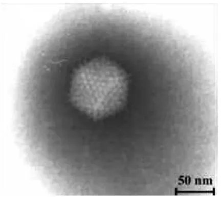
Fig. 1.Adenovirus. Electron micrographs of enteric viruses after negative staining
Ryc. 1.Adenowirus. Widok z mikroskopu elek− tronowego. Barwienie negatywne
Fig. 2.Structure of adenovirus scheme. The locations of the capsid and cement components are reasonably well defined. In contrast, the disposition of the core compo− nents and the viral DNA is largely conjectural [9]
Ryc. 2.Struktura adenowirusa – schemat.
Umiejscowienie składników kapsydu i elementów spa− jających jest dobrze zdefiniowane. Rozmieszczenie składników rdzenia i wirusowego DNA w większości stanowi przypuszczenie [9]
capsid proteins białka kapsydu
core proteins białka rdzenia
cement proteins białka spajające
hexon heksan
fibre włókno
era (ca. 4%) [12]. Filament polypeptides show high homology, and most differences were noted in loops L1and L2.
Each penton (vertex) has a projecting fiber with a terminal knob 10–37 nm in length [3, 6]. Domains on the knob bind appropriate cell recep− tors [8, 9]. They have various additional functions, such as allowing the virus to enter the cell, take part in intracellular transport, and viral growth and accumulation in the cell [13]. Studies of the amino−acid homology of the filaments in AdVs of one or more sub−genera showed that in the case of high homology (above 90%), adenoviruses inter− act with cells similarly and cause similar types of infection in a particular tissue [14].
Properties
Adenoviruses are able to replicate in the envi− ronment of the gastrointestinal tract. This means that adenoviruses are resistant to proteolytic enzymes [2]. They are stable to pH 6–9 and the virions are not sensitive to treatment with lipid solvents. A low con− centration of chloride does not inactivate the virus. They are quickly inactivated at 56°C [3].
Pathogenesis
Adenoviruses have been found in many verte− brates. Mammalian adenoviruses were isolated from humans, cattle, sheep, horses, pigs, monkeys, dogs, and rodents. Owing to the large size of the Adenoviridae family, individual serotypes show characteristic trophism to different tissues [3]. They infect postmitotic cells, even highly differen− tiated tissue such as skeletal muscle, lungs, brain, and heart [9]. The most common are adenoviral infections of the pulmonary system and gastroin− testinal tract. They cause exudative pharyngitis and may cause acute respiratory disease epidemics in adults. These involve febrile laryngitis and con− junctivitis [2, 8], keratitis and conjunctivitis,
necrotizing enterocolitis, pharyngeal−conjunctival fever, and hemorrhagic cystitis [7]. Less frequent are testitis, nephritis, arthritis, myocarditis, and pericarditis. Infections of the gastrointestinal tract are especially frequent in children and include gas− troenteritis, mesenteric lymphadenitis, agonal intussusception, hepatitis, and appendicitis [2, 3, 5, 14, 15]. New molecular diagnostics showed that adenoviruses are involved in bronchopulmonary dysplasia and chronic obturative lung diseases. [16]. Adenoviruses sometimes cause neurological dis− orders. Several cases of acute flaccid paralysis due to adenovirus B and encephalitis have been found in children (Tab. 2) [5].
During the incubation of an acute adenovirus infection one observes increased nasal secretion, pharyngalgia, feeling of increased cold or warmth, chills, fatigue, and increased body temperature (38–39°C). Other symptoms include pharyngeal redness, dry cough, and sometimes jugular lymph node enlargement. Symptoms of mucositis domi-nate in laryngitis and conjunctivitis. Keratitis and conjunctivitis may be accompanied by unilateral or bilateral papulovesicular lesions and can some-times lead to corneal damage and scars [2]. In intestinal infections, damage may be mild, involv-ing mostly symptoms of gastroenteritis.
It is widely known that most adenovirus serotypes occur endemically in the environment and may by responsible for the onset of epidemics in a human population [2]. Serological surveys car− ried out in different age groups and regions indi− cated a high dissemination of certain AdV serotypes [3]. Most patients had been infected by at least one AdV serotype when they were below 15 years of age [17]. They cause ca. 5% of the acute respiratory infections in children under 5 years of age and are responsible for ca. 10% of pediatric pneumonia cases [7]. The most common factor of over 80% of pediatric infections is aden− ovirus sub−genus C [5]. Usually they cause asymp− tomatic infections or only minor symptoms (cough,
Fig. 3.The conserved and vari− able regions of the hexon gene (authors’ design)
Ryc. 3.Region zmienny i regiony konserwatywne w obrębie hexon genu (projekt autorów)
hexon gen
highly conserved region variable middle region highly conserved region
region konserwatywny region zmienny region konserwatywny
L1, L2, L4 – genus− and subgenus−specific determinants (hypervariable regions) L1, L2, L4 – determinanty sero− i podrodzajowo typowe (regiony zmienne)
nasal obstruction), i.e. no clear clinical picture [3]. They cause epidemics quite rarely, although they are common in conditions of crowding, e.g. among recruits, swimming pool users, and in hospitals [2, 3]. At the highest risk of infection are transplant recipients and other immunosuppressed patients, in whom morbidity is 20–50%. The AdV sub−genera C, B and A cause 90% of adenoviral infections in transplant patients, frequently with severe conse− quences [18]. These patients can be infected by adenoviral strains in the environment or those orig− inating from the transplant donor, especially due to activated latent virus [15]. It has been shown that adenovirus can survive in lymphoid tissue or kid− ney for many years after the primary infection [12, 17]. Most of these diseases are clinically ambiguous or mild, self−limiting disorders, but there are severe, even fatal diseases in very young or very old or immunosuppressed patients [11]. The etiological proportions of individual serotypes depend on seasonal and endemic changes as well as on the environment and region [3].
Mechanism of Infection
Adenovirus spreads directly through respirato− ry droplets. It is suggested that they also spread via water in swimming pools and through the fecal−oral Table 2.Clinical syndromes of adenovirus (own schedule)
Tabela 2.Zespoły kliniczne powodowane przez adenowirusy – zestawienie (tabela własna)
Clinical syndrome Most common serotypes
(Zespół kliniczny) (Najczęstsze serotypy)
Pharyngitis 1, 2, 3, 5, 7
(Zapalenie gardła)
Pharyngoconjunctival fever 3, 7
(Gorączka gardłowo−spojówkowa)
Acute respiratory disease of recruits 3, 4, 7, 14, 21 (Ostra choroba oddechowa u rekrutów)
Pneumonia 1, 2, 3, 7
(Zapalenie płuc)
Acute follicular conjunctivitis 3, 4, 11 (Ostre pęcherzykowe zapalenie spojówki)
Epidemic keratoconjunctivitis 8, 8, 19
(Epidemiczne zapalenie spojówki i rogówki)
Pertussis−like syndrome 5
(Syndrom krztuścopodobny)
Acute hemorrhagic cystitis 3, 7, 11, 21
(Ostre krwotoczne zapalenie pęcherza moczowego)
Gastroenteritis 12, 18, 31, 40, 41
(Zapalenie żołądka i jelit)
Intussusception 1, 2, 5
(Wgłębienie agonalne jelita)
Meningitis, encephalitis 3, 7
(Zapalenie opon, zapalenie mózgu)
Fig. 4.Epithelial cell nucleus with virions that are closely packed in crystalline arrays (arrays of dark inclusions). Electron micrograph [19]
route or unsterile surgical instruments [3]. During relatively short incubation (5–10 days) the virus replicates in the larynx, conjunctiva, and small intestine [2]. Adenoviral infection has two phases. The early phase starts with absorption of the virion into the host cell via receptors and fiber protein. The virus uncoats and the penton base migrates into the nucleus, where the process of dsDNA replication takes place. Adenovirus is replicated in the cell’s cytoplasm, but viral DNA is replicated inside the nucleus of the cell [9]. During the process of transcription, early and late mRNA are produced and later unstructured proteins [2]. The phase lasts quite long, i.e. 6–8 h. The early processes modulate cell functions: they initiate the transcription and translation of late genes which provide for the pro− duction of many structural proteins allowing for the formation and growth of the molecules of the nascent virions. The course of the second phase is quicker and lasts ca. 4 h [9]. The nascent viruses gather in the nucleus in an amount large enough to form crystal aggregates (Fig. 4) [2, 19]. Cells show three effects of adenoviral infection. In lytic infec− tion the cell dies, while in latent infection the virus remains inside the host cell without causing its death. Most frequently these are lymphoid cells, i.e. tonsils, adenoid hypertrophy, and Peyer’s patches. In some cases the transformation may be onco− genic, i.e. the virus replicates and does not cause death of the cell. This phenomenon has only been observed in hamsters, but one should consider this when analyzing infections in humans [2, 9].
It is widely known that the host immune sys− tem plays a significant role in limiting viral spread. An anti−adenovirus defense mechanism is the release of defensin−type proteins by epithelial cells. Certain tissues are capable of producing complex chemokines that activate neutrophils and induce inflammatory response. The primary defense includes macrophages, complement sys− tem, and NK cell activation and it plays an impor− tant role in controlling adenoviral infections. Another strategy of defense against viruses is the infected cells’ capability to change its metabolism to an apoptosis pathway [9]. In adenoviral infec− tions the organism’s defense is limited due to the high number of serotypes (51 different AdV serotypes) and high antigenic variation that con− tribute to epidemic infections [20].
Adenoviruses
in Immunosuppression
Stem−cell and bone−marrow transplant patients are at a higher risk of viral infection. Adenoviruses are recognized as factors responsible for morbidity and mortality in this population [12, 21]. They usu−
This is similar to allogenic transplant from MUDs (matched unrelated donors) or MMFD (mis− matched family donors) [21]. Literature data describe many co−infections. They indicate that adenoviral infection in transplant patients coexists with BKV and CMV infections. Coexisting infec− tions may involve diagnostic difficulties and more severe course of disease [18, 28]. Progress in rapid and quantitative detection methods and careful determination of serotypes and strains may provide important information crucial for infection supervi− sion and control [21, 29].
Diagnostics
Recognition of acute respiratory diseases based only on clinical diagnosis is extremely diffi− cult due to symptoms that are identical in viral and bacterial infections. Considering the epidemiolog− ical, bacteriological, and virological aspects is cru− cial in making a full and proper diagnosis.
Adenoviral Culture
and Isolation
The gold standard for detecting adenoviruses is their isolation and rapid identification in clinical samples. This method is the reference for all other diagnostic methods and is an indication for disease etiology [2, 7]. Adenoviruses can be isolated from nasopharyngeal swabs, conjunctival secretion, sputum, and stool. The material for diagnosis may be cornal washings, scrapings, bioptates, and autopsy material [3, 7]. Antibiotics are added to the aspirated material, then a cell line is infected and between days 2 and 21 after infection the cyto− pathic effects (CPE) in the cells are observed. In adenoviral in vitrocultures, the following lines are used: HeLa (cervical cancer cells), HEK (human embryonic kidney cells), Hep−2 (laryngeal can− cer), and A549 (lung cancer cells) [2, 3, 9]. When the virus proliferates, clusters of rounded cells resembling bunches of grapes are observed [2]. The virus proliferating in the cell culture can be identified by the complement−binding method or using a neutralization reaction. The weaknesses of these methods are their long duration (days or weeks) and high cost [3]. A negative result does not rule out infection due to their limited sensitiv− ity. On the other hand, a positive culture result does not prove invasive disease, as adenoviruses may remain latent in lymphoid tissue [25]. Studies have shown that the CPE method is effective in different cell lines for detecting infection in target organs in symptomatic disease, but are not sensi− tive enough to detect a small number of viral mol− ecules circulating in the peripheral blood [12].
Microscopic
and Serological Methods
The characteristic morphology of adenovirus− es enables their detection in stool samples using an electron microscope. Immunofluorescent micro− scopy offers the opportunity to detect antigens against adenovirus, e.g. in nasopharyngeal swabs. The method is highly sensitive and specific. It allows revealing adenoviral particles in the nuclei of alveolar epithelial cells, plasma cells, and alve− olar macrophages. Therefore it is recommended for rapid preclinical diagnostics. Microscopic methods are especially recommended in hospital laboratories and should be widely used in virolog− ical laboratories. They enable observing all cyto− pathic effects and degenerative changes in cultures and organ specimens [1, 3, 30].
Serological methods detect respective epi− topes located on the adenoviral molecular surface in clinical material and antigens after viral prolif− eration in sensitive cells (culture) and detecting and identifying specific antibodies against the virus in serum as a result of the immunological reaction to infection [20]. Type−specific identifica− tion is based on serum neutralization (SN) and/or hemagglutination inhibition (HI) [11].
The serum neutralization method (SN) is neu− tralization of the virus’s infectivity by antibodies and enables assessment of the titer of neutralizing antibodies in the serum [3]. The type−specific determinants of AdV recognized by SN are locat− ed primarily on the surface of the hexon cap− somere, but determinants are also present on the fiber and on the penton proteins. Antigenic domains have been classified into seven hyper− variable regions, HVR1–6. HVR1–6 are the logical focus for the molecular typing of AdV [11]. Examination should take place in the acute phase and 2–3 weeks later. The result is reliable if at least a fourfold increase in antibodies against a certain type of viral titer is noted. This unequivocally con− firms adenoviral infection [3].
Immunomorphological
Methods
Immunoenzymatic (EIA), radioimmunologic (RIA) and immunofluorescent (IFA) (direct and indirect) tests enable the detection of virus or its antibodies as well as antibodies against adenovirus [3]. One of the antigen−antibody reaction ingredi− ents is bound with a specific marker, i.e. an enzyme, radioisotope or fluorochrom.
Serological tests are time−consuming and do not yield reliable results for all 51 human adenovi− ral serotypes. These tests have several flaws typi− cal of serological reactions, i.e. various sensitivity and specificity, lack of reactivity before serocon− version, or diagnostic difficulties due to adenovi− ral variability [20]. Using these tests in immuno− suppressed patients is limited due to immune sys− tem deficiency [21, 29]. They are repeatable and yield comparable results and are rapid and easy to perform [3, 6].
The histochemical method is a different and rarely performed test for assessing adenoviral infec− tion. AdV is detected in tissue specimens embedded in paraffin blocks and specific morphological changes are observed. Immunohistochemistry can be combined with in situhybridization [12].
Conventional diagnostic methods do not meet current requirements, which include a short length of time required for diagnosis. The amount of obtained diagnostic material is sometimes not enough to use these methods. Therefore, highly sensitive and specific molecular techniques are more widely used.
Molecular Techniques
Nucleic Acid Probes:
in situ
Hybridization Using Radioactively
Labeled and Non−Radioactively
Labeled Genetic Probes
Nucleic acid probes are short, single−stranded segments of DNA that have a known sequence complementary to a particular adenoviral DNA sequence and are hybridized with AdV single− stranded DNA. Identification is made by labeling the probe with radioisotopes and other markers that help visualize the hybrid [20].
Polymerase Chain Reaction
Polymerase chain reaction techniques for detecting adenoviral genetic material: PCR allows the amplification of specific target adenoviral DNA sequences with the use of complement
primers and DNA polymerase. The primers are specific for a certain DNA sequence and allow the amplification of the specific sequence of interest. The product is obtained on an electrophoresis agarose gel and the DNA is stained with ethidium bromide in UV light [7, 31]. PCR detection indi− cates AdV nucleic acid and gives no information on viral infectivity, which is the main goal for clin− icians [32].
Restriction Enzyme Assay
Restriction enzyme assay is cutting viral DNA into 4− to 7−nucleotide−long fragments with the appropriate endonuclease and electrophoretic sep− aration of these fragments in gel. Addition of ethidium bromide allows visualization of the DNA fragments in UV light [6].
Real−Time PCR
One of the methods that has recently gained growing interest is real−time PCR (RQ−PCR). It is a type of polymerase chain reaction for detecting and assessing specific sequences of nucleic acids. Analyzing the increase in the amount of the prod− uct enables full monitoring of PCR kinetics. During amplification, the fluorescent signal corre− lates with the primary material in the sample. The number of adenoviral copies in the sample from the patient can be calculated [33]. The advantage of real−time PCR is determination of minimal amounts of nucleic acid. Its high sensitivity allows detection of as few as 10 copies of the virus. The results can be calculated in clinically useful units, e.g. the number of copies of virus in 1 ml of low− −concentration cell fluid per 1 cell or 1 g of stool. Another advantage of the method is the detection and determination of one DNA sequence present in a sample containing a large number of other molecules [34]. The method is rapid and reliable. It is performed in closed automated systems, so the possibility of contaminating the probe in the labo− ratory is minimal as is false diagnosis [25, 33]. The RQ−PCR technique is especially useful in immunodeficient patients as the quantitative deter− mination of AdV in blood may give information on the risk of dissemination of the infection [12, 25, 35]. The course of the disease and the efficacy of the treatment can be monitored [23, 33].
tions and high genome variability cause problems in routine diagnostics. It is suggested that in the future this method will allow simple identification, genotyping and quantitative estimation of many viral DNA molecules in one rapid reaction [33].
Treatment and Prophylaxis
Adenoviral infections are treated with cido− fovir, gancyklovir and ribavirin or the recently introduced cytotoxic T lymphocytes. The last
method is, however, limited due to the epitope variation recognized by the immune system among serotypes. Immunological therapy may be dependent on the type of AdV [25, 29]. Proper prophylaxis against infection and the spread of adenoviral infection must be stressed, especially in children and in large places of assembly. Prophylaxis involves mainly maintaining personal hygiene, chlorinating swimming pools, steriliza− tion/disinfection of otolaryngological instruments, using disposable instruments, and administering vaccines (in select cases) [2, 15].
References
[1] Arcangeletti MC, De Conto F, Pinardi F, Medici MC, Valcavi P, Ferraglia F, Motta F, Covan S, Calderaro A, Chezzi C, Dettori G:Electron microscopy as a reliable tool for rapid and conventional detection of enteric viral agents: a five−year experience report. Acta Biomed 2005, 76, 165–170.
[2] Collier L, Oxford J: Namnażanie wirusów w laboratorium. Zakażenia górnych dróg oddechowych i oczu wywołane przez adenowirusy, koronawirusy i rinowirusy. Wirusy wywołujące zapalenie żołądka i jelit. In: Wirusologia. Ed.: Łuczak M, Wydawnictwo Lekarskie PZWL, Warsaw 1996, 47–57, 129–134, 176.
[3] Końtoch M, Blasković D:Diagnostyka wirusologiczna. Adenoviridae. Wirusologia lekarska. Państwowy Zakład Wydawnictw Lekarskich, Warszawa 1991, wyd. 3 zm., 154–179, 200–204.
[4] Stone D, Furthmann A, Sandig V, Lieber A:The complete nucleotide sequence, genome organization, and ori− gin of human adenovirus type 11. Virology 2003, 309, 152–165.
[5] de Azevedo JP, Nascimento LR, Cortinovis MC, Oliveira SS, da Costa EV, da Silva EE:Characterization of species B adenoviruses isolated from fecal specimens taken from poliomyelitis−suspected cases. J Clin Virol 2004, 31, 248–252.
[6] Hierholzer JC:Adenoviruses in the immunocompromised host. Clin Microbiol Rev 1992, 5, 262–274.
[7] Zaremba M, Borowski J: Wirusologia lekarska. Mikrobiologia lekarska. Wydawnictwo Lekarskie PZWL, Warsaw 1997, 529–530, 571–584.
[8] Mei YF, Lindman K, Wadell G:Human adenoviruses of subgenera B, C and E with various tropisms differ in both binding to and replication in the epithelial A549 and 293 cells. Virology 2002, 295, 30–43.
[9] Russell WC:Update on adenovirus and its vectors. J Gen Virol 2000, 81, 2573–2604.
[10] Pring−Akerblom P, Trijssenaar FE, Adrian T:Hexon sequence of adenovirus type 7 and comparison with other serotypes of subgenus B. Res Virol 1995, 146, 383–388.
[11] Lu X, Erdman DD:Molecular typing of human adenoviruses by PCR and sequencing of a partial region of the hexon gene. Arch Virol 2006, 151, 1587–1602.
[12] Gu Z, Belzer SW, Gibson CS, Bankowski MJ, Hayden RT:Multiplexed, real−time PCR for quantitative detec− tion of human adenovirus. J Clin Microbiol 2003, 41, 4636–4641.
[13] Defer C, Belin MT, Caillet−Boudin ML, Boulanger P:Human adenovirus−host cell interactions: comparative study with members of subgroups B and C. J Virol 1990, 64, 3661–3673.
[14] Pring−Akerblom P, Heim A, Trijssenaar FE:Conserved sequences in the fibers of epidemic keratoconjunctivi− tis associated human adenoviruses. Arch Virol 1997, 142, 205–211.
[15] Gray GC:Adenovirus transmission – worthy of our attention. J Infect Dis 2006, 194, 871–873.
[16] Gray GC, Setterquist SF, Jirsa SJ, DesJardin LE, Erdman DD:Emergent strain of human adenovirus endem− ic in Iowa. Emerg Infect Dis 2005, 11, 127–128.
[17] Shirali GS, Ni J, Chinnock RE, Johnston JK, Rosenthal GL, Bowles NE, Towbin JA:Association of viral genome with graft loss in children after cardiac transplantation. N Engl J Med 2001, 344, 1498–1503.
[18] Leruez−Ville M, Chardin−Ouachèe M, Neven B, Picard C, Le Guinche I, Fischer A, Rouzioux C, Blanche S:
Description of an adenovirus A31 outbreak in a paediatric haematology unit. Bone Marrow Transplant 2006, 38, 23–28.
[19] Parizhskaya M, Walpusk J, Mazariegos G, Jaffe R:Enteric adenovirus infection in pediatric small bowel trans− plant recipients. Pediatr Dev Pathol 2001, 4, 122–128.
[20] Polz−Dacewicz M:Diagnostyka zakażeń wirusowych. Diagnosta Laboratoryjny 2006, 3, 11, 6–10.
[21] Lion T, Baumgartinger R, Watzinger F, Matthes−Martin S, Suda M, Preuner S, Futterknecht B, Lawitschka A, Peters C, Potschger U, Gadner H:Molecular monitoring of adenovirus in peripheral blood after allogeneic bone marrow transplantation permits early diagnosis of disseminated disease. Blood 2003, 102, 1114–1120.
[22] Hale GA, Heslop HE, Krance RA, Brenner MA, Jayawardene D, Srivastava DK, Patrick CC:Adenovirus infection after bone marrow transplantation. Bone Marrow Transplant 1999, 23, 277–282.
[24] Venard V, Carret A, Corsaro D, Bordigoni P, le Faou A:Genotyping of adenoviruses isolated in an outbreak in a bone marrow transplant unit shows that diverse strains are involved. J Hosp Infect 2000, 44, 71–74.
[25] Seidemann K, Heim A, Pfister ED, Köditz H, Beilken A, Sander A, Melter M, Sykora KW, Sasse M, Wessel A:
Monitoring of adenovirus infection in pediatric transplant recipients by quantitative PCR: report of six cases and review of the literature. Am J Transplant 2004, 4, 2102–2108.
[26] Chakrabarti S, Mautner V, Osman H, Collingham KE, Fegan CD, Klapper PE, Moss PA, Milligan DW:
Adenovirus infection following allogeneic stem cell transplantation: incidence and outcome in relation to graft manipulation, immunosuppression, and immune recovery. Blood 2002, 100, 1619–1627.
[27] Echavarria M, Forman M, Ticehurst J, Dumler JS, Charache P:PCR method for detection of adenoviruses in urine of healthy and human immunodeficiency virus−infected individuals. J Clin Microbiol 1998, 36, 3323–3326.
[28] Akiyama H, Kurosu T, Sakashita C, Inoue T, Mori Si, Ohashi K, Tanikawa S, Sakamaki H, Onozawa Y, Chen Q, Zheng H, Kitamura T:Adenovirus is a key pathogen in hemorrhagic cystitis associated with bone mar− row transplantation. Clin Infect Dis 2001, 32, 1325–1330.
[29] Ebner K, Rauch M, Preuner S, Lion T:Typing of human adenoviruses in specimens from immunosuppressed patients by PCR−fragment length analysis and real−time quantitative PCR. J Clin Microbiol 2006, 44, 2808–2815.
[30] Kawai T, Fujiwara T, Aoyama Y, Aizawa Y, Yamada Y:Diffuse interstitial fibrosing pneumonitis and aden− ovirus infection. Chest 1976, 69, 692–694.
[31] Powledge TM:The polymerase chain reaction. Adv Physiol Educ 2004, 28, 44–50.
[32] Ko G, Jothikumar N, Hill VR, Sobsey MD:Rapid detection of infectious adenoviruses by mRNA real−time RT−PCR. J Virol Methods 2005, 127, 148–153.
[33] Valasek MA, Repa JJ:The power of real time PCR. Adv Physiol Educ 2005, 29, 151–159.
[34] Mackay IM, Arden KE, Nitsche A:Real−time PCR in virology. Nucleic Acids Res 2002, 1292–1305.
[35] Edwards K, Logan J, Saunders N:Applications of real−time PCR in clinical microbiology. Real−time PCR. An essential guide. Horizon Bioscience, Wymondham 2004, 11, 286–293.
Address for correspondence:
Iwona Bil Ślężna 96/34 53−301 Wrocław Poland
Tel.: +48 603 833 969
E−mail: [email protected]
Conflict of interest: None declared