I
ZABELAŁ
ACZMAŃSKA1, K
AROLINAP
ESZ1, Ł
UKASZŁ
ACZMAŃSKI2Application of Selected Methods Based on the Polymerase
Chain Reaction in Medical Molecular Diagnostics
Wykorzystanie wybranych metod opartych na reakcji polimerazy
do diagnostyki molekularnej chorób genetycznych
1Department of Genetics, Wroclaw Medical University, Poland
2Department of Endocrinology and Diabetology, Wroclaw Medical University, Poland
Adv Clin Exp Med 2009, 18, 1, 85–92 ISSN 1230−025X
REVIEWS
Abstract
A breakthrough in molecular biology came with the discovery of the polymerase chain reaction. It allowed the amplification of DNA fragments in vitroin an efficient, rapid, and inexpensive way. Furthermore, it is so versatile that the polymerase chain reaction technique has been used for a multitude of purposes and been found to be applic− able in a variety of different disciplines. The authors present the most frequently used laboratory methods based on the polymerase reaction and discuss their application in diagnosing genetic disorders (Adv Clin Exp Med 2009, 18, 1, 85–92).
Key words: polymerase reaction, diagnostic methods.
Streszczenie
Rozwój diagnostyki genetycznej jest ściśle związany z odkryciem łańcuchowej reakcji polimerazy (PCR). Pozwala ona na szybką, wydajną i tanią amplifikacjęin vitro fragmentów DNA. Reakcja prowadzona przez polimerazę stała się pon− adto podstawą do stworzenia i rozwoju wielu różnych metod powszechnie stosowanych w laboratoriach. Autorzy przedstawiają najczęściej wykorzystywane metody biologii molekularnej oparte na reakcji polimerazy oraz opisują ich zastosowanie w diagnostyce chorób uwarunkowanych genetycznie (Adv Clin Exp Med 2009, 18, 1, 85–92).
Słowa kluczowe: reakcja polimerazy, metody diagnostyczne.
The synthesis of deoxyribonucleic acid cat− alyzed by DNA polymerase (the Klenow fragment of the enzyme) is one of the most crucial reactions in molecular biology. Its adoption in in vitrolabo− ratory use has contributed to the universal and rou− tine application of the polymerase chain reaction (PCR) in research studies on living organisms and in forensics, and medical diagnostics. This paper discusses the application of PCR in diagnosing genetic disorders.
PCR and PCR−Based
Methods
The polymerase chain reaction, first described by Kary Mullis [1], gave its originator the Nobel Prize for chemistry in 1993. Since then, this sim−
ple technique has become a fundamental and stan− dard method in molecular biology. At present, this enzymatic reaction, catalyzed in vitro by a ther− mostable enzyme, polymerase, is the only method that enables obtaining several million or more copies of a critical DNA fragment chosen from the whole genome. The enzyme which catalyses the polymerization of deoxynucleotides in vitrois iso− lated from thermophilic bacteria, originally from
Thermus aquaticus (Taq). Like other DNA poly− merases, it needs a primer to begin the reaction. Therefore, to start copying the double−stranded DNA, two oligonucleotides, a forward and a reverse, are necessary. Because of the specificity in nucleotide pairing, primers about 20 nucleotides long are enough to anneal at one target DNA site, selecting between a huge number of other sequences. PCR is a three−step reaction performed
in an automatic cycler, which is able to cool and heat the reaction mixture rapidly in a small tube. A typical PCR run begins with 3–5 minutes of ini− tial denaturation at 94–95°C, which is necessary to destroy the tertiary structure of the genomic DNA. After that, reaction in regular three−step cycles takes place: 1) denaturation at 90–94°C, necessary to melt the template double−stranded DNA to sin− gle strands, 2) annealing at the temperature of primer hybridization when the primers are matched to the complementary sequences, which initiates the polymerase chain reaction, and 3) elongation/extension at 72°C, optimal for the Taq polymerase to read the template from the 3’ to 5’ ends and extend a new complementary strand from the 5’ to 3’ ends using deoxynucleotide triphosphates. In each cycle, the number of dsDNA fragments is doubled. After about 30–35 cycles and 5 minutes of a final extension at 72°C, several million or more copies are synthesized by the polymerase from one copy of the target sequence.
This simple procedure has allowed the devel− opment of a variety of PCR−based methods both in research and in medical diagnostics, especially for diagnosing genetic disorders that result from numerical and structural chromosome aberrations, deletions or amplifications of genes or gene frag− ments, point mutations, or abnormal methylation.
ASA−PCR
One of the simplest and least expensive meth− ods of detecting DNA point mutations is allele− specific amplification PCR (ASA−PCR) [2]. In this technique, three primers are used to amplify a fragment of a critical gene and simultaneously identify a change in the DNA sequence. One of the reverse primers is complementary to the wild−type allele and a second to the mutated allele. The for− ward primer is the same for both alleles [3]. The difference between the reverse primers is at the 3’ end, in which the critical single−nucleotide polymorphism (SNP) or mutation is located. Due to the unique conditions of the reaction, amplifica− tion of the wild−type and mutated alleles is only possible between their specific reverse and com− mon forward primers. In order to reach such a high affinity of both reverse primers, the reaction con− ditions must be precisely determined and restric− tively adhered to.
Because one of the reverse primers is comple−
mentary to the DNA sequence about 20
nucleotides upstream and produces a longer prod− uct, the difference in the lengths of the PCR prod− ucts, visible on an agarose gel, allows distinguish−
ing between the mutated and wild−type alleles. One band for homozygotes and two bands for het− erozygotes are present [3].
Examples of Clinical
Application
ASA−PCR is used to detect point mutations in the BRCA1 (breast cancer 1) gene. The BRCA1
mutations predispose their carriers to the develop− ment of breast and/or ovarian cancer. Iden− tification of this DNA defect enables the introduc− tion of preventive surveillance programs for peo− ple at high risk of cancer development. In recent years, many mutations in BRCA1gene have been identified in different populations. Four founder mutations in BRCA1in the Polish population have been reported: 5382insC, C61G (300 T/A) and 4153delA [4], and 185delAG [5]. The most fre− quently used diagnostic test in Poland based on ASA−PCR and PCR−RFLP was described by Lubinski et al. [6]. There is also another multiplex diagnostic test for the mutations 5382insC in exon
20 and 185delAG in exon 2 of BRCA1 and
6147delT in exon 11 of BRCA2 (breast cancer 2) with three sets of primers described by Chan et al. [7].
Fig. 1. ASA−PCR. M – mass marker, HO1 – homozy− gote 1, HT – heterozygote, HO2 – homozygote 2. Description in the text
PCR−RFLP
and ACRS−PCR−RFLP
In the PCR−RFLP (PCR−restriction fragment length polymorphism) method, PCR amplification products are cleaved into shorter fragments by a specific endonuclease that recognizes a specific DNA sequence. The restriction enzyme usually cleaves the DNA sequence at a symmetric frag− ment called the palindrome sequence, for example 5’−GAT↓C−3’. Any alteration in this sequence makes it unrecognizable for a given enzyme but, on the other hand, an alteration in a non−palindromic sequence may result in the creation of a digestion site. Cleavage of the PCR product produces two or more shorter fragments that can be visualized on an agarose gel. In the commonly used agarose elec− trophoresis, restricted and unrestricted PCR frag− ments differing in no less than 20–25 nucleotides are necessary for detection. The variety of com− mercially available restriction enzymes and the low cost and equipment requirements make PCR−RFLP a widely applicable method.
However, point mutations are usually distrib− uted randomly over the genome, which means that
a critical point mutation predisposing to or causing a genetic disorder rarely appears within a palin− drome sequence. Therefore the PCR−RFLP method has been modified to enable the detection of point mutations that not only destroy/create a palindrome sequence, but which are also present in other sequences. In ACRS−PCR (amplification− created restriction site or artificially created/con− structed restriction site PCR), one of the primers is designed in such a way that it creates a new “muta− tion” in the PCR product [8]. This alteration may create or abolish a palindromic sequence itself or together with the critical mutation. After that, RFLP may be applied. It is possible to distinguish between wild−type and mutated alleles using poly− acrylamide or agarose gel electrophoresis provid− ed that the examined point mutation or SNP is pre− sent about 20–25 nucleotides from the 5’ or 3’ ends of the PCR product.
Examples of Clinical Application
PCR−RFLP has been adopted to diagnose a variety of monogenic disorders, such as Rett syn− drome [9, 10], or as an alternative method to ASA− −PCR in the detection of BRCA1mutations (C61G, 5382insC, 185delAG) [4, 11]. Rett syndrome is a cause of mental retardation in girls and is char− acterized by regression of skills after a period of apparently normal development until 6–18 months of age. Verbal communication, gait, and purpose− ful hand usage are severely impaired and stereo− typical hand movements such as washing, wring− ing, or clapping appear. In the majority of cases this disorder is caused by a point mutation in the
MeCP2gene, usually a missense mutation [10].
MS−PCR
MS−PCR (methylation−specific PCR) is a method which allows evaluating the methylation pattern in CpG islands. Methylation is an essential process for epigenetic gene expression regulation. Hypermethylation causes down−regulation of genes and hypomethylation up−regulation. Examples of this phenomenon include X chromosome inactiva− tion and tumor suppressor or mismatch−repair gene silencing in cancer or regulation of expression of imprinted genes [12]. This method is based on the conversion of all unmethylated cytosines to uracils, while methylated cytosines remain unmodified dur− ing sodium bisulfide treatment of DNA. Two sets of primers that distinguish between methylated and unmethylated (unmodified and modified, respec− tively) fragments are used. The result is indicated by the presence or absence of the PCR products. Fig. 2. PCR−RFLP. M – mass marker, HO1 – homozy−
gote 1, HT – heterozygote, HO2 – homozygote 2. Description in the text
Examples of Clinical
Application
MS−PCR is used for the detection of promoter region hypermethylation of the tumor suppressor genes p16/CDKN2, 14−3−3 sigma DAP kinase, and p15 [12–14]. It can also be applied for the diagnosis of Prader−Willi and Angelman syn− dromes, which may be caused by a lack of a pater− nal or maternal pattern of CpG island methylation in the region of 15q11−13, respectively [15, 16].
MLPA
Multiplex ligation−dependent probe amplifica− tion (MLPA) was developed by Microbiology Research Center Holland in 2002 and is now com− monly applied for the analysis of large exon dele− tions and amplifications. This method enables 1) the detection of large deletions/amplifications in one allele (which is impossible using PCR) [17, 18], 2) the identification of small changes such as SNPs or point mutations [19], 3) finding the break− point sites in deleted DNA fragments [20], and 4) methylation pattern analysis [21]. MLPA also allows mRNA analysis [22]. Furthermore, it can be employed as a routine technique for large groups of patients and up to 45 sequences can be examined in one reaction.
In this method, pairs of specific probes that hybridize to critical DNA fragments are used. Only precise hybridization of both probes of a pair to the complementary sequence allows them to be joined by a ligase in the following step. The prod− uct of the probe ligation then serves as a target sequence for PCR amplification. PCR products fluorescently labeled by one of the common primers are separated according to their size in a sequence−type electrophoresis device, for exam− ple a sequencer. Analysis of the peaks representing the intensities of the products’ fluorescence obtained after separation and fluorescence detec− tion allows evaluating the amount of alteration in the examined DNA fragment compared to the con− trol probe [23].
Examples of Clinical
Application
Commercially available MLPA kits have been applied in many laboratories for the diagnosis of selected critical mutations that may be found in hereditary cancer syndromes, such as hereditary breast/ovarian cancer (BRCA1), hereditary non− polyposis colorectal cancer (MLH1, MSH2,
MSH6, PMS2), neurofibromatosis (NF2), and retinoblastoma (RB1), for pre− and postnatal analyses such as aneuploidy detection and sub− telomeric deletions, and for the diagnosis of a vari− ety of clinical syndromes, such as Rett syndrome (MeCP2), cystic fibrosis (CFTR), and Hirschsprung’s disease (RET, ZFHX1B, EDN3,
GDNF). So far, MLPA may be applied only in research studies as it has not yet been certified by the responsible committees and its results must be confirmed by another, alternative method.
Real−Time PCR
Real−time PCR (or quantitative real−time PCR – qRT−PCR) is not only used for the detection, but most of all for the quantification of a small amount of specific DNA or (after a reverse transcription reaction) RNA sequences. Therefore it is used in gene expression analysis as well as viral/bacterial copy number quantification, SNP or methylation pattern estimation, and the detection of mosaicism. Real−time PCR, as its name implies, allows observing the progress of PCR in real time and measures the amount of the reaction products in its exponential phase, when the copy number of PCR products correlates with the starting amount of the DNA target sequence [24]. This method is based on the measurement of a reporter molecule’s fluo− rescence level, which increases during the time of the reaction [25, 26]. Several different fluorescent probes (reporters) are used in real−time PCR: dou− ble−stranded DNA binding dyes (SYBR−green I and II), hydrolysis probes (5’ nuclease probes: TaqMan Probes), and hybridization probes (mole− cular beacons, sunrise primers, and scorpion primers) [27].
The most popular reporter is SYBR−green I or II because of its low price and simplicity of appli− cation. SYBR−green fluorescence is very low in solution but strong when intercalating dsDNA [28]. SYBR−green−based real−time PCR requires very rigorous optimization of the reaction condi− tions because it is impossible to distinguish between specific PCR products and nonspecific amplifications or a primer−dimer complex [29].
fluorescence resonance energy transfer) [30, 31]. TaqMan probes are complementary to the middle part of the PCR product. When DNA polymerase is replicating using the PCR product as a template, it cleaves, as a 5’ exonuclease, the 5’ end of the reporter and releases the fluorescent dye, which is far from the quencher and can emit fluorescence. The accumulation of the free fluorescent reporter dye correlates with the increase in the number of the PCR products. The important thing is that the probe attaches only to its complementary sequence of the PCR product, so fluorescence is present only if specific amplification occurs. Another vari-ant of reporter-quencher probes, including molecu-lar beacons, are sunrise and scorpion primers, which are designed in such a way that enable keeping the reporter and quencher together. When the reaction is initiated, the quencher and reporter are disconnected, which generates fluorescence during amplification.
Examples of Clinical
Application
Real−time PCR may be used for the detection and quantification of the BCR/abl fusion tran− scripts (present in the Philadelphia chromosome) in chronic myeloid leukemia (CML), which has implications for therapy and prediction of response to treatment, drug resistance, and disease monitoring in Ph−positive CML [32].
Sequencing
Sequencing, i.e. determining the DNA sequence, is the ultimate and most precise tech− nique for DNA analysis. It may be applied as a method for the detection of small DNA altera− tions, but also as a confirmation technique for PCR−RFLP or ASA−PCR because of its accuracy. DNA sequencing has enabled determining the whole sequence of the human genome, and has contributed to finding the genetic principles of sus− ceptibility to human diseases (the complete human DNA sequence as well as those of an increasing number of other organisms are currently known) [33, 34]. The automation of DNA sequence analy− sis using a sequencer and the application of fluo− rescent labeling enabled reductions in the costs and time of analysis. It is possible to analyze up to 96 templates per run using a 96−capillary sequencer.
Sequencing by Sanger’s method
The Sanger method of DNA sequencing is undoubtedly the most frequently used and, since
1977, many various refinements of the protocol have been developed, rather than inventing new technologies [34, 35]. The Sanger technique is based on DNA synthesis by DNA polymerase with the incorporation of either dNTP or ddNTP (dideoxynucleotide) triphosphate analogues which, because of their chemical structure, are the last nucleotides in the newly formatted oligomer. The population of specifically terminated products is afterwards separated using high−resolution elec− trophoresis [34]. Its application in terminating the polymerase reaction of fluorescently labeled dideoxynucleotides has allowed enclosing the reaction in one tube and makes the method non− radioactive and safe.
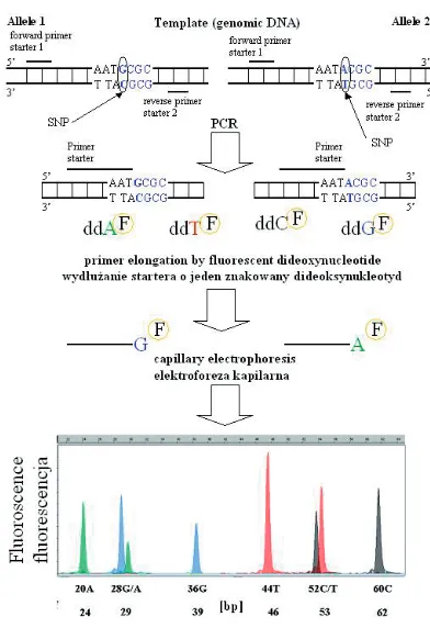
Minisequencing
Minisequencing is used for the detection of single−nucleotide polymorphisms or point muta− tions. The first step is the amplification of about 200 bp of target DNA using PCR. Afterwards, PCR product purification to remove primers and unincorporated dNTPs is necessary. There are three possible methods: 1) shrimp alkaline phos− phatase (SAP) and exonuclease I (ExoI) treatment, 2) PCR product cleaning on a minicolumn, 3) separation of the PCR product by agarose gel electrophoresis and elution from the gel using a minicolumn.
The minisequencing method is based on the incorporation of a single fluorescently labeled dideoxynucleotide at the 3’ end of a special oligonuclotide which is complementary to the sequence located one nucleotide before the exam− ined polymorphic site [36]. The incorporation of one of four ddNTPs labeled by differently colored dyes depends on the genotype. Furthermore, the products of the reaction are separated by capillary electrophoresis. The color of the one (for homozy− gote) or two (for heterozygote) signals obtained allows identifying the incorporated ddNTP and, furthermore, the complementary deoxynucleotide in the DNA sequence. If multi−length oligonu− cleotides are used, several different polymor− phisms/mutations in one multiplex reaction can be detected [36].
Pyrosequencing
Pyrosequencing is a technique which uses four different enzymes for reading short DNA sequences (at least 20 nucleotides) in real time. Six reactions with four enzymes take place in one tube: 1) the Klenow fragment of the DNA poly− merase catalyses the polymerization of the added nucleotide (one of four at a time) with the DNA template if it is complementary, 2) ATP sulfurylase uses as a substrate APS (adenosine 5’–phospho− sulfate) and an inorganic pyrophosphate (PPi) released by the polymerase, producing ATP, 3) and 4) luciferase uses ATP, D−luciferin, and an oxygen molecule (O2) to produce light in two reactions, and 5) and 6) between the addition of different nucleotides, apyrase is added to remove unincor− porated dNTPs and ATPs and therefore prevent light production in the next cycle, which starts without any additional dNTPs except for the one provided by the sequencer:
1) (DNA)n+ dNTP→(DNA)n+1+ PPi
2) PPi + APS →ATP + SO42− 3) luciferase + D−luciferin + ATP→
luciferase−luciferin−AMP + PPi 4) luciferase−luciferin−AMP + O2→
luciferase + oxyluciferin + AMP + CO2+ LIGHT
5) ATP→AMP + 2Pi 6) dNTP→dNMP + 2Pi
In this way, light is produced only when adding a certain nucleotide and incorporation occurs (Fig. 4) [34, 38].
Pyrosequencing is applied for the detection of known and unknown mutations and SNPs with a definite position as well as taq sequencing and microorganism typing. An unquestionable advan− tage of pyrosequencing is the possibility of automation, economical reagent use, and multiple probe sequencing (96 to 384) in one process [34]. It is particularly important in studies with a large number of DNA alterations and/or a large number of tested samples.
Since the second half of the 20th century, a variety of applications of the in vitropolymerase chain reaction have been discovered. The speci− ficity of this technique makes it one of the most important and frequently used in biomedical investigations and other biological analyses. There are many other techniques based on the poly− merase chain reaction that can be applied in labo− ratory practice apart from those described in this paper. The authors presented those methods that are the most interesting in their opinion and/or commonly used.
Fig. 3. SNaPshot. ddAF– fluorescent labeled
dideoxyadenosine triphosphate, ddTF– fluorescent
labeled dideoxythymidine triphosphate, ddCF– fluo−
rescent labeled dideoxycytidine triphosphate, ddGF– fluorescent labeled dideoxyguanosine triphos−
phate. Description in the text
Ryc. 3. SNaPshot. ddAF– znakowany fluorescen−
cyjnie trifosforan dideoksyadenozyny, ddTF– zna−
kowany fluorescencyjnie trifosforan dideoksytymi− dyny, ddCF– znakowany fluorescencyjnie trifosforan
dideoksycytydyny, ddGF– znakowany fluorescen−
cyjnie trifosforan dideoksyguanozyny. Opis w tekście
Fig. 4. Pyrosequencing. dCTP, dTTP, dATP – deoxynucleotide triphosphates, PPi – pyrophosphate, Pi – phosphate, APS – adenosine−5’−phosphosulfate, ATP – Adenosine triphosphate
Acknowledgements
The authors would like thank Prof. Maria M. Sąsiadek for the help with the manuscript.
References
[1] Saiki RK, Scharf S, Faloona F, Mullis KB, Horn GT, Erlich HA, Arnheim N:Enzymatic amplification of beta− globin genomic sequences and restriction site analysis for diagnosis of sickle cell anemia. Science 1985, 20, 230, 1350–1354.
[2] Sommer SS, Groszbach AR, Bottema CD:PCR amplification of specific alleles (PASA) is a general method for rapidly detecting known single−base changes. Biotechniques 1992, 12, 82–87.
[3] Okayama H, Curiel DT, Brantly ML, Holmes MD, Crystal RG:Rapid, nonradioactive detection of mutations in the human genome by allele−specific amplification. J Lab Clin Med 1989, 114, 105–113.
[4] Lubiński J, Górski B, Huzarski T, Byrski T, Gronwald J, Serrano−Fernández P, Domagała W, Chosia M, Uciński M, Grzybowska E, Lange D, Maka B, Mackiewicz A, Karczewska A, Bręborowicz J, Lamperska K, Stawicka M, Gozdecka−Grodecka S, Bębenek M, Sorokin D, Wojnar A, Haus O, Sir J, Mierzwa T, Niepsuj S, Gugała K, Góźdź S, Sygut J, Kozak−Klonowska B, Musiatowicz B, Posmyk M, Kordek R, Morawiec M, Zambrano O, Waśko B, Fudali L, Skret J, Surdyka D, Urbański K, Mituś J, Ryś J, Szwiec M, Rozmiarek A, Dziuba I, Wandzel P, Wiśniowski R, Szczylik C, Kozak A, Kozłowski W, Narod SA:BRCA1−positive breast cancers in young women from Poland. Breast Cancer Res Treat 2006, 99, 71–76.
[5] Grzybowska E, Siemińska M, Zientek H, Kalinowska E, Michalska J, Utracka−Hutka B, Rogozińska− −Szczepka J, Kaźmierczak−Maciejewska M:Germline mutations in the BRCA1gene predisposing to breast and ovarian cancers in Upper Silesia population. Acta Biochim Pol 2002, 49, 351–356.
[6] Janiszewska H, Haus O, Lauda−Swieciak A, Pasińska M, Laskowski R, Szymański W, Górski B, Lubiński J: Frequency of three BRCA1gene founder mutations in breast/ovarian cancer families from the Pomerania−Kujawy region of Poland. Clin Genet 2003, 64, 502–508.
[7] Chan PC, Wong BY, Ozcelik H, Cole DE:Simple and rapid detection of BRCA1and BRCA2mutations by mul− tiplex mutagenically separated PCR. Clin Chem 1999, 45, 1285–1287.
[8] Jacobson DR, Moskovits T:Rapid, nonradioactive screening for activating ras oncogene mutations using PCR− primer introduced restriction analysis (PCR−PIRA). PCR Methods Appl 1991, 1, 146–148.
[9] Balmer D, Arredondo J, Samaco RC, LaSalle JM:MECP2 mutations in Rett syndrome adversely affect lym− phocyte growth, but do not affect imprinted gene expression in blood or brain. Hum Genet 2002, 110, 545–552. [10] Matijevic T, Knezevic J, Slavica M, Pavelic J: Rett syndrome: from the gene to the disease. Eur Neurol 2009,
61, 3–10.
[11] Górski B, Cybulski C, Huzarski T et al.:Breast cancer predisposing alleles in Poland. Breast Cancer Res Treat 2005, 92, 19–24.
[12] Herman JG, Graff JR, Myöhänen S, Nelkin BD, Baylin SB:Methylation−specific PCR: a novel PCR assay for methylation status of CpG islands. Proc Natl Acad Sci U S A 1996, 93, 9821–9826.
[13] Gutierrez MI, Siraj AK, Bhargava M, Ozbek U, Banavali S, Chaudhary MA, El Solh H, Bhatia K: Concurrent methylation of multiple genes in childhood ALL: Correlation with phenotype and molecular subgroup. Leukemia 2003, 17, 1845–1850.
[14] Hayslip J, Montero A: Tumor suppressor gene methylation in follicular lymphoma: a comprehensive review. Mol Cancer 2006, 6, 44–45.
[15] Nicholls RD, Knoll JH, Butler MG, Karam S, Lalande M:Genetic imprinting suggested by maternal het− erodisomy in nondeletion Prader–Willi syndrome. Nature 1989, 16, 342, 281–285.
[16] Sutcliffe JS, Nakao M, Christian S, Orstavik KH, Tommerup N, Ledbetter DH, Beaudet AL:Deletions of a differentially methylated CpG island at the SNRPNgene define a putative imprinting control region. Nat Genet 1994, 8, 52–58.
[17] Gille JJ, Hogervorst FB, Pals G, Wijnen JT, van Schooten RJ, Dommering CJ, Meijer GA, Craanen ME, Nederlof PM, de Jong D, McElgunn CJ, Schouten JP, Menko FH:Genomic deletions of MSH2and MLH1in colorectal cancer families detected by a novel mutation detection approach. Br J Cancer 2002, 87, 892–897. [18] Hogervorst FB, Nederlof PM, Gille JJ, McElgunn CJ, Grippeling M, Pruntel R, Regnerus R, van Welsem
T, van Spaendonk R, Menko FH, Kluijt I, Dommering C, Verhoef S, Schouten JP, van’t Veer LJ, Pals G: Large genomic deletions and duplications in the BRCA1gene identified by a novel quantitative method. Cancer Res 2003, 63, 1449–1453.
[19] Belogianni I, Apessos A, Mihalatos M, Razi E, Labropoulos S, Petounis A, Gaki V, Keramopoulos A, Pandis N, Kyriacou K, Hadjisavvas A, Kosmidis P, Yannoukakos D, Nasioulas G:Characterization of a novel large dele− tion and single point mutations in the BRCA1gene in a Greek cohort of families with suspected hereditary breast cancer. BMC Cancer 2004, 4, 61.
[20] Nakagawa H, Hampel H, de la Chapelle A:Identification and characterization of genomic rearrangements of
MSH2and MLH1in Lynch syndrome (HNPCC) by novel techniques. Hum Mutat 2003, 22, 258.
[21] Nygren AO, Ameziane N, Duarte HM, Vijzelaar RN, Waisfisz Q, Hess CJ, Schouten JP, Errami A: Methylation−specific MLPA (MS−MLPA): simultaneous detection of CpG methylation and copy number changes of up to 40 sequences. Nucleic Acids Res 2005, 33, 128.
[23] Schouten JP, McElgunn CJ, Waaijer R, Zwijnenburg D, Diepvens F, Pals G:Relative quantification of 40 nucleic acid sequences by multiplex ligation−dependent probe amplification. Nucleic Acids Res 2002, 30, 57. [24] Higuchi R, Dollinger G, Walsh PS, Griffith R: Simultaneous amplification and detection of specific DNA
sequences. Biotechnology (N Y) 1992, 10, 413–417.
[25] Lee LG, Connell CR, Bloch W:Allelic discrimination by nick−translation PCR with fluorogenic probes. Nucleic Acids Res 1993, 21, 3761–3766.
[26] Livak KJ, Flood SJ, Marmaro J, Giusti W, Deetz K:Oligonucleotides with fluorescent dyes at opposite ends provide a quenched probe system useful for detecting PCR product and nucleic acid hybridization. PCR Methods Appl 1995, 4, 357–362.
[27] Valasek MA, Repa JJ:The power of real−time PCR. Adv Physiol Educ 2005, 29, 151–159.
[28] Morrison TB, Weis JJ, Wittwer CT:Quantification of low−copy transcripts by continuous SYBR Green I mon− itoring during amplification. Biotechniques 1998, 24, 954–958.
[29] Vandesompele J, De Paepe A, Speleman F:Elimination of primer−dimer artifacts and genomic coamplification using a two−step SYBR green I real−time RT−PCR. Anal Biochem 2002, 303, 95–98.
[30] Hiyoshi M, Hosoi S:Assay of DNA denaturation by polymerase chain reaction−driven fluorescent label incorpo− ration and fluorescence resonance energy transfer. Anal Biochem 1994, 221, 306–311.
[31] Chen Q, Lentz BR:Fluorescence resonance energy transfer study of shape changes in membrane−bound bovine prothrombin and meizothrombin. Biochemistry 1997, 36, 4701–4711.
[32] Lee WI, Kantarjian H, Glassman A, Talpaz M, Lee MS:Quantitative measurement of BCR/abltranscripts using real−time polymerase chain reaction. Ann Oncol 2002, 13, 781–788.
[33] Venter JC, Adams MD, Myers EW et al.: The sequence of the human genome. Science 2001, 291, 1304–1351. [34] Ahmadian A, Ehn M, Hober S: Pyrosequencing: history, biochemistry and future. Clin Chim Acta 2006, 363,
83–94.
[35] Shendure J, Mitra RD, Varma C, Church GM:Advanced sequencing technologies: methods and goals. Nat Rev Genet 2004, 5, 335–344.
[36] Pastinen T, Partanen J, Syvänen AC:Multiplex, fluorescent, solid−phase minisequencing for efficient screen− ing of DNA sequence variation. Clin Chem 1996, 42, 1391–1397.
[37] Fiorentino F, Magli MC, Podini D, Ferraretti AP, Nuccitelli A, Vitale N, Baldi M, Gianaroli L:The minise− quencing method: an alternative strategy for preimplantation genetic diagnosis of single gene disorders. Mol Hum Reprod 2003, 9, 399–410.
[38] Ronaghi M, Uhlén M, Nyrén P:A sequencing method based on real−time pyrophosphate. Science 1998, 281, 363–365.
Address for correspondence:
Izabela Łaczmańska Department of Genetics Wroclaw Medical University Marcinkowskiego 1
50−368 Wroclaw Poland
Tel. +48 71 784 12 56 E−mail: [email protected]
Conflict of interest: None declared